Discard the supernatants and re- suspend ... C. parvum oocyst wall antigens or any potential cross-re- activity. ..... Patty Reeh and Larry Mitchell, Water Program,.
Am. J. Trop. Med. Hyg., 65(1), 2001, pp. 1–9 Copyright 䉷 2001 by The American Society of Tropical Medicine and Hygiene
DEVELOPMENT AND APPLICATION OF A QUANTITATIVE, SPECIFIC ASSAY FOR CRYPTOSPORIDIUM PARVUM OOCYST DETECTION IN HIGH-TURBIDITY ENVIRONMENTAL WATER SAMPLES YEUK-MUI LEE, PATRICK W. JOHNSON, JEFFERY L. CALL, MICHAEL J. ARROWOOD, BRUCE W. FURNESS, SARAH C. PICHETTE, KATHERINE K. GRADY, PATTY REEH, LARRY MITCHELL, DAVID BERGMIRE-SWEAT, WILLIAM R. MACKENZIE, AND VICTOR C. W. TSANG Immunology and Epidemiology Branches, Division of Parasitic Diseases, National Center for Infectious Disease, Centers for Disease Control and Prevention, Atlanta, Georgia; Water Program, Texas Natural Resource Conservation Commission, Austin, Texas; Medical Parasitology Section, Texas Department of Health and Infectious Disease Epidemiology and Surveillance Division, Texas Department of Health, Austin, Texas
Abstract. Chlorine-resistant Cryptosporidium parvum oocysts in drinking water play an important role in the epidemiology of cryptosporidiosis. Current methods of detecting these organisms in water are insensitive, laborintensive, highly subjective, and severely limited by sample turbidity. We describe here an alternative technique utilizing electrochemiluminescence (ECL) technology for detecting C. parvum oocysts in environmental water samples. This method is quantitative, reproducible, and requires only minimal sample processing. Currently, the ECL assay can detect as few as one oocyst in one milliliter of concentrated test sample with sample turbidity of up to 10,000 nephelometric turbidity units. Water and sewer samples collected during a cryptosporidiosis outbreak were tested by ECL assay. Cryptosporidium parvum oocysts were found in the source water at the time of outbreak, and a sharply decreasing level of oocysts in sewer samples was observed over a three-month period following the outbreak. situ hydridization-confocal laser scanning microscopy,21,22 polymerase chain reaction,23–27 immunomagnetic separation,28 and laser scanning with video microscopy.29 Although most of these assays are adequate for analysis of oocysts in low-turbidity water samples, they might not be satisfactory for analysis of oocysts in samples with high turbidity. In this communication, we describe the development of an electrochemiluminescence (ECL) assay for detecting and enumerating C. parvum oocysts in environmental water samples. The ECL offers high signal-to-background noise ratios and has the advantage over other chemiluminescence techniques of being initiated by voltage potential, thus providing better controlled luminescence.30 Our assay uses solubilized oocyst wall antigens as target analyte, allowing detection of antigens from oocysts in highly turbid samples. The application of the ECL assay in investigating a cryptosporidiosis outbreak is also documented.
INTRODUCTION
Cryptosporidium parvum is a protozoan parasite that causes gastrointestinal illness in humans. For immunocompromised individuals, these infections can lead to chronic or life-threatening diarrhea.1–3 Cryptosporidium oocysts are often present in raw and treated drinking water, even when the drinking water meets United States Environmental Protection Agency (EPA) treatment standards.4–6 In fact, several epidemics of cryptosporidiosis in industrialized countries have been linked to contaminated drinking water; the most notable of these occurred in 1993 in Milwaukee7 and caused an estimated 400,000 cases of gastrointestinal illness. Cryptosporidium oocysts are resistant to routine water treatments and chemical disinfectants,8 and low numbers of viable oocysts can cause infection.9–11 The EPA developed a comprehensive nationwide monitoring program termed the Information Collection Rule (ICR) in 1996,12 which requires routine monitoring for these protozoa, other infectious agents, and contaminants. The current method endorsed by the American Society for Testing and Materials (ASTM) for detecting Cryptosporidium oocysts in water supplies is laborintensive and time-consuming. This method involves multiple steps, including filtration and centrifugation to concentrate and purify oocysts, followed by immunofluorescence microscopy to identify and enumerate the oocysts present in test samples.13 The limitations of this procedure include nonspecific antibody binding, poor oocyst recovery, and long assay times.14 Microscopy also seriously limits the volume of the sample to be assayed to approximately 50 l. Difficulties in detecting and enumerating oocysts in environmental samples are further compounded by the composition and quantity of sediment presents in these samples. Various alternative technologies have been investigated in efforts to provide more effective detection of waterborne Cryptosporidium.15 These include flow cytometry,16 ultraviolet-visible spectroscopy,17 enzyme-linked immunosorbent assays,18,19 use of charge-coupled devices,20 fluorescence in
MATERIALS AND METHODS
Sources and purification of oocysts. Cryptosporidium parvum oocysts of Iowa bovine origin and OW3 monoclonal antibodies (MAbs) specific for C. parvum oocyst wall antigens were produced as previously described.31–33 Cryptosporidium muris and C. serpentis were obtained from Lihua Xiao (Centers for Disease Control and Prevention [CDC]). Cryptosporidium baileyi oocysts were kindly supplied by Byron Blagburn (Auburn University, Auburn, AL). Giardia duodenalis, Eimeria nieschulzi, and Eimeria spp. were a generous gift from Ron Fayer (United States Department of Agriculture, Washington, DC). Oocysts were supplied as purified preparations stored at 4⬚C either in antibiotic solution (1,000 U/ml of Penicillin and 1,000 micrograms/ml streptomycin) or 2.5% potassium dichromate. Oocysts and organisms used in this study were less than six months old. Various algal cultures were obtained from Marshall Darley (University of Georgia, Athens, GA). The concentration of the oocyst stock solution and algal cultures was determined
1
2
LEE AND OTHERS
by hemacytometer counting using an Olympus BX50 microscope (Olympus Optical Co. Ltd., Japan) with a 40H/1.3 plan-neofluor objective and 10⫻ eyepieces. Briefly, two lower concentrations of the oocyst stock solution were prepared by dilution, and three hemacytometer numerations were performed on each diluted stock solution. The final concentration of the stock solution was calculated from the average concentration of the six numerations. The coefficient of variation of these six numerations was within 10%. For experiments using spiked oocyst samples, serial 10-fold dilutions of the stock solutions were made to prepare the 100⫻ seeded oocyst solutions (102/ml to 106/ml). Conjugation of OW3 MAb to ruthenium. The OW3 MAb (IgM class) derived from hybridoma OW3 was one of the first MAbs developed specifically for a C. parvum oocyst wall surface determinant,31,33 and it was used in a commercial immunofluorescence assay (IFA) for detection of C. parvum.34 This antibody was selected over others because of its specificity for C. parvum oocysts and for its availability. The OW3 MAb was purified from hybridoma cell supernatant by precipitation with 45% ammonium sulfate followed by size exclusion chromatography using Pharmacia (Piscataway, NJ) Superose-6 media. N-Hydroxy-succinimide-Ru(bpy)32⫹ ester (TAG-NHS) (Igen Corp., Gaithersburg, MD) was dissolved in dimethylsulfoxide for 15 min at a concentration of 1.42 ⫻ 10⫺4 mM. Purified OW3 MAb in 0.01 M NaPO4 was combined with dissolved TAG-NHS at a 1:34 antibody to ruthenium molar reaction ratio and incubated in a glass reaction vial at room temperature for 60 min in the dark on an orbital shaker. Following coupling, unreacted active groups on TAG were capped by incubating with 0.2 M TrisHCl for 15 min. Unbound TAG was removed by desalting using a Pharmacia FAST-desalting column pre-equilibrated with 0.05 M Tris-HCl, 0.5 M NaCl, pH 8.0. A final Ru2⫹-to-antibody-molar incorporation ratio was determined by measuring the protein concentration of the conjugate and its absorbance at 455 nm and then calculated according to reagent protocol. The molar-incorporationratio for Ru(bpy)32⫹-labeled OW3 antibody was 14.4 labels per antibody. All protein concentrations were determined by Bradford protein assay (Coomassie blue, Bio-Rad Corp., Hercules, CA) against a series of standard protein values (Sigma Chemical Co., St. Louis, MO). A working concentration of 10⫺4 mg/ml of Ru2⫹-MAb conjugate in ECL diluent (0.05 M Tris-HCl, 0.5 M NaCl, 1% bovine serum albumin (BSA), 0.7% Tween 20, pH 8.0) was used for each ECL assay. Preparation of sediment samples and oocyst-spiked samples. Clay-rich soil, three feet below the ground surface in an area undisturbed by urban development for more than 50 years, was obtained to be used as a reference sample. This sample originated in the watershed impacting the Chattahoochee River basin, Georgia. When oocysts were spiked into environmental samples especially with high clay content, low recoveries of C. parvum oocysts were observed. Therefore, clay-rich soil was selected as reference sediment sample to optimize the ECL assay to achieve maximum oocyst recovery when oocysts were seeded. The reference sediment sample was prepared in 0.05 M Tris-HCl containing 0.05% NaN3, pH 8.0. The nephelometric turbidity units (NTU) were measured using a Turbidimeter (Model # 11520;
Industrial Chemical Measurement, Hillsboro, OR). Reference sediment samples with wide ranges of turbidity were prepared either by concentration or dilution. Other environmental sediment samples were obtained from lake, reservoir, and river water sites and processed as follows. Briefly, a large volume of a water sample was filtered through a 1.0m-nominal-pore-size polypropylene yarn-wound cartridge filter (model V1A10U; Filterite Corp., Timmonium, MD). Retained oocysts and debris were eluted from the filter with an eluting solution (phosphate-buffered saline containing 0.1% sodium dodecyl sulfate [SDS], 0.1% Tween 80, and 0.01% Antifoam A), and concentrated by centrifugation at 1,050 ⫻ g for 10 min. The pellet volume was estimated, resuspended in an equal volume of 2.5% potassium dichromate, and stored at 4⬚C. After being collected and processed, all sediment samples were further analyzed for Cryptosporidium using the ICR method35 based on the ASTM method.13 Archived environmental sediment samples containing no detectable oocysts were used in the study. Environmental sediment samples were prepared by mixing one part of the concentrated samples with 10 parts of 0.05 M Tris-HCl, pH 8.0. The samples were centrifuged at 1,050 ⫻ g for 15 min, and the supernatant was discarded. Pellets were resuspended in 100 parts of 0.05 M Tris-HCl, pH 8.0 and the turbidity of samples was measured using the Turbidimeter. Samples with an equivalent turbidity of 10,000 NTU were prepared by concentration of the low-turbidity samples. For oocystspiked samples containing 1–10,000 oocysts, 10 l of various 100⫻ seeded solutions (102/ml to 106/ml) were added to the sediment samples and allowed to stand at room temperature for 1 hr before processing for antigen solubilization. Solubilization of oocyst wall antigens. Zwittergent 3-14 (Calbiochem-Novabiochem International, La Jolla, CA) was used to solubilize the oocyst wall antigens, and gelatin (teleostean gelatin, Sigma Chemical Co.) was used to enhance oocyst recovery. Specific epitopes of the oocyst wall antigen targeted by MAb OW3 were solubilized and detected by the ECL assay. To solubilize the C. parvum oocysts in environmental samples, 1.0-ml test samples were placed in 1.7-ml siliconized tubes, and centrifuged at 21,000 ⫻ g for 5 min at 4⬚C. The supernatants were removed and the pellets were resuspended in 1.0 ml of solubilization buffer (SB: 0.02% Zwittergent 3-14, 1.0% [v/v] gelatin, 0.05 M Tris-HCl, pH 8.0). The tubes were then capped and placed in a boiling water bath for 60 min. After treatment, samples were centrifuged at 21,000 ⫻ g for 10 min at 4⬚C, and the solubilized antigen (SA) supernatants were removed for ECL assay. Electrochemiluminescence immunoassay. Since the composition and turbidity of the tested environmental samples could not be pre-determined, an IgM assay was used in addition to the OW3 assay to determine the background of individual test samples. For magnetic beads used in the OW3 assay, M450 rat antimouse IgM magnetic beads obtained from Dynal, Inc. (Oslo, Norway) were incubated at a concentration of 107 beads/ml (0.075 mg/ml) with 0.01 mg/ml of purified OW3 antibody. For the IgM assay, nonspecific beads were prepared by incubating M450 rat anti-mouse IgM magnetic beads with IgM antibodies from a mouse myeloma (Calbiochem-Novabiochem International) at the concentration mentioned earlier.
QUANTITATIVE ASSAY FOR DETECTION OF C. PARVUM OOCYSTS
Incubations were performed in disposable 12 ⫻ 75-mm borosilicate glass culture tubes on an orbital shaker (⬃ 800 rpm) for 60 min at room temperature. Immunomagnetic separation (IMS) was carried out by placing the bead solutions onto a magnetic rack (MPC-6; Dynal, Inc.) for 2 min, the magnetic beads were concentrated and the solutions containing unbound MAb were removed. Magnetic beads were washed twice with 2 ml of ECL diluent, and resuspended in ECL diluent to a working concentration of 2.5 ⫻ 106 beads/ ml (0.018 mg/ml). Positive controls were prepared as 100⫻ stock solutions containing 100 to 1,000,000 oocysts in SB. The negative control contained SB only. All controls were processed following the solubilization procedure. In the ECL assay, 10 l of the solubilized concentrated positive or negative stock solution was diluted to 1,000 l with ECL diluent to make up the required positive (1 to 10,000 oocysts) and negative controls. To determine the presence of oocysts in test samples, the OW3 assay was carried out by incubating 900 l of SA with 100 l of OW3 beads in a disposable 12 ⫻ 75-mm borosilicate glass culture tube on an orbital shaker for 2 hr at room temperature. All samples were assayed in duplicate. Negative and positive controls with known numbers of oocysts (1–104) were assayed in the same manner. After incubation, IMS was used to isolate target antigens and remove unbound materials. Three hundred microliters of Ru2⫹-labeled antibodies were added to each tube, the samples were returned to the orbital shaker for 30 min, and then assayed according to instrument specifications using an Origen analyzer (Igen Corp.). For the IgM assay to determine the background of test samples, SA obtained from test samples were assayed in the same manner as the OW3 assay except that SA was incubated with the IgM beads. To enumerate the number of C. parvum oocysts in test samples, net ECL counts were calculated by subtracting the ECL counts obtained in the IgM assay from the ECL counts obtained in the OW3 assay. A standard curve was constructed by using the net ECL counts obtained from positive buffer samples, and the number of oocysts in the test samples was calculated from the curve. The protocol for the ECL assay is shown in Table 1. Specificity of the ECL assay. To determine the specificity of the ECL assay in detecting and enumerating C. parvum oocysts, organisms and oocysts from various Cryptosporidium species, and other protozoan parasites that may be present in environmental water samples were examined. The organisms tested included Cryptosporidium parvum, Cryptosporidium baileyi, Cryptosporidium muris, Cryptosporidium serpentis, Giardia duodenalis, Eimeria nieschulzi, and Eimeria spp. Various algal cultures and Cyclospora spp. were also tested by the ECL assay. Oocysts or other organisms were seeded into reference sediment samples with an equivalent turbidity of 10,000 NTU, and the samples were then solubilized. Nine-hundred microliters of SA containing one to 10,000 organisms were assayed for the presence of C. parvum oocyst wall antigens or any potential cross-reactivity. Examination of environmental samples related to epidemiologic investigations. Ten liters of water or sewage was collected for each environmental sample, aliquotted into cen-
3
TABLE 1 Protocol for electrochemiluminescence (ECL) detection of Cryptosporidium parvum oocysts in environmental samples 1. Divide test samples into two 1.0-ml aliquots and centrifuge samples at 21,000 ⫻ g for 2 min. Discard the supernatants and resuspend the pellets in 1.0 ml of solubilization buffer. 2. Solubilize samples in a boiling water bath for 1 hr. Centrifuge the test samples at 21,000 ⫻ g for 10 min. Incubate 900 l of solubilized antigen supernatant from one of the aliquot test samples with 100 l of OW3 beads. Another 900 l of solubilized antigen supernatant from another aliquot is incubated with IgM beads. A batch of standard positive and negative controls is assayed in the same manner. 3. Place the beads-solution on a shaker at room temperature for 2 hr. Following incubation, immunomagnetic separation is carried out to remove unbound antigen, 100 l of conjugated antibodies and 200 l ECL of diluent are added to the beads, and samples are incubated at room temperature for 30 min with shaking. 4. Samples are then placed on the Igen analyzer and assayed. The net ECL of the test sample is determined by subtracting the ECL count obtained from the IgM assay from the ECL count obtained from the OW3 assay. 5. A standard curve is constructed using the net ECL counts obtained from positive buffer samples. The number of oocysts present in the test samples is calculated from the curve.
trifuge bottles that were precoated with 1% BSA, and concentrated by centrifugation at 25,000 ⫻ g for 10 min. The pooled pellet was resuspended in 0.05M Tris buffer to a final volume of 10 ml. For sewage samples, the final volume of concentrated samples was 100 ml. The concentrated samples were then assayed for the presence of C. parvum oocysts. RESULTS
Because of the difficulties in obtaining large volumes of environmental samples with consistent composition, reference sediment samples were used to optimize the solubilization procedure and configuration of ECL assays. Optimization of the solubilization procedure. Cryptosporidium parvum oocyst walls are notoriously resistant to various types of chemical disinfectants.8 Detergents (Zwittergent, Tween 20, Triton X-100, NP 40, and SDS) and denaturing reagents (urea and guanidine hydrochloride) were tested for their abilities to solubilize the oocyst wall antigens. Signal-to-background-noise ratio (S:N ratio) was obtained by dividing the net ECL counts generated from oocystspiked samples by the net ECL counts generated from oocyst-free samples. All members of the Zwittergent family efficiently solubilized the antigens specifically recognized by OW3 MAb. Among the members of the Zwittergent family tested in our assay, Zwittergent 3-14 was the preferred detergent based on its higher S:N ratio. The optimal concentration of this detergent was determined by solubilizing and assaying 10,000 oocysts in SB containing various concentrations (0–5%) of Zwittergent 3-14. Since the highest S:N ratio was obtained from the sample treated with 0.02% Zwittergent 3-14, this concentration was considered optimal for oocyst solubilization (Figure 1A). Reagents used in the SB were evaluated for their ability to enhance solubilization of target antigens. Tris-HCl (0.05 M) was used as a buffering agent in all comparisons except
4
LEE AND OTHERS
FIGURE 2. Determination of the limit of sample turbidity assayed in electrochemiluminescence assays. Ten thousand oocysts were seeded in control buffer samples (⬍ 0.5 nephelometric turbidity units [NTU]) and reference sediment samples at various turbidities (650– 60,000 NTU). The samples were solubilized and assayed for oocyst signal. Error bars indicate standard deviations.
FIGURE 1. Optimization of the solubilization procedure. Ten thousand oocysts were incubated in solubilization buffer with selected parameters for 1 hr, and the degree of solubility of oocyst antigen was determined by the electrochemiluminescence (ECL) assay. A, titration of Zwittergent 3-14 detergent. B, effect of 1% teleostean gelatin on oocyst signal recovery in reference sediment samples with a turbidity of 10,000 nephelometric turbidity units. Error bars indicate standard deviations. Ref. ⫽ reference.
that of pH. Parameters included in the assessment of solubilization were ionic strength (0–500 mM NaCl), pH (3.0– 11.0), and temperature (25–100⬚C). Briefly, known numbers of oocysts were incubated in SB with selected reagents for 1 hr, and the degree of solubility of oocyst antigens was determined by ECL assay as described earlier. The highest S:N ratio for 10,000 oocysts was generated from samples solubilized in SB containing 0 mM NaCl. Unlike other amphoteric surfactants, Zwittergent detergents retain their zwitterionic character over a broad pH range because of the presence of both a strongly basic quaternary ammonium ion and an acidic sulfonate ion of equal strength. Thus, the concentration of exogenous ions (Na⫹, Cl⫺) in SB had no profound effect on detergent performance (data not shown). To optimize pH during solubilization, stock buffers containing 1.0 M N-2-hydroxyethylpiperazine-N-2-ethanesulfonic acid (HEPES), 1.0 M boric acid, and 1.0 M Na2HPO4 were titrated to specified pH. Buffers (0.05 M) were prepared from stock buffers and used as base buffers for the Zwittergent 3-14 detergent. The pH stability of these SB was then verified. Results indicated that oocyst wall antigens were solubilized most efficiently in SB at pH 8.0. The effect of temperature on oocyst solubilization was also evaluated. An eight-fold increase in S:N ratios was observed at 100⬚C
compared with 25⬚C, indicating that high temperature facilitates the solubilization process. Additional studies demonstrated that oocysts stored in SB for four days at room temperature released negligible amounts of solubilized antigen (data not shown). Recovery of oocyst signals by gelatin. Previous studies showed that blocking reagents are required to prevent attachment of C. parvum oocysts to the inner surface of containers. We observed that the recovery of spiked oocysts was further compounded by nonspecific interactions between oocysts and sediment in reference sediment samples and environmental samples that were previously confirmed negative for C. parvum oocysts by IFA. Since our solubilization procedure includes a 60-min incubation at 100⬚C, most of the blocking reagents (e.g., BSA, casein, dry milk, ovalbumin) were denatured by this procedure, resulting in inhibition of oocyst detection in the ECL assay. As shown in Figure 1B, gelatin restored oocyst signals most efficiently in spiked reference sediment samples with an equivalent turbidity of 10,000 NTU. To determine the upper limit of sample turbidity that could be analyzed with the ECL assay, 10,000 oocysts were seeded into control buffer samples (⬍ 0.5 NTU) and reference sediment samples at various turbidities (650–60,000 NTU). The samples were then solubilized and assayed for oocyst signal. The detection rate was calculated as follows: assuming the seeded oocysts were fully recovered in buffer control samples, the oocyst signal obtained in tested samples was divided by the signal obtained from the buffer control and multiplied by 100%. Figure 2 shows that with a concentration of 1.0% (v/v) gelatin in SB, ⱖ 93% of the oocyst signal was obtained from reference sediment samples with turbidities ⱕ 10,000 NTU. Sensitivity of the ECL assay. Ten independent experiments were carried out to determine the sensitivity of the ECL assay. Buffer control and reference sediment samples with an equivalent of 10,000 NTU were seeded with 1– 10,000 C. parvum oocysts. Oocyst-free buffer control and reference sediment samples were also prepared. The samples were solubilized and assayed according to the ECL protocol. The sensitivity data are presented in Figure 3. The results
5
QUANTITATIVE ASSAY FOR DETECTION OF C. PARVUM OOCYSTS
TABLE 2 Specificity of the electrochemiluminescence (ECL) assay Specimens
FIGURE 3. Sensitivity of the electrochemiluminescence (ECL) assay. Cryptosporidium parvum oocysts were seeded in buffer control and reference sediment samples. Samples were treated with solublilization procedure and analyzed by the ECL assay. The results shown were calculated from 10 independent experiments. Error bars indicate standard deviations.
were calculated from the 10 independent experiments and the standard deviations of corresponding samples were also determined. Good correlations were noted between oocyst titration curves generated from sediment samples and buffers, and from sediment samples obtained from 10 independent studies. Correlation coefficients of the individual titration curves constructed from oocyst-spiked buffer and oocyst-spiked reference sediment samples were 0.992, 0.992, 0.990, 0.989, 0.993, 0.990, 0.991, 0.986, 0.987, 0.988, 0.991, 0.991, 0.993, 0.990, 0.992, 0.988, 0.990, 0.987, 0.990, 0.990, and 0.990. These data indicate that one oocyst can be readily detected by the ECL assay. Specificity of the ECL assay. To determine the specificity of the ECL assay for the detection of C. parvum oocysts, oocysts of various Cryptosporidium species and other protozoan parasites that may be present in environmental water samples were tested. The organisms tested included C. parvum, C. baileyi, C. muris, C. serpentis, G. duodenalis, E. nieschulzi, Eimeria spp., Cyclospora spp., and various algal cultures. Various number of organisms (1–10,000) were seeded into reference sediment samples with an equivalent turbidity of 10,000 NTU, and the samples were solubilized and assayed for the presence of C. parvum oocyst wall antigens. The results of these experiments are shown in Table 2. None of the tested organisms generated a net ECL count higher than those generated from one C. parvum oocyst, indicating that the ECL assay was highly specific for C. parvum oocysts. Additional analyses will be carried out to determine any cross-reactivity present with other Cryptosporidium spp. and other organisms. Use of the ECL assay with environmental samples. Environmental water samples obtained from lake, reservoir, and river water were used to evaluate the ECL assay. The samples were concentrated to a turbidity of 10,000 NTU. Various numbers of oocysts (0–10,000) were titrated into each of these samples in 1.0-ml aliquots, and samples were solubilized and assayed for the presence of oocyst wall antigens. Because of the limited volume of some of the environmental samples, 10 and 1,000 oocysts were titrated into six
ECL*
Protozoa Cryptosporidium parvum Cryptosporidium baileyi Cryptosporidium muris Cryptosporidium serpentis Cyanobacter anabaena Cyclospora spp. Eimeria nieschulzi Eimeria spp. Giardia duodenalis
⫹ ⫺ ⫺ ⫺ ⫺ ⫺ ⫺ ⫺ ⫺
Algae Bacillariphyceae asterionella Bacillariphyceae nitzschia Chlorophyta chaetophora Chlorophyta chelamydomanas Chlorophyta chlorococcum Chlorophyta closterium Chlorophyta euastrum Chlorophyta klebsormidium Chlorophyta pandorina Chlorophyta pediastrum Chlorophyta scenedesmus Glaucocystophyta cyanophora Xanthophyceae tribonema
⫺ ⫺ ⫺ ⫺ ⫺ ⫺ ⫺ ⫺ ⫺ ⫺ ⫺ ⫺ ⫺
* 10,000 organisms were tested in each ECL assay.
of 10 samples. Both the OW3 and IgM assays were performed on these samples. The net ECL counts of these samples and the buffer controls are shown in Figure 4A. The net ECL counts obtained from the buffer controls and tested samples ranged from 95.5 to 130 in samples spiked with 10 oocysts, and from 1,004 to 1,109 in samples spiked with 1,000 oocysts. Assuming that the seeded oocysts were fully recovered in buffer control samples, an oocyst signal detection rate ⱖ 93% was obtained in all tested samples when compared with the oocyst signals generated from buffer control samples. Various numbers of oocysts (1–10,000) were titrated into each of four remaining environmental samples (reference #145, #150, #225, and #226) and the buffer control. Both the OW3 and IgM assays were carried out. As shown in Figure 4B, a good correlation was observed between the oocyst titration curves generated by the buffer control and four tested samples. The correlation coefficients of these samples were 0.965, 0.972, 0.969, and 0.967, respectively. Examination of the Brushy Creek outbreak samples. A lightning strike during a thunderstorm on July 13, 1998 damaged the controls of a sewage lift station near the Brushy Creek Municipal Utility District (MUD) in Texas. Approximately 167,000 gallons of raw sewage flowed into Brushy Creek through underground fissures in the creek bed and into the aquifer near five municipal utility district wells that served drinking water to 60% of the unincorporated community (approximately 6,000 residents). Chlorinated finished water from the MUD wells tested prior to distribution remained consistently negative for fecal coliforms. However, on July 21, 1998, raw water samples tested by the Texas Natural Resource Conservation Commission (TNRCC) were reported positive for Escherichia coli in four of five MUD wells. All five wells were then shut down. From July 16
6
LEE AND OTHERS
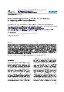
FIGURE 5. Detection of Cryptosporidium parvum oocysts in source water samples collected from the epidemic area during and after the cryptosporidiosis outbreak in the Brushy Creek Municipal Utility District (MUD) in Texas. Ten-liter samples were concentrated by centrifugation and assayed by the electrochemiluminescence assay.
FIGURE 4. Comparison of oocyst recovery in seeded environmental water samples with a turbidity of 10,000 nephelometric turbidity units. Samples were tested by the OW3 and IgM assays. Results are shown as net electrochemiluminescence counts. A, samples were seeded with either 10 or 1,000 oocysts. B, titration of oocysts (1–10,000) in various environmental samples and buffer controls. Error bars indicate standard deviations. Ref. ⫽ reference.
through August 1, epidemiologic studies conducted by the Texas Department of Health (TDH) estimated that approximately 1,300–1,500 residents became ill, and C. parvum oocysts were found by ova and parasite examinations in 89 stool specimens from individuals seeking medical attention.36 Environmental samples from the epidemic area were collected by the TNRCC, and the ECL assay was used to determine the presence of C. parvum oocysts. On August 5, 10 liters of water were collected from each of four MUD wells (#2–#5) that previously tested positive for E. coli, and additional samples were collected on August 25 and October 26. Each of the 10-liter samples was concentrated by multiple centrifugations, and pellets were pooled and resuspended in 0.05 M Tris buffer to a final volume of 10 ml. Cryptosporidium parvum oocysts were detected by the ECL assay and reported as the number of oocysts in one liter of test sample. Oocysts were found in water samples collected from the presumed contaminated wells three weeks after the sewage spill; well #2 and well #4 had 160 and 115 C. parvum oocysts, respectively, while 15 and four oocysts were detected in well #3 and well #5, respectively. In the August 25 sampling, two and three oocysts were found in samples obtained from well #2 and well #3, respectively. No oocysts
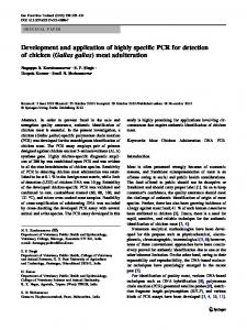
were detected in any of samples collected on October 26 (Figure 5). Sewage samples were also collected from the primary sewer collection points in the Brushy Creek MUD. Five 10liter samples were collected from August 5 to October 28, 1998. Samples were processed and assayed for C. parvum oocysts in a similar manner as described for water samples except that final volume of each concentrated sample was 100 ml. Cryptosporidium parvum oocysts were reported as the number in one liter of test sample. More than 300,000 C. parvum oocysts were detected in the first sampling on August 5. A significant decrease in oocyst number was observed in the subsequent samples: 71,229 oocysts, 9,903 oocysts, and 2,225 oocysts were found in the sewage samples collected on September 25, October 6, and October 15, respectively. In the final sampling on October 28, 7,122 oocysts were detected, as shown in Figure 6. The data generated from these environmental samples provide evidence that C. parvum oocysts were present in well water during the outbreak, and that the number of oocysts decreased in the three-month period following the outbreak.
FIGURE 6. Detection of Cryptosporidium parvum oocysts in raw sewage collected from the Brushy Creek Municipal Utility District in Texas from August 5 to October 28, 1998. Ten-liter samples were concentrated by centrifugation and assayed by the electrochemiluminescence assay.
QUANTITATIVE ASSAY FOR DETECTION OF C. PARVUM OOCYSTS
A similar trend was also found in sewage samples. Although the volume of sewage sampled was small and the representativeness of each sewage sample is unknown, the sample collected on August 5 appeared to reflect the severity of the outbreak in the Brushy Creek MUD community. An average of 6,417 oocysts per liter of sample was obtained from the three samplings collected between October 6 and October 28, and this might represent the endemic level of C. parvum oocysts in primary sewage in Brushy Creek. Therefore, a large number of individuals may have been infected with C. parvum and shed oocysts to constitute a 48-fold increase in oocysts level in the sewage during the outbreak. DISCUSSION
In recent years, there has been a dramatic increase in the incidence of waterborne disease outbreaks caused by the protozoan parasite C. parvum.7,37 Regulatory agencies are concerned that source and finished water is screened for these organisms, but a major obstacle is the lack of reliable methodologies and baseline information on oocyst prevalence in various water sources. A reliable, reproducible, simple detection method with a short assay time to enumerate viable/infectious oocysts would be the most ideal tool to prevent drinking water-related cryptosprodiosis. Until such a method is developed, a detection assay for the entire oocyst with enhanced sensitivity and simplicity is needed. Because of the low levels of C. parvum in water sources, the main objective of our study was to develop a detection assay that could enumerate oocysts in concentrated water samples of potentially high turbidity. In our initial studies, immunomagnetic beads coupled with OW3 MAb were used to capture intact C. parvum oocysts in test samples. We could enumerate oocysts present in spiked buffer samples (from 1 to 100,000 oocysts per sample), but experienced difficulties in detecting oocysts in environmental samples with turbidities greater than 5,000 NTU. This was presumably due to a nonspecific interaction between oocysts and the sediment present in test samples, and the disruption of antigen-antibody binding by the sediment. Therefore, solubilized C. parvum antigens became an attractive alternative for oocyst detection, since immunomagnetic capture of target analytes is more probable than capture of whole organism in complex samples. Data generated in this study demonstrated the advantages of this assay over direct microscopic analysis for the detection of C. parvum oocysts in environmental samples. For IFA testing with the ICR rule, in which a Percoll-sucrose gradient centrifugation procedure is required to remove contaminating debris prior to microscopic examination, a significant loss of oocysts occurs during this procedure and a considerable amount of time is required for sample examination. Our assay used Zwittergent 3-14 detergent to solubilize oocyst wall antigens, allowing detection of C. parvum oocysts without tedious sample preparation. The composition and quantity of sediment present in concentrated water samples play an important role in the recovery of C. parvum oocyst. In the ECL assay, recovery of oocysts seeded into environmental samples was enhanced by including gelatin in the SB. This addition facilitated recovery of at least 93% of the oocyst signals in the available test samples with an
7
equivalent turbidity up to 10,000 NTU compared with the signals obtained from buffer controls with turbidities less than 0.5 NTU. The actual mechanism of interaction between gelatin and sediment samples is not well defined, but it is effective in this assay. Bovine serum albumin and other nonspecific blocking reagents failed to restore oocyst antigen signal recovery when included in a solubilization procedure that did not require a high temperature. Further studies are planned to obtain a better understanding of this interaction as it relates to the physico-chemical properties of sediments. Such information may lead to even greater recoveries of oocyst signals in high turbidity samples (NTU ⬎ 10,000). From previous studies and outbreak data, the number of C. parvum oocysts present in environmental water samples is known to be sporadic and extremely low.7,38 Concentration of a large sample volume is typically required prior to oocyst detection, which in turn can lead to decreased reliability of current methods for detection of C. parvum oocysts. Our assay can detect oocysts in samples with high turbidity. Thus, concentrated samples from a large volume of water can be assayed without a significant loss of oocyst signals. With this tool, efforts will be directed to evaluate the efficiencies of available filtration systems for oocyst recovery. A filtration system that is capable of filtering a large volume of environmental water (⬎ 100 liters for source water or ⬎ 1,000 liters for finished water) with high oocyst recovery will be a valuable addition to the ECL assay and will increase the sensitivity of detection of C. parvum. The ECL assay was used to detect C. parvum oocysts in various environmental samples collected from several epidemiologic investigations in the past year. In the 1998 Brushy Creek cryptosporidiosis outbreak,36 C. parvum oocysts were detected in all well water samples collected three weeks after a sewage spill led to contamination of the wells. A significant decrease in oocyst numbers was observed in the samples collected at later time points. Concentrated water samples were prepared by centrifugation to minimize oocyst loss during sample preparation, thus allowing oocyst enumeration by ECL assay in highly turbid samples. Our assay detected as few as two oocysts in a one-liter well water sample collected six weeks after the spill, indicating that the assay is highly sensitive and capable of enumerating low numbers of oocysts in test samples. In addition, C. parvum oocysts in primary sewage can also be detected by the ECL assay. In the Brushy Creek cryptosporidiosis outbreak, a 48-fold increase in oocyst level in the primary sewage was observed three weeks after the spill, which casts light on the impact of the outbreak on the community. Waste-water treatment plants routinely release treated water back into rivers and other water sources, and communities located downstream may draw their drinking water from these sites. The ECL assay may be useful in monitoring the effectiveness of waste-water treatment on C. parvum oocysts to minimize potential source water contamination. Therefore, an assay that can enumerate oocysts in sewage may play an important role in reducing the risk of waterborne cryptosporidiosis. Water treatment plant operators use surrogate markers such as turbidity and particle counting to evaluate their plants’ ability to remove C. parvum oocysts from source
8
LEE AND OTHERS
water. Unfortunately, there is no consistent correlation between the turbidity of water and the number of C. parvum oocysts.4,39 The ECL assay can be used to assess plant efficiency on water treatment by measuring C. parvum concentrations in source and finished water and at intermediate points in the treatment process. Additionally, this assay makes it possible to evaluate the strengths and limitations of surrogate markers, such as turbidity, in detecting C. parvum oocysts. We are presently optimizing the ECL assay in combination with a filter/concentration system for point-of-use analyses by water treatment facilities. To date, a guideline for water quality regarding C. parvum oocysts has not been established, and the numbers of oocysts in drinking water required to develop disease in healthy individuals has not been conclusively defined. Difficulties in determining the prevalence of cryptosporidiosis in communities are compounded by the following: medical attention is infrequently sought by persons with diarrhea, physicians infrequently order stool testing for patients with diarrhea, the vast majority of laboratories do not routinely test stools for Cryptosporidium, and the sensitivity of available detection assays for Cryptosporidium in stool is poor. The ECL assay can detect oocysts in source water and in sewage samples with high turbidity. Although further studies are needed to understand the dynamics of oocyst shedding in large populations, the ECL assay of sewage may provide a surveillance tool to estimate the level of Cryptosporidium infection in a community. Additionally, in the setting of an outbreak, this assay could be used to correlate the prevalence of C. parvum oocysts in source and finished water with the prevalence of C. parvum infection in a community. The average incubation period for C. parvum infection is seven days with a range of 2–14 days. Following detection of a transient increase in C. parvum oocysts in finished water, one would expect to observe a progressive increase in oocyst levels found in sewage that would likely peak 7–14 days after the finished water peak. The goal is that in both non-outbreak and outbreak settings, we can correlate the frequency and amplitude of the finished water peaks with the phase-shifted peaks in sewage, and the magnitude of finished water peaks on diarrheal disease incidences in community. This may provide valuable information for setting water quality standards, and to establish important baseline data on the occurrence of C. parvum oocysts in water and its correlation with the incidence of diarrheal illness. Acknowledgments: We thank the following individuals at CDC who provided great support in various stages of the assay development: Lihua Xiao, Frank Steurer, Kimberly Donaldson, and Long-Ti Xie. Financial support: This work was supported by the Opportunistic Infections Working Group, CDC. Authors’ addresses: Yeuk-Mui Lee, Patrick W. Johnson, Jeffery L. Call, Michael J. Arrowood, Bruce W. Furness, Sarah C. Pichette, Katherine K. Grady William R. MacKenzie, and Victor C. W. Tsang, Immunology and Epidemiology Branches, Division of Parasitic Diseases, National Center for Infectious Disease, Centers for Disease Control and Prevention, Mailstop F-13, 4770 Buford Highway, Atlanta, GA 30341. Patty Reeh and Larry Mitchell, Water Program, Texas Natural Resource Conservation Commission, Austin, TX 78758. David Bergmire-Sweat, Medical Parasitology Section, Texas Department of Health and Infectious Disease Epidemiology and Surveillance Division, Texas Department of Health, Austin, TX 78758.
REFERENCES
1. Berkelman RL, 1994. Emerging infectious diseases in the United States. J Infect Dis 170: 272–277. 2. Cook GC, 1987. Opportunistic parasitic infections associated with the acquired immune deficiency syndrome (AIDS): parasitology, clinical presentation, diagnosis, and management. Q J Med 65: 967–983. 3. Petersen C, 1992. Cryptosporidiosis in patients infected with the human immunodeficiency virus. Clin Infect Dis 15: 903–909. 4. LeChevallier MW, Norton WD, Lee RG, 1991. Giardia and Cryptosporidium spp. in filtered drinking water supplies. Appl Environ Microbiol 57: 2617–2621. 5. Ongerth JE, Stibbs HH, 1987. Identification of Cryptosporidium oocysts in river water. Appl Environ Microbiol 53: 672–676. 6. Rose JB, Cifrino A, Madore MS, Gerba CP, Sterling CR, Arrowood MJ, 1986. Detection of Cryptosporidium from wastewater and freshwater environments. Water Sci Technol 18: 233–239. 7. MacKenzie WR, Hoxie NJ, Proctor ME, Gradus MS, Blair KA, Peterson DE, Katmierczak JJ, Addiss DG, Fox KR, Rose JB, Davis JP, 1994. A massive outbreak in Milwaukee of Cryptosporidium infection transmitted through the public water supply. N Engl J Med 331: 161–167. 8. Black EK, Finch GR, Taghi-Kilani R, Belosevic M, 1996. Comparison of assays for Cryptosporidium parvum oocysts viability after chemical disinfection. FEMS Microbiol Lett 135: 187–189. 9. Chappell CL, Okhuysen PC, Sterling CR, DuPont HL, 1996. Cryptosporidium parvum: intensity of infection and oocyst excretion patterns in healthy volunteers. J Infect Dis 173: 232–236. 10. DuPont HL, Chappell CL, Sterling CR, Okhuysen PC, Rose JB, Jakubowski W, 1995. The infectivity of Cryptosporidium parvum in healthy volunteers. N Engl J Med 332: 855–859. 11. Miller RA, Bronsdon MA, Morton WR, 1990. Experimental cryptosporidiosis in a primate model. J Infect Dis 161: 312– 315. 12. United States Environmental Protection Agency (USEPA), 1996. National primary drinking water regulations: monitoring requirements for public drinking water supplies; final rule. Fed Register 61: 24354–24388. 13. American Society for Testing and Materials, 1991. Proposed test method for Giardia cysts and Cryptosporidium oocysts in low-turbidity water by fluorescent antibody procedure. American Society for Testing and Materials Standards 11.01. Philadelphia: American Society for Testing and Materials, 925– 935. 14. Nieminski EC, Schaefer FW III, Ongerth JE, 1995. Comparison of two methods for detection of Giardia cysts and Cryptosporidium oocysts in water. Appl Environ Microbiol 61: 1714–1719. 15. Jakubowski W, Boutros S, Faber W, Ghiorse W, LeChevallier M, Rose JB, Schaub S, Singh A, Stewart M, 1996. Status of environmental methods for Cryptosporidium. A report prepared by Technical Task E, CDC Working Group on Waterborne Cryptosporidiosis. J Am Water Works Assoc 88: 107– 121. 16. Vesey G, Hutton P, Champion A, Ashbolt N, Williams KC, Warton A, Veal D, 1994. Application of flow cytometric methods for the routine detection of Cryptosporidium and Giardia in water. Cytometry 16: 1–6. 17. Pattern K, Bacon C, Rose JB, Garcia-Rubio L, 1994. Rapid methods for on-line detection of Cryptosporidium oocysts and Giardia cysts. Proceedings of the Water Quality Technology Conference. San Francisco: American Water Works Association, 555–567. 18. Chapman PA, Rush BA, McLauchlin J, 1990. An enzyme immunoassay for detecting Cryptosporidium in faecal and environmental samples. J Med Microbiol 32: 233–237. 19. de la Cruz AA, Sivaganesan M, 1994. Detection of Giardia and Cryptosporidium spp. in source water samples by commercial enzyme-immunoassay kits. Proceedings of the Water Quality
QUANTITATIVE ASSAY FOR DETECTION OF C. PARVUM OOCYSTS
20.
21.
22.
23.
24.
25.
26.
27.
28.
29.
Technology Conference. San Francisco: American Water Works Association, 543–554. Campell AT, Robertson LJ, Smith HV, 1993. Novel methodology for the detection of Cryptosporidium parvum: a comparison of cooled charge coupled devices (CCD) and flow cytometry. Water Sci Technol 27: 89–92. Amann RI, Ludwig W, Schleifer KH, 1995. Phylogenetic identification and in situ detection of individual microbial cells without cultivation. Microbiol Rev 59: 143–169. Lawrence D Jr, Korber R, Hoyle BD, Costerton JW, Caldwell DE, 1991. Optical sectioning of microbial biofilms. J Bacteriol 173: 6558–6567. Johnson DW, Pieniazek NJ, Griffin DW, Misener L, Rose JB, 1995. Development of a PCR protocol for sensitive detection of Cryptosporidium oocysts in water samples. Appl Environ Microbiol 61: 3849–3855. Rochelle PA, De Leon R, Stewart MH, Wolfe RL, 1997. Comparison of primers and optimization of PCR conditions for detection of Cryptosporidium parvum and Giardia lamblia in water. Appl Environ Microbiol 63: 106–114. Rochelle PA, Ferguson DM, Hanojo TJ, De Leon R, Stewart MH, Wolfe RL, 1996. Development of a rapid detection procedure for Cryptosporidium, using in vitro cell culture combined with PCR. J Eukaryot Microbiol 43: 72S. Wagner-Wiening D, Kimmig P, 1995. Detection of viable Cryptosporidium parvum oocysts by PCR. Appl Environ Microbiol 61: 4514–4516. Webster KA, Pow JDE, Giles M, Catchpole J, Woodward MJ, 1993. Detection of Cryptosporidium parvum using a specific polymerase chain reaction. Vet Parasitol 50: 35–44. Bukhari S, McCuin RM, Fricker CR, Clancy JL, 1998. Immunomagnetic separation of Cryptosporidium parvum from source water samples of various turbidities. Appl Environ Microbiol 64: 4495–4499. Anguish LJ, Ghiorse WC, 1997. Computer-assisted laser scanning and video microscopy for analysis of Cryptosporidium
30.
31.
32.
33.
34. 35.
36.
37. 38.
39.
9
parvum oocysts in soil, sediment, and feces. Appl Environ Microbiol 63: 724–733. Gatto-Menking DI, Yu H, Bruno JB, Guode MT, Miller M, Zulich AW, 1995. Sensitive detection of biotoxoids and bacterial spores using an immunomagnetic electrochemiluminesence sensor. Biosens Bioelectron 10: 501–507. Arrowood MJ, Donaldson K, 1996. Improved purification methods for calf-derived Cryptosporidium parvum oocysts using discontinuous sucrose and cesium chloride gradients. J Eukaryot Microbiol 43: 89S. Arrowood MJ, Sterling CR, 1989. Comparison of conventional staining methods and monoclonal antibody-base methods for Cryptosporidium oocyst detection. J Clin Microbiol 27: 1490–1495. Garcia LS, Brewer TC, Bruckner DA, 1987. Fluorescence detection of Cryptosporidium oocysts in human fecal specimens by using monoclonal antibodies. J Clin Microbiol 25: 1119– 1121. Arrowood MJ, 1997. Diagnosis. Fayer R, ed. Cryptosporidium and Cryptosporidiosis. Boca Raton, FL: CRC Press, 43–64. United States Environmental Protection Agency, 1996. ICR (Information Collection Rule) Microbial Laboratory Manual. Washington, DC: Office of Research and Development, Section VII-1–VII-39. Bergmire-Sweat D, Morgan J, Wilson K, Von Alt K, Marengo L, Bennett T, Lee YM, Tsang VC, MacKenzie WR, Furness B, 1999. Cryptosporidiosis at Brushy Creek: describing the epidemiology and causes of a large outbreak in Texas, 1998. Proceedings of the Water Quality Technology Conference. San Francisco: American Water Works Association. MacKenzie WR, Kazmierczak JJ, Davis JP, 1995. An outbreak of cryptosporidiosis associated with a resort swimming pool. Epidemiol Infect 115: 545–553. Millard PS, Gensheimer KF, Addiss DG, Sosin DM, Beckett GA, Houck-Jankoski A, Hudson A, 1994. An outbreak of cryptosporidiosis from fresh-pressed apple cider. JAMA 272: 1592–1596. Juranek DD, MacKenzie WR, 1998. Drinking water turbidity and gastrointestinal illness. Epidemiology 9: 228–230.