Journal of Tlze~irericalMrdrcme,Vol. 3, pp. 125.142 Reprints available dmctly from the publisher Photocopyng permmed by license only
02001 OPA (Orer\ed\ Publ~shersAssoclatmn) N V. Publ~shedby hcense under the Cordon and Breach Science h b h r h r r i ~mprint.
Development and Evaluation of a Stochastic Simulation of Cartilage Bone Osteogenesis R.P. CHURCH and C.M. LANGTON* Centre,for Metabolic Bone Disease, Hull and East Yorkshire Hospitals Trust and University of Hull, Hull HU3 2RW UK (Received June 13, 2000; Infinal form July 11, 2000)
Both long and irregular bones tend to form via endochondral ossification and are referred to as cartilage bones. Based upon the hypothesis that bone grows and forms as a semi-deterrninistic lsemi-chaotic system, it should be possible to accurately model the osteogenesis of cartlage bones using a stochastic simulation. A thorough review of the literature has been undertaken enabling the cell types and tissues to be identified and a set of simulation rules to be established. The operation of the simulation has been evaluated longitudinally, reporting bone and blood vessel structures as the simulation develops to completion with fusion of the epiphyses; the simulation variability has been assessed by repeat runs using the same default conditions; and the effect of independently modifying key simulation parameters has been studied. This is thought to be the first report of a stochastic simulation of cartilage bone osteogenesis. The developed structures accurately follow the growth and form of irregular cartilage bones such as the vertebrae or calcaneus. The future of the simulation is now dependent primarily upon its potential utility in the field of bone metabolism and disease. Keywords: osteogenesis, simulation, cartilage bone
INTRODUCTION
sensus, cancellous bone has a minimum porosity of
30%. Cortical bone principally serves a biomechanical support function for organs, muscles etc. whereas cancellous bone serves as a biomechanical 'shock-absorber' and a focus of high metabolic activity. There are three basic types of bone shape within the skeleton - long bones, which are approximately cylindrical in shape, with a "head" on each end (e.g. tibia); flat bones, which are not in general flat but slightly rounded in shape (e.g. cranium); and irregular bones, which are formed of shapes that do not agree
Bone: tissue, types and formation Bone tissue consists of a collagen matrix onto which calcium hydroxyapatite is adsorbed. The bone tissue is organised into two structures, cortical and cancellous (or trabecular). Cortical bone is dense and compact, whereas cancellous bone consists of a complex open-celled porous framework of rod-and plate-like trabeculae: interspersed with bone marrow. By con-
* Correspondence Address: Dr C M Langton Centre for Metabolic Bone Disease Hull Royal Infirmary Hull HU3 2RW UK Tel. 44 1482 675311 Fax. 44 1482 675301 Email:
[email protected] 125
126
R.P. CHURCH and C.M. LANGTON
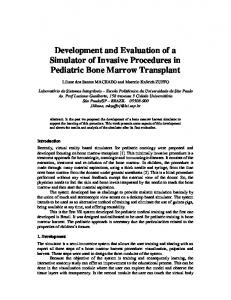
with either of the two above descriptions (e.g. vertebrae and calcanei). In a long bone, illustrated in Figure 1, cortical bone is found covering the whole outer surface but mainly at the central section (diaphysis), essentially formed as a hollow cylinder surrounding the medullary cavity. Cancellous bone is present towards the ends (metaphyses and epiphyses). Flat bones essentially consist of a layer of cancellous bone sandwiched between layers of cortical bone. Irregular bones consist predominantly of cancellous bone (up to 95%) with a thin cortical shell. A thin layer of periosteum covers the surface of all bones, consisting of resting osteogenic cells. Bones are developed and formed in two ways intermembranous ossification, where the bone forms directly from the mesenchyme via differentiation into osteoblasts, and endochondral ossification, where a cartilage model is produced initially, which is then calcified by hypertrophication of chondrocytes and ossified. As a general rule, flat bones tend to form via intermembranous ossification, and are therefore commonly referred to as membrane bones, whereas long and irregular bones tend to form via endochondral ossification and are referred to as cartilage bones. This paper describes the stochastic simulation of bone development and formation (osteogenesis) of cartilage bones.
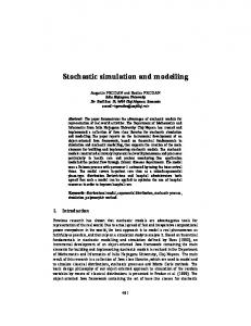
DETERMINISTIC AND CHAOTIC MECHANISMS OF CANCELLOUS BONE OSTEOGENESIS If one considers the structure of cancellous bone, illustrated in Figure 2, although there appears to be an underlying deterministic pattern as predicted by Wolffs law6, where the general trajectory of the trabeculae follow the principal lines of stress experienced by the bone, there is still a significant degree of variability and randomness. This is supported by recent work demonstrating that there is a strong relationship between mechanical and fractal properties of cancellous bone135and evidence that realistic structures simulating those of cancellous bone may be created using fractal modelling4. This leads us to propose
Endosteum A
Cancellous bone
FIGURE 1 Diagramatic representation of the bony tissues asaociated with a typical long bone. Note that irregular cartilage bones such as the vertebra and calcaneus do not have a diaphysis section
that cancellous bone grows and forms as a semi-deterministic lsemi-chaotic system and hence could be accurately modelled using a stochastic simulation. Complementary to this, we have recently described a dynamic stochastic simulation of cancellous bone resorption2 based upon the concept of the basic multicellular unit (BMU), where net resorption (-AB.BMU) was considered at bonelmarrow surfaces. The simulation was applied to a simple 2D matrix lattice structure representing vertebral cancellous bone, consisting of square bone or marrow elements of approximately 20pm in dimension. The simulation considered the probability that any surface element would be activated into a BMU, and if activated, the length of the resorption cavity. The mechanical stiffness of each resultant structure was calculated by finite element analysis. As an example of its potential clinical utility, the simulation has been used to investigate the recovery in cancellous bone stiffness resulting from a stochastic simulation of anabolic treatment following varying degrees of resorption3. The simulation demonstrated that during osteoporosis, the stiffness of cancellous bone decreases at twice the rate that bone density is lost. When the original density was regained due to anabolic treatment, the stiffness of the structure was lower than the original stiffness, the deviation being linearly proportional to the degree of initial resorption prior to anabolic simulation. Hence, a significant increase in cancellous bone density is required in order to regain the original stiffness.
STOCHASTIC SIMULATION OF OSTEOGENESIS
a) Iliac Crest
b) Femalral Head
c) Lumbzw Spine
FIGURE 2 3D micro-CT reconstructions of human cancellous bone from a) ilia'c crest, b)femoral head, and c) lumbar spine. The complex trabecular patterns follow the principle line of stress endured yet maintain a high degree of variability and randomness. (Images provided bj Professor P: Ruegseggel; University of Zurich)
Formation of cartilage bones Cartilage consists of a dense network of collagenous fibres (with some elastic fibres) surrounded by a ground substance in the form of a gel. Cartilage bones form first by the formation of cartilage, then by the
127
ossification of the formed cartilage. A flow-chart describing cartilage bone osteogenesis is shown in Figure 3, to which the following description of the various development processes should be referred. 1. Initially, mesenchymal (undifferentiated 'building block' cells) cells condense into the shape of a particular "model" of the bone. These cells are largely undifferentiated and have oval nuclei and fused cytoplasm. 2. The condensed mesenchymal cells then differentiate into chondroblasts (cartilage forming cells) and the intercellular matrix is laid down - this forms a stiff gel that accounts for the stiffness and flexibility of cartilage. When the chondroblasts have laid down matrix surrounding themselves, they differentiate into chondrocytes (cartilage cells). 3. The cells on the outside of the model form the perichondrium, which has two layers. The outer layer differentiates into fibroblasts, which produce collagen fibres. The inner layer remains as undifferentiated mesenchymal cells. The perichondrium forms a transition from the cartilage tissue to the tissue surrounding it. 4. Cartilage grows in two directions, along the length and breadth of the model. Lengthways growth is due to interstitial growth (from within) by mitosis of the cells close to the ends of the model - there are closely defined growth areas where this takes place. Thickening is due to appositional growth (on the surface) by mitosis of the mesenchymal cells on the inside edge of the perichondrium, some of which then differentiate into chondrocytes. 5. As the model grows from the centre outwards, the oldest chondrocytes are those at the centre of the model. These mature chondrocytes hypertrophy, enlarging the spaces in the ground material that they occupy, and then begin to produce alkaline phosphatase. This causes the cartilage to calcify (crystals of calcium phosphate build up on the organic matrix). However, as the minerals and nutrients for the chondrocytes cannot diffuse through the calcified cartilage in the same way
R.P. CHURCH and C.M. LANGTON
1. Condensation in shape of bone 2. Cells differentiate into chondroblasts which lay down cartilage and differentiate into chondrocytes. 3. Perichondrium forms
4a. Inner layer of perichondnum 5. Cells in centre of model start to mature. Hypertrophication occurs and phosphatase is produced. Calcification takes place. Cells die, leaving cavities.
cartilage mitosis occurs, causing the cartilage to
I 6. Blood vessels invade perichondrium. Mesenchymal cells differentlate into osteoblasts. Periosteal ossification. Ring of bone forms at the centre of cartilage model.
I
,, ' , ,
Head of bone moves upwards.
I
osteoblasts invade cavities. Trabecular bone forms in
1
10a. Diaphyseal wall grows and thickens in long bones.
I 11. Bone growth completed.
'
outwards. Cartilage produced at epiphyseal end of growth plate and ossified at the metaphyseal end
Removal of unnecesary trabecuae.
7
I
Remodelling Phase
FIGURE 3 Flow chart defining the various stages of cartilage bone osteogenesis. Note that the current simulation commences at stage 4 and that there are three concurrent pathways
that they can through ordinary cartilage, the chondrocytes die. This then leaves large cavities with calcified cartilage surrounding them, which slowly decays in the absence of the chondrocyte system to maintain it.
6. As the model enlarges, blood vessels invade the perichondrium at the centre of the model. At this point, the inner layer of mesenchymal cells then differentiates into osteoblasts and osteogenic cells. These cause a layer of bone to be laid down on the
STOCHASTIC SIMULATION OF OSTEOGENESIS
.
Mitosis takes place here, along with formation of matrix, expanding the model
-
Growth zone
Hypertrophic chondrocyte zone
3
-
\
Hypertrophicationof these cells takes place
Calcification of the matrix leading to death of these chondrocytes producing void
- Osteoclasts and osteoblasts Area of osteogenesis
invade
- forming bone
FIGURE 4 Diagramatic representation of cartilage bone formation (adapted from Ham and Leeson, Histology, 1961)
inside of the perichondrium, leading to a ring of bone (aroundthe centre of the model. This means that the membrane that was the perichondrium is now covering bone, and therefore it is referred to as the periosteum. It still retains its basic two-layer structure. 7. The c,avities left by the death of the chondrocytes are invaded by blood vessels, osteoblasts and osteoclasts from the periosteum. The osteoclasts remove some of the calcified cartilage and the osteoblasts lay down a layer of bone on top of the cartilage that remains. This leads to the characteristic open framework of cancellous bone. 8. Periosteal ossification keeps the end of the periosteal bone level with the hypertrophied cartilage. Blood vessels running longitudinally along the edges of the model are surrounded with bone and Haversian systems are formed. The older remain-
ing cartilage cells, those closest to the calcified cartilage already formed and converted into bone, undergo the process of hypertrophication, calcification and death. The cavities remaining are then invaded by osteoblasts from closer to the centre of the bone, causing ossification. Thus the boney part of the model expands. 9. Conversion of cartilage to bone starts at two other secondary centres of ossification in the epiphysis, as shown in Figure 4. A growth plate of cartilage forms between the cancellous bone of the metaphysis and that of the epiphysis, as shown in Figure 1. The growth plate can be split into four sections - resting cartilage adjacent to the epiphysis; proliferating cartilage where the cells are undergoing mitosis, enlarging the model; maturing cartilage, where the cartilage is ageing and becoming hypertrophied; and calcified cartilage,
R.P. CHURCH and C.M. LANGTON
130
where the cartilage has become calcified due to the action of phosphatase enzyme. Thus at the epiphyseal end of the growth plate, new cartilage is being produced and at the metaphyseal end, it is being converted into bone. The bone grows in width by deposition on the outside and resorption on the inside. As the epiphysis grows, the " h e a d of the bone moves further along the structure, again by absorption and deposition. 10. Deposition continues on the outer surface of the pereosteal bone. Resorption also takes place on the inner wall due to osteoclastic activity, but at a slower rate, which leads to a thickening of the periosteal bone and an increase in the bone radius overall. As the periosteal bone expands, many of the central trabeculae loose their mechanical function supporting the bone and are removed by osteoclastic activity as they are no longer necessary.
11. When growth of the bone is complete, the growth plate is completely ossified and the metaphysis and epiphysis fuse. The only cartilage that now remains is that fom~ingthe joint at the end of the bone.
METHOD The stochastic simulation of osteogenesis was implemerited in MATLAB (version 5.0, Mathworks Inc., Natick, US).
Fundamental basis of the stochastic simulation The chronological starting point for the simulation was chosen to be the initial cartilage phase of a generic bone. The model is based around a 2-dimensional matrix where square elements are assigned values that represent the substance or cell present within a particular element. A small piece of cartilage is initially present in a mostly empty matrix and grows to fill it. The size of the various components within the model is crucial. Thus it is necessary to have a list of the components involved so that their elemental sizes may be defined. Table I describes the various compo-
nents incorporated within the simulation along with their corresponding size. The data indicates that an elemental spatial resolution of 20pm would provide an acceptable compromise between physiological accuracy with memory requirements and simulation running time, noting that a sample of cartilage l c m by 5cm would contain 1.25 million elements. It is acknowledged that the cells involved are not square in reality, but this deviation will not detract from the validity of the simulation. The elemental size attributed to each cell is indicated in Table I. The operation of the model is based upon the contents of each element. Each component within the model is therefore allocated a specific code, indicated in Table I. The code for an empty space is 0, cell codes lie between 1 and 9, and tissues (bone, cartilage, etc) codes are between 10 and 13. The codes for the chondrocytes, osteoblasts and osteoclasts vary across a non-integer range. This is because the codes include the reciprocal of the age of the cell (in cycles). Using this arrangement, a range of ages that goes as far upwards as necessary may be stored without having to create another array or use a large range of data to store the ages. For a chondrocyte, the value is simply the reciprocal of the age; for the osteoblasts and osteoclasts, the reciprocal of the age is added to the starting value. A second matrix of the same dimensions is used to contain the structure of the blood vessels, using a similar coding system where void is zero; a blood vessel developing up, down, left or right is coded 1 to 4 respectively; a stable blood vessel being coded 5 . Thus it is possible to have, for example, bone and a blood vessel in the same place. This is necessary as the blood vessels and capillaries grow through the tissue to supply blood to the different cells present.
Cartilage implementation Cartilage consists of cells arranged in strips parallel to the length of the model. The cells are closer together, parallel to the length of the bone than they are in the two directions perpendicular to it. The space between the cells is filled with an intercellular matrix of collagen and ground substance.
STOCHASTIC SIMULATION OF OSTEOGENESIS
131
TABLE I Description of simulation components, along with their real size, elemental dimension within the simulation matrix, along with simulation coding and image colour
Size
Component
Elernent.7
Code
Colour
1x1
O