265 Journal of Food Protection, Vol. 68, No. 2, 2005, Pages 265–272
Development and Evaluation of an On-Line Hide Decontamination Procedure for Use in a Commercial Beef Processing Plant† JOSEPH M. BOSILEVAC,1* XIANGWU NOU,1 MATTHEW S. OSBORN,2 DELL M. ALLEN,2 MOHAMMAD KOOHMARAIE1 1U.S.
AND
Department of Agriculture, Agricultural Research Service, Roman L. Hruska U.S. Meat Animal Research Center, P.O. Box 166, Spur 18D, Clay Center, Nebraska 68933-0166; and 2Excel Corporation, 151 North Main Street, Wichita, Kansas 67202, USA MS 04-311: Received 9 July 2004/Accepted 20 September 2004
ABSTRACT The hides of cattle are the source of Escherichia coli O157:H7 that contaminates beef carcasses during commercial beef processing. Therefore, effective interventions that reduce hide contamination should reduce subsequent carcass contamination. The first objective of this study was to identify the most effective reagents for decontamination of beef hides. Cattle hides draped over barrels were used for in vitro experiments to compare the efficacy of washes using 1.6% sodium hydroxide, 4% trisodium phosphate, 4% chlorofoam, or 4% phosphoric acid, each followed by a rinse step using either water or acidified (pH 7.0) chlorine at 200 or 500 ppm. All treatments using a water rinse reduced hide coliform counts by 1.5 to 2.5 log CFU/ 100 cm2. Compared with water rinses, 200 and 500 ppm acidified chlorine rinses increased efficacy by approximately 1.0 and 2.0 log CFU/100 cm2, respectively. Vacuuming of the treated areas to remove excess liquid improved hide cleanliness by an average of an additional 1.0 log CFU/100 cm2. The second objective was to evaluate the use of an on-line hide-wash cabinet that used a sodium hydroxide wash and a chlorinated (1 ppm) water rinse. Hides sampled before entering and after exiting the cabinet had aerobic plate counts and Enterobacteriaceae counts that were reduced by 2.1 and 3.4 log CFU/100 cm2, respectively, and the prevalence of E. coli O157 on hides was reduced from 44 to 17% when the cabinet was in use. Preevisceration carcass aerobic plate counts and Enterobacteriaceae counts were both reduced by 0.8 log CFU/100 cm2, and the prevalence of E. coli O157 on preevisceration carcasses was reduced from 17 to 2% when the cabinet was in use. These results support decontamination of hides as an effective means to reduce pathogen contamination of cattle carcasses during processing.
Cattle hides are major sources of beef carcass contamination that occurs during processing (3, 5, 16). During the hide removal process, pathogens such as Escherichia coli O157:H7 and Salmonella are transferred from the hide, which carries high concentrations, to the carcass (1, 3, 5, 16). The current hazard analysis and critical control point plans (25) implemented in most beef processing plants in the United States focus on decontamination of the carcass by a combination of intervention strategies, including steam vacuuming, acid rinses, steam, and hot water spray (11). Such antimicrobial interventions combined with strict hygiene practices have significantly improved the microbial quality of beef carcasses in processing plants (1–3, 10). However, occasional process failures result in higher levels of contamination that cannot be effectively removed with current carcass interventions. Processes that effectively clean the hides before hide removal are successful in lowering carcass microbial contamination (5, 16). In an evaluation of chemical dehairing, * Author for correspondence. Tel: 402-762-4225; Fax: 402-762-4149; E-mail:
[email protected]. † Names are necessary to report factually on available data; however, the U.S. Department of Agriculture neither guarantees nor warrants the standard of the product, and the use of the name by the U.S. Department of Agriculture implies no approval of the product to the exclusion of others that may also be suitable.
Nou et al. first showed that the prevalence of E. coli O157: H7 on the carcass was almost eliminated when bacterial contamination of the hide was greatly reduced before hide removal (16). In subsequent studies with cetylpyridinium chloride (CPC), water washing followed by antimicrobial washes prior to slaughter also was highly effective in reducing carcass contamination during hide removal (5). CPC, however, is not yet approved for use inside beef processing plants. Other chemicals and antimicrobial compounds such as sodium hydroxide, trisodium phosphate, acidified chlorine, and phosphoric acid are approved for use in processing plants and are effective for carcass and boneless beef trim decontamination (2, 9, 12, 18–20). The use of these compounds on hides has not been evaluated. Therefore, the first objective of the present study was to evaluate the use of these antimicrobials as wash steps to reduce hide contamination in vitro. Because these materials do not have a neutral pH, a rinse also was needed to remove the residual antimicrobial to minimize exposure risks for plant personnel. Rinse compounds evaluated were water and acidified chlorine, which also has antimicrobial effects. Wash and rinse steps left excess liquids on the hides that could easily run onto the carcass when the hide was opened. To prevent excess liquid on the hide from contaminating the carcass, a vacuuming step to remove liquids also was
266
BOSILEVAC ET AL.
J. Food Prot., Vol. 68, No. 2
TABLE 1. Effect of various wash and rinse combinations and vacuuming on coliform counts of hidesa Coliform countd
Washb
Rinsec
n
Control
Treated
Treated 1 vacuumede
Water Chloroform Phosphoric acid Sodium hydroxide Trisodium phosphate Water Chloroform Phosphoric acid Sodium hydroxide Chloroform Phosphoric acid Sodium hydroxide
Water Water Water Water Water AC-200 AC-200 AC-200 AC-200 AC-500 AC-500 AC-500
30 30 30 30 30 30 24 24 24 21 21 21
5.9 6.2 5.8 5.7 5.9 5.7 5.9 5.9 6.0 5.9 5.6 5.9
4.3 2.6 3.3 4.2 4.4 2.8 3.2 2.3 3.2 2.0 1.5 2.2
2.3 2.6 2.3 1.8 3.4 2.8 2.0 1.6 2.2 1.5 0.2 2.0
a
Values represent the mean log CFU/100 cm2. Standard error of the mean ranged from 0.16 to 0.20. The minimum significant difference (P , 0.05) between any two values was 0.5 log CFU/100 cm2 (i.e., when the difference between any two values is $0.5, the difference is significant). b Wash steps consisted of 20-s end-to-end passes using high pressure (1,200 lb in22) application of water, 4% chloroform, 4% phosphoric acid, 1.6% sodium hydroxide, or 4% trisodium phosphate. c Rinse steps consisted of water or acidified chlorine at 200 ppm (AC-200) or 500 ppm (AC-500) applied at high pressure in 20-s endto-end passes. A 10-s dwell time occurred between wash and rinse applications. d Samples were serially diluted to a predetermined range in buffered peptone water and plated to Petrifilm E. coli or coliform count plates. e Alternating halves of each treated hide were vacuumed to remove excess liquid and visible contamination before the sample was collected.
evaluated to determine whether it increased or decreased the effectiveness of the decontamination procedure. The second objective of this study was to evaluate the practical application, inside the processing plant, of a hide wash and rinse using a specially designed on-line hide decontamination wash cabinet. The efficacy of the hide-wash cabinet was determined by measuring the reduction in bacterial indicators and the prevalence of E. coli O157 on hides and preevisceration carcasses before any carcass interventions were applied. MATERIALS AND METHODS Experiment 1: comparison of wash and rinse chemicals for reducing hide contamination. At a beef processing plant, whole pulled hides were draped over barrels to simulate hide-on carcasses for evaluation of hide decontamination treatments. Sample sponges do not pick up the same level of bacteria from dry hides as they do from wet hides (21). Therefore, a low-pressure hand pump sprayer was used to briefly (3 s) spray water on control sample sites to prevent controls from underrepresenting counts. After control samples were obtained, the appropriate wash and rinse combinations were applied to the hides. Twelve treatment combinations were evaluated with and without an additional vacuuming step in an incomplete block design. Wash steps used one of the following: water, 1.6% sodium hydroxide (Birco Corporation, Henderson, Colo.), 4% chlorofoam (Tergo Industries Limited, New Lynn, Auckland, New Zealand), 4% trisodium phosphate (Simple Green, Huntington Harbour, Calif.), or 4% phosphoric acid (Birco). Rinse steps used either water or acidified chlorine at concentrations of 200 or 500 ppm. The wash plus rinse combinations evaluated are shown in Table 1. Water used for washing and for diluting the antimicrobials was from a local potable water
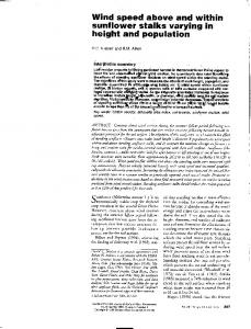
tap. The sodium hydroxide, trisodium phosphate, and phosphoric acid were diluted to working concentrations according to the directions from the manufacturers. Chlorofoam is a chlorinated alkaline detergent. According to the specification of the manufacturer, when prepared at 4%, chlorofoam has a pH of 12 and 1,200 ppm free chlorine. The acidified chlorine used in the rinse steps was prepared by diluting sodium hypochlorite (household bleach) and adjusting it to a pH of 7.0 with glacial acetic acid. The total chlorine content of the acidified chlorine was calculated to be 200 or 500 ppm. The free chlorine of the acidified chlorine solutions was not determined. The wash and rinse were applied using an electric pressurized sprayer (Karcher, Duluth, Ga.) with a rotating nozzle that was used at the maximum setting (1,200 lb in22). The distance between the nozzle and hide was kept at 65 cm during sprayings. The sprayer heated the dispensed liquid to 608C before spraying. Each wash and rinse application lasted 20 s and consisted of four passes across the hide, end to end. There was a 20-s delay between wash and rinse steps. Each treatment was applied to one side of 10 hides. Alternating halves of each side were vacuumed with a steam vacuum (Kentmaster Manufacturing Company, Inc., Monrovia, Calif.) without steam to remove residual liquid and visible contamination. Prior to treatment, nine sequential blocks of approximately 500 cm2 (16 by 31 cm) were marked on each side of each hide, and samples were taken from blocks 1, 5, and 9 for untreated controls. Following the wash and rinse steps, 3 samples were collected from blocks 2, 3, and 4 or from blocks 6, 7, and 8, for a total of 6 treated samples (3 vacuumed, 3 not vacuumed) per side and 30 samples per treatment (Fig. 1). Treated samples were obtained after a 30-s dwell period following rinse. Enterobacteriaceae, coliform, and E. coli counts were determined for all samples.
J. Food Prot., Vol. 68, No. 2
ON-LINE HIDE DECONTAMINATION
267
source is termed a lot, and the prevalence of E. coli O157:H7 on hides can vary greatly among lots. Therefore, to measure the direct effect of the hide intervention and not the lot-to-lot variation, each animal was tagged and sampled before the hide intervention and again after the intervention. When the hide decontamination cabinet was in operation, hide samples were collected from every 12th animal before it entered the cabinet and from the same animal after it exited the cabinet at a point approximately 10 min after the vacuuming step when the hide was in the process of being opened. When the hide cabinet was not in operation, hides were only sampled at the before-cabinet location. Carcass samples were collected from the corresponding preevisceration carcass of each animal from which a hide sample had been collected. Every sixth or fourth intervening carcasses was sampled, so that two or three carcasses were sampled for every hide sampled. This design was used to increase carcass sample numbers because our previous experience at this particular plant suggested that hide-to-carcass transfer was generally low and that a larger number of carcasses than hides would be needed for valid statistical conclusions to be drawn.
FIGURE 1. Hide sampling design in experiment 1. Prior to treatment, nine sequential blocks of approximately 500 cm2 (16 by 31 cm) were marked on each side of each hide, and 100-cm2 control (c) samples were taken from blocks 1, 5, and 9. Each wash 1 rinse treatment was applied to one side of 10 hides, and alternating halves of each side were vacuumed (v) with a Kentmaster steam vacuum without steam to remove residual liquid and visible contamination. Following the wash 1 rinse and vacuuming steps, 100-cm2 samples were collected from blocks 2, 3, 4, 6, 7, and 8, for a total of 6 treated samples (3 vacuumed, 3 not vacuumed) per side and 30 samples per treatment.
Sampling. All hide samples were collected using SpeciSponge Whirl-Pak bags (Nasco, Fort Atkinson, Wis.) from 100cm2 areas. For experiment 1, samples were taken from the block design as described using sponges wetted with 25 ml of DeyEngley neutralization broth (Becton Dickinson Microbiology Systems, Sparks, Md.) prepared at a 23 concentration (78 g/liter). For experiment 2, hide samples were collected from the brisket area with sample sponges wetted with 25 ml of buffered peptone water (Becton Dickinson Microbiology Systems) for samples collected before intervention or with 25 ml of Dey-Engley neutralization broth prepared at 13 concentration (39 g/liter) for samples collected after intervention. Samples were collected using 10 bidirectional strokes of the sponge, which was turned over halfway through the process. Carcass samples were collected from analhock or brisket-and-plate 4,000-cm2 areas using Speci-Sponge bags containing 25 ml of buffered peptone water. Carcass samples were collected in a manner similar to that for hide samples using bidirectional strokes and turning the sponge over halfway through the process. Fresh latex gloves were used for each sample. All samples were transported to the laboratory on ice and processed within 24 h.
Experiment 2: evaluation of on-line hide decontamination. A hide decontamination cabinet (Chad Co., Olathe, Kans.) that was designed to apply wash and rinse to double-shackled cattle immediately following stunning and exsanguination was evaluated. The cattle were oriented so that the portions of the hide that would be initially cut through to begin the hide removal process (i.e., hide-opening pattern lines) were directly perpendicular to the washing headers. The wash portion of the cabinet delivered 1.0 to 1.5% sodium hydroxide plus a proprietary surfactant from a mixing tank. The wash was delivered at 658C and 700 lb in22 with a flow rate of 350 to 400 gal/min. A dwell time of approximately 10 s occurred before the rinse section of the cabinet delivered sodium hypochlorite (household bleach) sufficient to supply 1 ppm free residual chlorine in the mixing tank. The rinse was applied at 358C and 700 lb in22 with a flow rate of 200 to 250 gal/min. As the hanging double-shackled cattle exited the cabinet, they were vacuumed using a steam vacuum (Kentmaster) without steam on the hide-opening pattern lines along the inside hind shanks, anus and cod/udder region and down to the brisket region. At a beef processing plant, cattle are received from a number of sources and sellers. Each group of cattle from an individual
Bacterial counts. Aerobic plate counts (APC), Enterobacteriaceae counts (EBC), E. coli counts, and total coliform counts were determined using Petrifilm count plates (3M Microbiology, St. Paul, Minn.) and a Bactometer (bioMe´rieux, Hazelwood, Mo.). In experiment 1, a 1-ml aliquot from each sample was serially diluted to a predetermined range in buffered peptone water. One milliliter of each dilution was plated to Petrifilm E. coli–coliform count plates and Petrifilm Enterobacteriaceae count plates for determination of coliforms, E. coli, and Enterobacteriaceae counts. Petrifilm plates were incubated for 16 h at 378C, and colonies were counted manually. The APC and EBC of hide and carcass samples in experiment 2 were performed by impedance measurements of 1-ml samples in the Bactometer. Each sample first was diluted 10-fold in GPM-Plus (General Purpose Medium-Plus, bioMe´rieux) supplemented with 18 g/liter dextrose (for a final concentration of 2% dextrose) and EM (Entero Medium, bioMe´rieux). Samples prepared in GPM-Plus were used for APC determination and samples prepared in EM were used in EBC determination. The Bactometer incubated samples for 16 h at 378C while measuring the initial detection time (IDT) for each sample. IDTs were converted to log CFU per milliliter using standard curves derived for each test. The standard curves were determined
268
BOSILEVAC ET AL.
by performing quadratic regression analysis of IDTs and log CFU per milliliter, which had been determined using Petrifilm aerobic count plates for APC or Petrifilm Enterobacteriaceae count plates for EBC. E. coli O157 detection. The procedure for detection of E. coli O157 consisted of enrichment, immunomagnetic separation, and plating as described previously (4), with minor modifications. All sample bags were enriched by the addition of 75 ml of tryptic soy broth (Becton Dickinson Microbiology Systems) immediately after the aliquot for APC and EBC was collected. Once all sample bags received tryptic soy broth, they were placed as a group into a programmable incubator for enrichment. The program for enrichment was 2 h at 258C to allow cell recovery and then 6 h at 428C to selectively enrich the growth of E. coli O157:H7. Enrichment was followed by refrigeration for 6 to 10 h at 58C to hold samples until immunomagnetic separation. One milliliter of enriched culture was used for immunomagnetic separation with anti–E. coli O157 DynaBeads (Dynal, Lake Success, N.Y.) following the manufacturer’s instructions. Bacterial cells bound to the beads were plated on sorbitol MacConkey agar (Becton Dickinson Microbiology Systems) supplemented with 0.05 mg/liter cefixime and 2.5 mg/liter potassium tellurite (Dynal) and on Rainbow agar (Biolog, Hayward, Calif.) supplemented with 10 mg/ liter novobiocin (Sigma, St. Louis, Mo.) and 8 mg/liter potassium tellurite (Sigma). The plates were incubated at 378C for 16 h, and suspect colonies (i.e., sorbitol negative on supplemented sorbitol MacConkey agar or characteristic blue on supplemented Rainbow agar) were confirmed as E. coli O157 using DrySpot O157 latex agglutination tests (Oxoid, Ogdensburg, N.Y.). The suspect colonies that tested positive with latex agglutination were considered to be E. coli O157:H7 because more than 90% of similar isolates had been confirmed as E. coli O157:H7 in earlier studies (3) but are referred to here as E. coli O157 to avoid misinterpretation of the level of strain identification. Statistical analyses. Data were analyzed with an analysis of variance (ANOVA) using the general linear models procedures of SAS (SAS Institute, Inc., Cary, N.C.). For significant main effects and interactions (P , 0.05), least squares mean separation was accomplished using the PDIFF option (a pairwise t test). Data for coliforms, E. coli, and EBC in experiment 1 were log transformed before the ANOVA using a 3 (treatment: control, treated, or treated 1 vacuuming) by 12 (wash-rinse combination) factorial arrangement of a repeated measures design. The model included the main effects of treatment. APC and EBC of hides and carcasses in experiment 2 were analyzed by ANOVA for a completely randomized design with the main effect of treatment (control and treated). Pairwise comparisons of frequencies of E. coli O157 detection were made using PROC FREQ and Mantel-Haenszel chisquare analysis (SAS).
RESULTS AND DISCUSSION Experiment 1. We first examined water, chlorofoam, phosphoric acid, sodium hydroxide, and trisodium phosphate for use during the wash step followed by a water rinse (Table 1). Neither trisodium phosphate nor sodium hydroxide performed any better than water as a wash step (P . 0.05) when followed by a water rinse without vacuuming. Trisodium phosphate, sodium hydroxide, and water reduced hide coliforms by 1.5, 1.5, and 1.6 log CFU/100 cm2, respectively, whereas a wash step that used chlorofoam or phosphoric acid significantly reduced hide coliforms (P , 0.05) by 2.4 and 2.5 log CFU/100 cm2, respectively. When
J. Food Prot., Vol. 68, No. 2
these treatment combinations were followed by vacuum removal of excess liquid and visible contamination, hide coliforms were reduced (P , 0.05) by an additional 1.0 to 1.2 log CFU/100 cm2 with trisodium phosphate, chlorofoam, and phosphoric acid and by an additional 2.0 to 2.4 log CFU/100 cm2 with water and sodium hydroxide. In the wash 1 rinse 1 vacuuming treatment with a water rinse, trisodium phosphate did not perform as well as did the other four compounds tested. The pathogen reductions by trisodium phosphate were significant but not as large as those for other compounds, including water. The reason for this reduced effect is unclear, because trisodium phosphate has been successful as an antimicrobial on many substrates other than hide (9, 18–20). Because of this outcome, trisodium phosphate was not included in the subsequent portions of experiment 1 that evaluated alternative rinses. The second and third sets of wash 1 rinse studies in experiment 1 included acidified chlorine in the rinse steps. Acidified chlorine was chosen because it is a commonly used sanitizing agent and antimicrobial. Used alone, acidified chlorine reduces contamination levels of beef trim and carcasses (8, 20). The acidified chlorine used as a rinse in our experiments was prepared at calculated concentrations of 200 and 500 ppm. The calculations were based on the total residual chlorine in the solution and the possible neutralizing effects of the acetic acid used to lower the pH of the solution were not considered. The free active chlorine in the final solutions was not determined; we assumed that the concentrations of active chlorine in our rinses were lower than calculated values. However, both solutions provided additional reductions of hide contamination compared with water, and the 500 ppm solution demonstrated greater efficacy than did the 200 ppm solution. Chlorine compounds can be rapidly neutralized by large amounts of organic matter on hides, but we suspected that the acidified chlorine rinses would produce additive effects because the interfering organic matter would be removed by the wash step. When acidified chlorine at 200 ppm was used as the rinse, additional reductions in hide coliforms (P , 0.05) of 1.1 to 1.3 log CFU/100 cm2 were recorded following washes of water, phosphoric acid, or sodium hydroxide. The efficacy of chlorofoam, a chlorinated detergent, was not enhanced (P . 0.05) by the acidified chlorine, suggesting that combinations of compounds with different active ingredients would provide greater antimicrobial effects. Applying the vacuuming step to the hides reduced hide coliforms further (P , 0.05) by 0.6 log CFU/100 cm2 for chlorofoam and phosphoric acid but by only 0.4 log CFU/100 cm2 for sodium hydroxide. Vacuuming made no difference (P . 0.05) following water 1 acidified chlorine. The use of the acidified chlorine rinse at 200 ppm with the vacuuming step improved the efficacy of phosphoric acid and chlorofoam (P , 0.05) but not that of water and sodium hydroxide over water rinsing. Reductions (P , 0.05) of 3.7 to 4.1 log CFU/100 cm2 were observed with chlorofoam, phosphoric acid, and sodium hydroxide when the rinse compound was 500 ppm acidified chlorine. The addition of the vacuuming step had no effect (P . 0.05) on sodium hydroxide and chlorofoam
J. Food Prot., Vol. 68, No. 2
but improved the effects of phosphoric acid by an additional 1.3 log CFU/100 cm2. Using 500 ppm instead of 200 ppm acidified chlorine improved the reduction of coliforms only by the phosphoric acid wash. The results of vacuuming were not consistent, possibly for several reasons. In some cases, vacuuming removed 1 to 2 log CFU/cm2 due to the removal of wash- and rinseloosened bacteria. In other cases, no significant reduction was achieved with vacuuming. Chlorofoam wash 1 water rinse and water wash 1 200 ppm acidified chlorine rinse did not produce reductions after vacuuming. In these cases, there may not have been a loosening of adherent bacteria by these treatments. The only observed reduction was that of the antimicrobial. For treatments that used 500 ppm acidified chlorine rinse after washes with either chlorofoam or sodium hydroxide, no significant reduction followed vacuuming, possibly because the antimicrobial and wash reduced the bacteria to such low numbers that very few bacteria remained to be vacuumed away. In addition to the measurement of coliform bacteria (Table 1), we also counted Enterobacteriaceae and E. coli. Because we did not know whether one group of bacteria would be better than another as an indicator of antimicrobial efficacy, all three groups were evaluated; only coliform results have been presented because E. coli is a coliform and coliforms are members of the Enterobacteriaceae. Because E. coli can make up a large proportion of either coliforms or Enterobacteriaceae, the results were not expected to be different, nor were they. There were no significant differences (P . 0.05) among counts of any of the bacterial groups for a given treatment, with one exception. The EBC and E. coli results for the vacuumed sodium hydroxide 1 water combination were different (P , 0.05); however, neither result was different from the result for coliform bacteria (P . 0.05) for the same treatment (data not shown). Reid et al. (22) examined the presence of foodborne pathogens on cattle hides. They found that the brisket area of the hide was frequently contaminated with pathogens and suggested this was a likely source of cross-contamination of beef during the dehiding process. McEvoy et al. (13) also observed a relationship between visual hide cleanliness and bacteria on carcasses and found that improving hygienic practices during dehiding of dirty animals resulted in significant reductions of bacteria on the carcass. Arthur et al. (1) described a direct relationship between the prevalence of E. coli O157 on hides and that on preevisceration carcasses. Therefore, effective decontamination of the hide should result in less contamination of the carcass following hide removal. Several hide interventions have been used with varying degrees of success. Hide decontamination before slaughter involving washes with and without antimicrobials such as lactic acid and chlorine (15) did not significantly decrease the bacterial counts on hides. Preslaughter washing of artificially surface-inoculated cattle hides also had no effect on hide-to-carcass transfer of pathogens despite washes of 1 and 3 min (7). Successful hide interventions include chemical dehairing (16), treatment with CPC (5), or treatment with subatmospheric pressure steam (14). These treatments have been effective in reducing hide
ON-LINE HIDE DECONTAMINATION
269
contamination, and for CPC and chemical dehairing, the hide treatment resulted in less contamination of the corresponding carcasses compared with carcasses that underwent conventional processing without hide treatment (5, 16). Some potential interventions may not be practical or may not be approved for in-plant use. Our goal was to test the effectiveness of various wash and rinse strategies applied to hides using antimicrobials that are currently approved for use in the processing plant for such things as equipment decontamination or carcass and trim interventions. Because sequential washing steps increase the efficacy of antimicrobial treatments (6), the antimicrobials were evaluated as a wash step followed by a rinse step. Rinses remove wash-loosened material and reduce the amount of antimicrobial that may later get on the carcass; they also reduce exposure of plant employees. Our results demonstrated that a number of options are readily available for reducing populations of bacteria on hides. The chemicals evaluated in this study are approved for use in processing plants as disinfectants and antimicrobials for equipment, carcasses, and trim; therefore, we cannot make direct comparisons with the results of their use on hides. The reductions we observed on hides were generally greater than those reported for carcass and trim interventions. Boneless beef trim interventions composed of 10% trisodium phosphate, 5% lactic acid, or 0.5% CPC reduced APC by 0.6 log CFU/g and reduced coliforms and E. coli by 0.6 to 0.8 log CFU/g (19, 24). Multiple-step interventions that used 5% acetic acid followed by 0.5% CPC, 200 ppm chlorine dioxide followed by 0.5% CPC, or 0.5% CPC followed by 10% trisodium phosphate reduced APC on boneless beef trim by 1.2, 1.8, and 0.9 log CFU/g, respectively (18). Treatment of beef trim with 200 and 1,000 ppm acidified chlorine has been described (20, 23). Others have reported using 1,200 ppm acidified chlorine to treat carcass surfaces (8). The bacterial reductions in these studies were consistently greater than 2.0 log CFU/g for E. coli and greater than 1.0 log CFU/g for APC. Our data indicate that water washing followed by water rinsing with subsequent vacuuming greatly reduced bacterial loads on hides. Including an antimicrobial in the water of the wash step increased the effectiveness of the treatment, but the improvement was largely negated by the vacuuming step. Substituting acidified chlorine for water in the rinse step also improved the effectiveness of the treatment, but this improvement also was largely negated by the vacuuming step, except at the highest concentrations of the acidified chlorine (500 ppm). The choice of compounds to use in an automated hide-washing cabinet involved consideration of cost, ease of implementation, and efficacy. Based on this combination of factors, a 1.5% sodium hydroxide wash followed by a rinse using recycled water containing 1 ppm chlorine was chosen for decontamination. The pattern lines were also vacuumed after treatment in the on-line hide-wash cabinet. Experiment 2. The effectiveness of cleaning the hide before dressing the carcass on the reduction of the bacterial load on carcasses under commercial processing conditions
270
BOSILEVAC ET AL.
J. Food Prot., Vol. 68, No. 2
TABLE 2. Effect of hide wash cabinet interventiona on hidesb and carcassesc APCd
Sample type
n
Intervention Hides before cabinet Hides after cabinet Carcasses
99 92 264
10.3 8.2 4.9
Af
98 251
10.2 5.7
A
B B
EBCd
E. coli O157e
7.0 3.6 1.6
A
44/99 (44%) B 15/92 (16%) C 5/264 (2%) B
7.1 2.4
A
B B
interventiong
No Hides Carcasses
A
A
86/98 (88%) 42/251 (17%)
A A
Intervention consisted of hide-wash cabinet applying a wash of 1 to 1.5% sodium hydroxide at 700 lb in22 and 658C followed by a rinse of 1 ppm residual chlorine applied at 700 lb in22 at 358C. The opening pattern lines of the hide were vacuumed to remove excess liquid and visible contamination. b Hides of stunned cattle were sampled before entering the wash cabinet and at a point after exit when intervention was in use. The first steps of opening the hide occurred at the second hide sampling point. c Carcasses were sampled pre-evisceration (before processing interventions). d APC and EBC were determined by impedance measurements using a bioMe ´ rieux Bactometer. Values are the mean log CFU per 100 cm2. Standard error of the mean was 0.1 for APC and EBC. e E. coli O157 values are the proportion of hides positive by culture (%). f Means within a bacterial and sample type (hide or carcass) with a common letter are not significantly different (P . 0.05). g Control samples were collected when the hide-wash cabinet was not in use. a
was evaluated. The participating processing plant had installed a hide decontamination wash cabinet. After cattle were stunned and exsanguinated, hides were washed before any portion of the hide was opened. The use of a hide-wash cabinet requires large volumes of water. At such large volumes, recirculation of the wash and rinse compounds is required to be cost effective. Sodium hydroxide at 1.5% was chosen as the wash because it does not lose activity as acids frequently do in a recirculating system (17). Chlorine at 1 ppm was used to clean the recirculated water used for the rinse step and was not intended to have an additional antimicrobial effect on the hides. As each animal exited the cabinet, plant personnel used a Kentmaster steam vacuum without steam to vacuum the hide along the hide opening pattern lines to remove excess liquid and visible contamination loosened by the wash 1 rinse process. An acidified chlorine rinse was not used because in experiment 1 it did not have any additional effect following a sodium hydroxide wash when vacuuming was in use. Because of the recycled nature of the water used in the wash and rinse steps, the APC at each step was determined to rule out potential recontamination of hides (data not shown). The sodium hydroxide wash had nondetectable APC at the lowest dilution (1021) examined. The 1 ppm chlorine rinse water had an APC of 3.5 log CFU/ml; however, compared with the APCs of hides before treatment (10.3 log CFU/ml) and after treatment (8.2 log CFU/ml), this amount was unlikely to contribute significantly more bacteria to the hide than was already present. The prevalence of E. coli O157:H7 on hides varies from lot to lot. Therefore, each animal was sampled before the hide intervention and again after the intervention to determine the actual effect of the individual hide intervention and not the lot-to-lot variation (Table 2). As has been reported previously (5, 16), hides and carcasses were visibly cleaner when the intervention was in use. These sub-
jective observations were confirmed by the APC and EBC of hides, which were reduced by 2.1 and 3.4 log CFU/100 cm2, respectively, by the hide-wash cabinet intervention. The prevalence of E. coli O157 on hides was reduced (P , 0.05) from 44 to 16% following passage through the hide-wash cabinet. The carcasses that corresponded to the sampled hides and the intervening carcasses were sampled when the hidewash cabinet was in operation, and the samples were compared with similarly collected samples from carcasses when the hide decontamination cabinet was not in use. The sodium hydroxide wash 1 water rinse and vacuuming of the hides resulted in a lower bacterial load on carcasses compared with conventional processing procedures. APC and EBC were each reduced by 0.8 log CFU/100 cm2 on preevisceration carcasses of the treatment group. When the online hide decontaminating cabinet was in use, E. coli O157: H7 prevalence on carcasses was 2%, whereas that of the control group was 17% (P , 0.05). There was a significant difference in the hide prevalence of E. coli O157 between the no-intervention (control) cattle and the before-treatment group of cattle (Table 2). These differences are unavoidable and are due to lot-to-lot and trip-to-trip variation in prevalence of E. coli O157 only. Efforts were made to avoid these differences in prevalence by dividing lots of cattle between treatments and control whenever possible and by sampling across as many lots as possible when dividing lots was not an option. The experiment was conducted in multiple segments spanning a period of 2 months, allowing us to assess the hide decontamination process on cattle from various production environments (i.e., feedlots). Comparison between the no-intervention cattle and the before-cabinet cattle revealed no differences (P . 0.05) in APCs and EBCs, but there were differences in the prevalence of E. coli O157. The difference in hide prevalence cannot account solely for the dif-
J. Food Prot., Vol. 68, No. 2
ferences in carcasses. An undeterminable portion of variation is likely due to carcass differences, but the major effect is the result of hide decontamination and lowering the hide prevalence from 44 to 16% before removal (16). We previously examined other hide decontamination methods and determined their effects on carcass cleanliness. CPC treatment of hides reduced the APC and EBC of carcasses by 1.5 and 1.1 log CFU/100 cm2, respectively, and reduced E. coli O157 prevalence on carcasses from 23 to 3% (5). In a similar fashion, chemical dehairing of the hide resulted in carcasses that had an APC and an EBC that were 2.0 and 1.8 log CFU/100 cm2 lower than controls, respectively. Only 1% of carcasses belonging to dehaired animals had detectable E. coli O157, whereas 50% of the untreated control group had detectable E. coli O157 (16). Use of the on-line hide-wash cabinet gave results comparable to those obtained with these other methods. The on-line hide-wash cabinet also has advantages over the other methods. The antimicrobials used in the hide-wash cabinet are approved for use inside the processing plant, and the hide-wash cabinet is a simpler system requiring less space and equipment than the chemical dehairing system. Procedures designed to clean cattle hides before carcass dressing can reduce the bacterial load on carcasses immediately after hide removal by about 1 log CFU/100 cm2 and can dramatically reduce the prevalence of E. coli O157:H7. Typically, the various antimicrobial interventions in beef processing plants have a combined effectiveness of 3- to 4-log reduction in bacterial load from preevisceration carcasses to carcasses chilling in the cooler (2, 3, 16). Combining an effective hide intervention with subsequent carcass interventions should further improve the safety of beef and beef products by virtually eliminating pathogen contamination, which is not possible with current carcass interventions alone. The reduction of contamination on the hide after treatment can be the focus of additional measures. Other compounds such as lactic acid or CPC (if approved) could be applied to the hide opening pattern lines while vacuuming. A process such as this could provide a greater reduction of bacteria in the hide opening areas and ultimately minimize hide-to-carcass transfer of contaminants. ACKNOWLEDGMENTS We are grateful to the participating processing plants and personnel for their cooperation. We thank Bruce Jasch and Gregory Smith for technical support and Carol Grummert for secretarial assistance.
ON-LINE HIDE DECONTAMINATION
4.
5.
6.
7.
8.
9.
10.
11.
12.
13.
14.
15.
16.
REFERENCES 1.
2.
3.
Arthur, T. M., J. M. Bosilevac, X. Nou, S. D. Shackelford, T. L. Wheeler, M. W. Kent, D. Jaroni, B. Pauling, D. M. Allen, and M. Koohmaraie. 2004. Escherichia coli O157 prevalence and enumeration of aerobic bacteria, Enterobacteriaceae, and Escherichia coli O157 at various steps in commercial beef processing plants. J. Food Prot. 67:658–665. Bacon, R. T., K. E. Belk, J. N. Sofos, R. P. Clayton, J. O. Reagan, and G. C. Smith. 2000. Microbial populations on animal hides and beef carcasses at different stages of slaughter in plants employing multiple-sequential interventions for decontamination. J. Food Prot. 63:1080–1086. Barkocy-Gallagher, G. A., T. M. Arthur, M. Rivera-Betancourt, X. Nou, S. D. Shackelford, T. L. Wheeler, and M. Koohmaraie. 2003.
17. 18.
19.
20.
271
Seasonal prevalence of Shiga toxin–producing Escherichia coli, including O157:H7 and non-O157 serotypes, and Salmonella in commercial beef cattle processing plants. J. Food Prot. 66:1978–1986. Barkocy-Gallagher, G. A., E. D. Berry, M. Rivera-Betancourt, T. M. Arthur, X. Nou, and M. Koohmaraie. 2002. Development of methods for the recovery of Escherichia coli O157:H7 and Salmonella from beef carcass sponge samples and bovine fecal and hide samples. J. Food Prot. 65:1527–1534. Bosilevac, J. M., T. M. Arthur, T. L. Wheeler, S. D. Shackelford, M. Rossman, J. O. Reagan, and M. Koohmaraie. 2004. Prevalence of Escherichia coli O157 and levels of aerobic bacteria and Enterobacteriaceae are reduced when hides are washed and treated with cetylpyridinium chloride at a commercial beef processing plant. J. Food Prot. 67:646–650. Bosilevac, J. M., T. L. Wheeler, M. Rivera-Betancourt, X. Nou, T. M. Arthur, S. D. Shackelford, M. P. Kent, D. Jaroni, M. Osborn, M. Rossman, J. O. Reagan, and M. Koohmaraie. 2004. Protocol for evaluating the efficacy of cetylpyridinium chloride as a beef hide intervention. J. Food Prot. 67:303–309. Byrne, C. M., D. J. Bolton, J. J. Sheridan, D. A. McDowell, and I. S. Blair. 2000. The effects of preslaughter washing on the reduction of Escherichia coli O157:H7 transfer from cattle hides to carcasses during slaughter. Lett. Appl. Microbiol. 30:142–145. Castillo, A., L. M. Lucia, G. K. Kemp, and G. R. Acuff. 1999. Reduction of Escherichia coli O157:H7 and Salmonella Typhimurium on beef carcass surfaces using acidified sodium chlorite. J. Food Prot. 62:580–584. Dickson, J. S., C. N. Cutter, and G. R. Siragusa. 1994. Antimicrobial effects of trisodium phosphate against bacteria attached to beef tissue. J. Food Prot. 57:952–955. Elder, R. O., J. E. Keen, G. R. Siragusa, G. A. Barkocy-Gallagher, M. Koohmaraie, and W. W. Laegreid. 2000. Correlation of enterohemorrhagic Escherichia coli O157 prevalence in feces, hides, and carcasses of beef cattle during processing. Proc. Natl. Acad. Sci. USA 97:2999–3003. Galland, J. C. 1997. Risks and prevention of contamination of beef carcasses during the slaughter process in the USA. Rev. Sci. Technol. 16:395–404. Kang, D. H., M. Koohmaraie, and G. R. Siragusa. 2001. Application of multiple antimicrobial interventions for microbial decontamination of commercial beef trim. J. Food Prot. 64:168–171. McEvoy, J. M., A. M. Doherty, M. Finnerty, J. J. Sheridan, L. McGuire, I. S. Blair, D. A. McDowell, and D. Harrington. 2000. The relationship between hide cleanliness and bacterial numbers on beef carcasses at a commercial abattoir. Lett. Appl. Microbiol. 30:390– 395. McEvoy, J. M., A. M. Doherty, J. J. Sheridan, I. S. Blair, and D. A. McDowell. 2001. Use of steam condensing at subatmospheric pressures to reduce Escherichia coli O157:H7 numbers on bovine hide. J. Food Prot. 64:1655–1660. Mies, P. D., B. R. Covington, K. B. Harris, L. M. Lucia, G. R. Acuff, and J. W. Savell. 2004. Decontamination of cattle hides prior to slaughter using washes with and without antimicrobial agents. J. Food Prot. 67:579–582. Nou, X., M. Rivera-Betancourt, J. M. Bosilevac, T. L. Wheeler, S. D. Shackelford, B. L. Gwartney, J. O. Reagan, and M. Koohmaraie. 2003. Effect of chemical dehairing on the prevalence of Escherichia coli O157:H7 and the levels of aerobic bacteria and Enterobacteriaceae on carcasses in a commercial beef processing plant. J. Food Prot. 66:2005–2009. Osborn, M. 2004. Personal communication. Pohlman, F. W., M. R. Stivarius, K. S. McElyea, Z. B. Johnson, and M. G. Johnson. 2002. Reduction of microorganisms in ground beef using multiple intervention technology. Meat Sci. 61:315–322. Pohlman, F. W., M. R. Stivarius, K. S. McElyea, and A. L. Waldroup. 2002. Reduction of E. coli, Salmonella typhimurium, coliforms, aerobic bacteria, and improvement of ground beef color using trisodium phosphate or cetylpyridinium chloride before grinding. Meat Sci. 60: 349–356. Ransom, J. R., K. E. Belk, J. N. Sofos, J. S. Stopforth, J. A. Scanga,
272
BOSILEVAC ET AL.
and G. C. Smith. 2003. Comparison of intervention technologies for reducing Escherichia coli O157:H7 on beef cuts and trimmings. Food Prot. Trends 23:24–34. 21. Reid, C.-A., S. M. Avery, M. L. Hutchison, and S. Buncic. 2002. Evaluation of sampling methods to assess the microbiological status of cattle hides. Food Control 13:405–410. 22. Reid, C.-A., A. Small, S. M. Avery, and S. Buncic. 2002. Presence of food-borne pathogens on cattle hides. Food Control 13:411–415. 23. Rourke, T. J., M. Guerra, G. K. Kemp, B. C. Tinsley, C. C. Warf, T. G. Richardson, P. L. Baxter, and K. R. Schneider. 2001. Evalua-
J. Food Prot., Vol. 68, No. 2
tions of acidified sodium chlorite for use on red meats. Available at: http://ift.confex.com/ift/2001/techprogram/paperp5893.htm. Accessed 1 February 2004. 24. Stivarius, M. R., F. W. Pohlman, K. S. McElyea, and A. L. Waldroup. 2002. Effects of hot water and lactic acid treatment of beef trimmings prior to grinding on microbial, instrumental color and sensory properties of ground beef during display. Meat Sci. 60:327–334. 25. U.S. Department of Agriculture, Food Safety and Inspection Service. 1996. Pathogen reduction; hazard analysis and critical control point (HACCP) systems, final rule. Fed. Regist. 61:38805–38989.