Papers in Press. First published June 15, 2006 as doi:10.1373/clinchem.2006.068445
Clinical Chemistry 52:8 000 – 000 (2006)
Endocrinology and Metabolism
Development and Performance Evaluation of a Tandem Mass Spectrometry Assay for 4 Adrenal Steroids Mark M. Kushnir,1* Alan L. Rockwood,1 William L. Roberts,1,2 Elizabeth G. Pattison,1 William E. Owen,1 Ashley M. Bunker,1 and A. Wayne Meikle1,2,3
Background: Congenital adrenal hyperplasia is a group of autosomal recessive disorders caused by a deficiency of one of 4 enzymes required for the synthesis of glucocorticoids, mineralocorticoids, and sex hormones. Analysis of 11-deoxycortisol (11DC), 17-hydroxyprogesterone (17OHP), 17-hydroxypregnenolone (17OHPr), and pregnenolone (Pr) in blood allows detection of these enzyme defects. Methods: The steroids were extracted from 200 L of serum or plasma by solid-phase extraction, derivatized to form oximes, and extracted again with methyl t-butyl ether. Instrumental analysis was performed on an API 4000 tandem mass spectrometer with electrospray ionization in positive mode and multiple reaction-monitoring acquisition. Results: The limits of detection were 0.025 g/L for 11DC, 17OHP, and Pr and 0.10 g/L for 17OHPr. The method was linear to 100 g/L for 11DC, 17OHP, and Pr, respectively, and to 40 g/L for 17OHPr. Within- and between-run (total) imprecision (CVs) were 72 h. Conclusions: The detection limit and selectivity of this method and its small sample volume requirement allow analysis of endogenous concentrations of adrenal ste-
1
roids in serum or plasma from children and adults. The method thus has an important potential role in the evaluation of the status of 4 of the enzymes involved in adrenal steroid biosynthesis. © 2006 American Association for Clinical Chemistry
Congenital adrenal hyperplasia (CAH)4 is a group of autosomal recessive disorders caused by a deficiency of one of 4 enzymes required for the synthesis of glucocorticoids, mineralocorticoids, and sex hormones (1–5 ). CAH may present with variety of clinical findings, such as virilization of the affected infants, signs of androgen excess, salt-wasting crisis secondary to aldosterone deficiency, or hormonal hypertension attributable to increased mineralocorticoids. The most common type of CAH is 21-hydroxylase deficiency, which is the result of sequence variations in the cytochrome P450, family 21, subfamily A, polypeptide 2 gene (CYP21A2;5 also known as CYP21) located on chromosome 6. Most of the remaining cases of CAH are caused by a deficiency of 11bhydroxylase, 17a-hydroxylase, or 3b-hydroxysteroid dehydrogenase. Evaluation of the concentrations of the steroid intermediates in the cholesterol pathway, i.e., 11-deoxycortisol (11DC; Reichstein’s substance S), 17hydroxyprogesterone (17OHP), 17-hydroxypregnenolone (17OHPr), and pregnenolone (Pr), allows detection of these enzyme defects (6 –9 ). If a person has deficiency of
4 Nonstandard abbreviations: CAH, congenital adrenal hyperplasia; 11DC, 11-deoxycortisol (Reichstein’s substance S); 17OHP, 17-hydroxyprogesterone; 17OHPr, 17-hydroxypregnenolone; Pr, pregnenolone; GC-MS, gas chromatography–mass spectrometry; LC-MS/MS, liquid chromatography–tandem mass spectrometry; MTBE, methyl t-butyl ether; SPE, solid-phase extraction; ESI, electrospray ionization; APCI, atmospheric pressure chemical ionization; and CID, collision-induced dissociation. 5 Human gene: CYP21A2, cytochrome P450, family 21, subfamily A, polypeptide 2.
ARUP Institute for Clinical and Experimental Pathology, Salt Lake City,
UT. Departments of 2 Pathology and 3 Medicine, University of Utah, Salt Lake City, UT. * Address correspondence to this author at: ARUP Institute for Clinical and Experimental Pathology, 500 Chipeta Way, Salt Lake City, UT 84108. Fax 801-584-5207; e-mail
[email protected]. Received February 8, 2006; accepted May 23, 2006. Previously published online at DOI: 10.1373/clinchem.2006.068445
1 Copyright © 2006 by The American Association for Clinical Chemistry
2
Kushnir et al.: LC-MS/MS Method for Adrenal Steroids
one of the enzymes, this leads to accumulation of the steroid precursors proximal to the enzyme defect. Simultaneous analysis of multiple steroids involved in the pathway helps in making a correct diagnosis because in some cases 17OHP can be increased in 11b-hydroxylase deficiency. Therefore, the enzyme defect can be confused with 21-hydroxylase deficiency if 11DC is not measured simultaneously. Testing for adrenal steroids is also useful for differential diagnosis of polycystic ovary syndrome and nonclassic forms of CAH, which may sometimes be indistinguishable by clinical presentation and sonography (6 ). To date, determination of 11DC, Pr, and 17OHPr is based solely on immunoassays and gas chromatography– mass spectrometry (GC-MS), which are available only in a few clinical laboratories (10 –14 ). The accuracy of immunoassays varies significantly because of cross-reactivity and matrix effects. immunoassays can have poor negative and positive predictive values, especially in preterm and acutely ill neonates (15–17 ). Because interfering substances and the degree of interference vary among commercially available immunoassays, reference intervals for adrenal steroids are often method-specific and cannot be used interchangeably (18 –23 ). Several GC-MS methods have been developed for measurement of adrenal steroids. These methods have good specificity, but they usually have low throughput and require large sample volumes (24, 25 ). The majority of the published tandem mass spectrometry (MS/MS) methods for measurement of adrenal steroids allow measurement of concentrations characteristic of pathologic conditions but are not sensitive enough to measure physiologic endogenous concentrations (26 –32 ). Lacey et al. (32 ) developed a liquid chromatography (LC)-MS/MS method for steroid profiling of newborns from dried blood spots collected on filter paper. Through measurement of 17OHP, androstenedione, and cortisol, the method allows evaluation of the status of the enzymes involved in the pathway and thus provides a better tool for diagnosis of CAH than does measurement of 17OHP alone. We have developed a sensitive and specific LCMS/MS assay for simultaneous measurement of endogenous concentrations of adrenal steroids that uses a small sample volume and allows evaluation of status of 4 of the enzymes involved in adrenal steroid biosynthesis. For the first time, reference intervals for males and females of different Tanner stages and ages were established by use of isotope-dilution LC-MS/MS.
Materials and Methods calibrators and reagents We purchased 11DC, 17OHP, 17OHPr, and Pr (all purities ⱖ98%) from Sigma. Deuterated analogs of the steroids (11DC-d2, 17OHP-d8, 17OHPr-d3, and Pr-d4) were purchased from Cambridge Isotope Laboratories. Stock solutions of 11DC, 17OHP, and Pr were prepared in methanol,
and 17OHPr was prepared in ethanol at a concentration of 1 g/L. Working combined calibrators and internal standards were prepared in an equivolume mixture of water and methanol at concentrations of 10 and 20 g/L, respectively. Adrenal steroid calibrators were prepared in 10 g/L bovine serum albumin at concentrations of 0.5, 1, 2.5, and 5.0 g/L6 (50, 100, 250, 500 ng/dL) with each set of samples. HPLC-grade water, methanol, methyl t-butyl ether (MTBE), and acetonitrile were obtained from Fisher Scientific. All other reagents were obtained from Sigma. Strata XTM solid-phase extraction (SPE) columns were purchased from Phenomenex.
sample preparation Aliquots (200 L) of calibrators, controls, or patient serum were transferred into glass tubes containing 2 mL of water. To each of these, we added 20 L of a working combined internal standard. The samples were extracted with SPE as follows: columns were preconditioned by consecutive addition of 2 mL of methanol and water; samples were applied and drained by gravity; columns were washed with 3 mL of water followed by 3 mL of 200 mL/L acetonitrile in water, and then dried for 15 min; finally, steroids were eluted with 3 mL of MTBE. The solvent was evaporated under nitrogen in a 35 °C water bath, and the dried residue was redissolved for derivatization in 300 L of an aqueous hydroxylamine solution (1.5 mol/L, pH 10). The tubes were vortex-mixed and incubated in a heating block at 90 °C for 30 min, and the derivatives were extracted with 2 mL of MTBE. The solvent was evaporated, the residue was reconstituted with 75 L of methanol–water (2:1 by volume), and the samples were analyzed on the LC/MS/MS instrument.
lc-ms/ms The method was validated on an API 4000 triple-quadrupole mass spectrometer (Applied Biosystems/MDS Sciex) equipped with a V spray ionization source operated at 400 °C. The system included an Agilent HPLC pump and oven (Series 1100) and a Perkin-Elmer 200 Series autosampler. The chromatographic separation was performed on a Phenomenex Synergy FusionTM RP (C18) column [50 ⫻ 2.0 mm (i.d.); particle size, 4 m]. The injection volume was 15 L, and the oven temperature was 50 °C. Mobile phase A was methanol, mobile phase B was 22 mmol/L aqueous formic acid. Mobile phase was delivered at a flow rate of 0.5 mL/min with the following program: 60% methanol for 2 min, linear gradient to 75% methanol between 2 and 3.5min, step gradient to 97% of methanol at 6 min, followed by reequilibration to initial conditions between 6.4 and 7.2 min. The autosampler injection syringe was
6 Throughout the manuscript, steroid concentrations are expressed in g/L; the conversion factors to nmol/L are 2.888, 3.028, 3.010, and 3.162 for 11-deoxycortisol, 17-hydroxyprogesterone, 17-hydroxypregnenolone, and pregnenolone, respectively.
Clinical Chemistry 52, No. 8, 2006
washed between the injections 12 times with methanol– water (4:1 by volume) containing 8.8 mmol/L trifluoroacetic acid. The Q1 and Q3 quadrupoles were tuned for unit resolution, and the mass spectrometer conditions were optimized for maximum signal intensity of each adrenal steroid. The instrument was operated in positiveion mode with an ion spray voltage of 4000 V and entrance potential of 10 V. MS/MS transitions used for the adrenal steroids and their internal standards and compound-specific voltages are listed in Table 1. Nitrogen was used as the gas in the collision cell. All data were acquired and processed with AnalystTM 1.4.1 software (Applied Biosystems/MDS Sciex). Quantitative calibration was performed with every batch of samples and was used in conjunction with the intensities of the transitions of internal standards to calculate concentrations in unknown samples. An API 3000 (Applied Biosystems/MDS Sciex) triple-quadrupole mass spectrometer was used to study relative sensitivities among different ion sources and for initial evaluation of the method.
assay performance characteristics We determined the method imprecision by analyzing 3 replicates per run of samples containing adrenal steroids at concentrations of 0.8, 4.6, 9.0, and 18.0 g/L in 1 run per day over a 5-day period. The samples were prepared in 10 g/L bovine serum albumin, aliquoted, and stored at ⫺70 °C until analysis. We evaluated method linearity by analyzing samples prepared with adrenal steroid concentrations between 1 and 100 g/L. We determined method sensitivity by analyzing samples prepared by serial 2-fold dilutions a sample containing 1.0 g/L adrenal steroids; the lowest evaluated concentration was 0.01 g/L. To
Table 1. Multiple-reaction monitoring transitions monitored (m/z) and compound-specific settings. Compound
11DC 11DC-d2 17OHPr
17OHPr-d3
17OHP 17OHP-d8 Pr Pr-d4 a
Mass transition, m/z
DP,a V
CE, V
377.23124.1 377.23112.1 379.23124.1 379.23112.1 348.23330.2 348.23312.2 348.23330.2 351.23333.2 351.23315.2 351.23333.2 361.23124.1 361.23112.1 369.23128.1 369.23115.1 332.2386.0 332.23300.2 336.2390.0 336.23304.2
60 60 60 60 35 35 35 35 35 35 60 60 60 60 60 60 60 60
42 42 42 42 5 20 3 5 20 3 45 45 45 45 40 30 40 30
DP, declustering potential; CE, collision energy.
3
determine the upper limit of linearity and limit of quantification for the assay, we used the following criteria: imprecision (CV) ⬍20%; a branching ratio of the mass transitions within 30% of the value set by the calibration; and measured values within 30% of the expected values. The limit of detection was defined as the lowest concentration at which chromatographic peaks of the adrenal steroids were present in both transitions at expected retention times. We determined the recovery by analyzing a patient sample containing 0.50 –1.50 g/L endogenous adrenal steroids to which we added 0.50 g/L each of 11DC, 17OHP, 17OHPr, and Pr. We compared the observed differences in the concentrations after the additions with the added concentrations. We determined the extraction efficiency of SPE by analyzing sample with combined internal standard added before and after SPE and comparing the observed concentrations. To determine cross-reactivity, we analyzed 43 steroids, steroid metabolites (see Table 1 in the Data Supplement that accompanies the online version of this article at http://www.clinchem.org/content/vol52/issue8/), and ⬎2000 random patient samples. A branching ratio of the transitions outside of established limits, broadening of the chromatographic peaks, split peaks, or an increase in the background were interpreted as potential interference. We evaluated ion suppression by analyzing extracted samples with adrenal steroid concentrations ⬍1.00 g/L injected in a flow of derivatized adrenal steroids prepared at a concentration of 0.50 g/L, infused by syringe at a flow rate of 10 L/min. A decrease in the intensity of the baseline in the mass transitions of the adrenal steroids was considered evidence of ion suppression.
reference intervals and specimen-type suitability and stability We collected serum samples for the reference interval study from apparently healthy volunteers after obtaining informed consent. Women on oral contraceptives or hormone replacement therapy were excluded from the study. Samples from children of Tanner stages 1 through 5 were collected after receipt of parental informed consent. The mean (median) ages of the adult volunteers were 32 (31.2), 52.5 (52), and 28.5 (37.5) years for premenopausal females, postmenopausal females, and males, respectively. The samples were stored at ⫺70 °C and thawed once before the analysis. The stability of the steroids under different storage conditions was evaluated. Portions of serum samples containing 0.90 –1.20 g/L 11DC, 17OHP, 17OHPr, and Pr were stored at room temperature, at refrigerator temperature (4 °C), and in a freezer (⫺20 °C). The samples were transferred into a ⫺70 °C freezer after 1, 3, 7, 14, 21, and 30 days of storage and analyzed in a single batch. Blood from a volunteer was collected in a serum gel separator tube, a sodium EDTA tube, and a sodium heparin tube for assessment of specimen-type suitability. All studies with
4
Kushnir et al.: LC-MS/MS Method for Adrenal Steroids
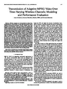
Fig. 1. Multiple-reaction monitoring chromatograms of extracted patient serum samples containing 0.21 g/L 11DC (A), 0.65 g/L 17OHPr (B), 0.19 g/L 17OHP (C), and 0.38 g/L Pr (D). Mass transitions (solid line, primary mass transition; dashed line, secondary mass transition): A, m/z 377.23124.1 and 3773112; B, m/z 3483330 and 3483312; C, m/z 3613124 and 3613112; D, m/z 332386 and 3323300.
samples from human subjects were approved by Institutional Review Board of the University of Utah.
method comparison We compared the LC-MS/MS method with an RIA for 17OHP (n ⫽ 87; Coat-a-count; Diagnostic Products Corporation) and an LC-MS/MS method (n ⫽ 10) of a commercial laboratory. Method comparisons for Pr (n ⫽ 58), 17OHPr (n ⫽ 35), and 11DC (n ⫽ 10) were performed with RIA methods of a commercial laboratory. To account for imprecision in both the comparison and the evaluated methods, the results were analyzed by Deming regression.
Results The ionization efficiency of nonderivatized adrenal steroids was studied in positive and negative ion modes with mobile phase containing various solvents and buffers and by use of electrospray ionization (ESI), heated nebulizer [atmospheric pressure chemical ionization (APCI)], and atmospheric pressure photoionization ion sources on an API3000 (Applied Biosystems/MDS Sciex) tandem mass spectrometer. Samples containing 10 g/L each of 11DC, 17OHP, 17OHPr, and Pr were injected in a flow of mobile phase (flow rate, 250 L/min). The absolute intensity of the [M⫹H]⫹ molecular ion of
5
Clinical Chemistry 52, No. 8, 2006
17OHP was highest with atmospheric pressure photoionization (toluene was used as a dopant) followed by APCI and ESI. The [M⫹H]⫹ molecular ion of 17OHP was observed with each ionization method, whereas Pr and 17OHPr were not detectable with any ionization technique, and the sensitivity for 11DC was approximately one third that of 17OHP. The poor ionization efficiency and collision-induced dissociation (CID) fragmentation of 11DC, 17OHPr, and Pr led us to evaluate the effect of derivatization on the sensitivity of MS/MS detection. The most promising derivatization of those evaluated was the oximation reaction, with hydroxylamine reacting with keto groups of the steroids to form oxime derivatives. Comparison of the signal intensity for nonderivatized steroids and the oxime derivatives in positive ion mode showed the best sensitivity with ESI. We found that ionization was more efficient with addition of formic acid to the mobile phase compared with the formate buffer (see Fig. 1 in the online Data Supplement). This was especially true for 17OHPr and Pr. The intensity of the molecular ion of 17OHPr oxime was also highly dependent on the declustering potential and the temperature in the ion source. The chromatograms of 2 multiple-reaction monitoring transitions for each compound in human serum are presented in Fig. 1 Total ion chromatograms of a sample from a healthy female patient and females with suspected 21-hydroxylase and 3b-hydroxysteroid dehydrogenase deficiencies are shown in Fig. 2 of the online Data Supplement. Experiments evaluating the ion suppression showed a decrease in the baseline at a retention time of 0.5 min, baseline recovery by 0.65 min, and no ion suppression at the retention times of the adrenal steroids (see Fig. 3 in the online Data Supplement). Concentrations of the steroids were determined by use of the first listed mass transition for each compound and its internal standard (Table 1). Additional mass transitions (listed second for each of the steroid and its internal standard) were used to assess the specificity of the detection. The calibrator containing 1.00 g/L each of the adrenal steroids was used to set a threshold for the branching ratio of the mass transitions for a batch of samples. The acceptance limit for the branching ratio was set at 30% for 11DC, 17OHP, and Pr and at 40% for 17OHPr when compared with the branching ratio set by the calibrators. Method performance characteristics are listed in Table 2, and the within-run, between-run, and total imprecision and recovery data obtained are shown in Table 2 of the online Data Supplement. The results of the method comparison are presented in Fig. 2. The branching ratios of the 2 mass transitions for 11DC, 17OHP, and Pr in all patient samples were within the acceptance limits. The branching ratio for 17OHPr was outside of the 40% limits in ⬃5% of samples from adults and up to 30% of samples from children. For the above
samples, an alternative mass transition (Table 1) was used for evaluation of the selectivity. Additional mass transitions were the same as the primary (m/z 3483330 for 17OHPr and m/z 3513333 for 17OHPr-d3) but were acquired at a collision energy 40% lower than the primary mass transitions. Steroids and metabolites of steroids (see Table 1 in the online Data Supplement) were evaluated for potential interference with the method. The steroids were analyzed at a concentration of 100 g/L, and the chromatograms were evaluated for the presence of peaks at the mass transitions used in the method. None of the evaluated steroids interfered with the method. Under the various storage conditions, 11DC and 17OHP were stable, but 17OHPr and Pr were not (see Fig. 4 in the online Data Supplement). No degradation of the steroids was observed after 3 freeze/thaw cycles. We observed no significant differences in analyte recovery for serum, EDTA plasma, or heparin plasma for 11DC, 17OHPr, and 17OHP, but the Pr concentration in the sample collected in sodium EDTA was 280% higher than the concentrations in serum and sodium-heparin plasma. Collection tubes containing EDTA should therefore not be used. The adrenal steroids appeared to be stable during 3 months of storage at ⫺20 °C. Results of analysis of the samples from volunteers grouped by Tanner stages and age are summarized in Tables 3 and 4. The main differences in the results grouped by Tanner stage between males and females were in the Pr concentration at Tanner stage 2, which was ⬃ 60% higher in females; 17OHP at Tanner stages 2 and 3, which was ⬃30% higher in females; and 11DC at Tanner stage 4 –5, which was ⬃40% higher in males than in females. In adult females (Table 4), 17OHPr concentrations were 50% lower and 17OHP concentrations were ⬃30% higher than in males.
Table 2. Summary of the method performance characteristics. 11DC
S/Na ratio Recovery,b % Absolute recovery of SPE,c % LOD,d g/L LOQ, g/L Upper limit of linearity, g/L
51 91.0 89 0.025 0.05 100
17OHP
17OHPr
Pr
12.6 97.2 100
8.5 99.3 98
69 96.5 100
0.025 0.05 100
0.10 0.25 40
0.025 0.05 100
a S/N, signal-to-noise ratio. Sample contained 0.50 g/L each of the adrenal steroids, and 20 pg was injected in HPLC column (n ⫽ 5). b Determined by analyzing serum samples with and without addition of 0.50 g/L each of the steroids (n ⫽ 2). c Determined by analyzing serum samples with internal standard added before and after SPE (n ⫽ 2). d LOD, limit of detection; LOQ, limit of quantification.
6
Kushnir et al.: LC-MS/MS Method for Adrenal Steroids
Fig. 2. Comparison of the evaluated method with RIAs (A–D) and LC-MS/MS (C) methods of commercial laboratories for 11DC (A), 17OHPr (B), 17OHP (C), and Pr (D). Regression statistics: (A), y ⫽ 1.41x ⫹ 0.08 g/L (r ⫽ 0.985; Sy兩x ⫽ 0.09 g/L; (B), y ⫽ 1.07x ⫺ 0.09 g/L (r ⫽ 0.955; Sy兩x ⫽ 0.76 g/L); (C), for RIA (‚), y ⫽ 1.76x ⫹ 0.42 g/L (r ⫽ 0.986; Sy兩x ⫽ 0.95 g/L); for LC-MS/MS (u), y ⫽ 1.01x ⫺ 0.009 g/L (r ⫽ 1.00; Sy兩x ⫽ 0.14 g/L); (D), y ⫽ 0.47x ⫹ 0.48 g/L (r ⫽ 0.310; Sy兩x ⫽ 0.54 g/L).
Discussion Despite structural similarities among adrenal steroids, the efficiencies of soft ionization methods (ESI and APCI) for their detection were very different. To date there have been no published LC-MS methods for quantification of endogenous concentrations of 17OHPr and Pr. This is most likely related to poor ionizability of the molecules caused by the position of the double bond within the structure. In 11DC and 17OHP, the double bond is located in ring A, whereas in Pr and 17OHPr it is located in ring B. The steroids included in the method contain 1 (17OHPr and Pr) and 2 (11DC and 17OHP) keto groups within the structure, which if derivatized may enhance detection (33 ). Improvements in sensitivity of detection of adrenal steroids were achieved through formation of oxime derivatives and optimization of the sample preparation and ionization. Formation of the double oxime derivatives of 11DC and 17OHPr requires more stringent conditions
(higher temperature and longer incubation time) than does formation of derivatives of 17OHP and Pr (results not presented). The greatest intensities of the molecular ions among the evaluated ionization techniques were for oxime derivatives of adrenal steroids with an ESI ion source and a mobile phase consisting of methanol and 0.022 mol/L formic acid (see Fig. 1 in the online Data Supplement). The oxime derivatives of adrenal steroids were efficiently ionized at relatively low voltage. Under these conditions, most other compounds present in serum would not be well ionized, giving greater specificity. The ease of ionization of the oxime derivatives can be explained by the presence of the resonance structures (34 ) promoted by the delocalization of charge within the molecule (see Scheme 1 in the online Data Supplement). In the oxime group, the nitrogen atom is bound to a carbon by a double bond. The angle between the nitrogen and the hydroxyl group is
Clinical Chemistry 52, No. 8, 2006
Table 3. Reference intervals (g/L) for 11DC, 17OHP, 17OHPr, and Pr by Tanner stage.a Females Tanner Tanner Tanner Tanner Males Tanner Tanner Tanner Tanner a
n
11DC
17OHPr
17OHP
Pr
1 2 3 4–5
181 76 98 104
⬍0.93 ⬍1.36 ⬍0.97 ⬍0.49
⬍2.35 ⬍3.67 ⬍4.30 0.26–4.12
⬍0.74 ⬍1.64 0.12–2.09 0.07–1.70
0.15–1.53 0.22–2.29 0.34–2.15 0.26–2.35
1 2 3 4–5
171 80 83 122
⬍1.05 ⬍1.06 ⬍1.11 ⬍0.81
⬍2.08 ⬍3.55 ⬍4.50 0.35–4.78
⬍0.62 ⬍1.04 ⬍1.51 0.20–1.73
0.13–1.55 0.12–1.43 0.16–2.14 0.19–2.01
Results are the nonparametric estimates of the central 95% interval.
⬃120 degrees; this leads to the formation of isomers that differ by the orientation of the ⫺OH group. Structures b and c (see Scheme 1 in the online Data Supplement) allow free rotation along the carbon–nitrogen bond, whereas the rotation between the atoms is restricted in structures a and d. We confirmed the presence of isomers by resolving the isomers of the analogous testosterone oxime with high-field asymmetric wave ion mobility LC-FAIMSMS/MS (35 ). In the adrenal steroid biosynthesis pathway, Pr is produced directly from cholesterol and is the precursor for all C18, C19, and C21 steroids. A large portion of Pr is present in blood in a conjugated form. All evaluated types of extraction other than direct SPE with samples at physiologic pH caused partial hydrolysis of Pr conjugates with consequent increases in the concentration of conjugated Pr compared with the concentration of unconjugated Pr. Chromatographic separation plays an important role in method performance. The Synergy Fusion column (C18 with imbedded polar end-capping) provided sufficient Table 4. Reference intervals (g/L) for 11DC, 17OHP, 17OHPr, and Pr by age.a Age, years
Females 7–9 10–11 12–13 14–15 16–17 18–51b Males 7–9 10–11 12–13 14–15 16–17 18–52 a b
n
11DC
17OHPr
17OHP
Pr
126 83 84 83 83 56
⬍0.93 ⬍1.06 ⬍1.34 ⬍1.05 ⬍0.47 ⬍0.41
⬍2.12 ⬍3.20 ⬍3.62 0.28–4.22 0.25–4.19 ⬍2.08
⬍0.71 ⬍0.89 ⬍1.65 0.12–2.08 ⬍1.79 0.10–1.98
0.14–1.50 0.15–1.97 0.22–2.20 0.23–2.14 0.22–2.29 0.17–1.30
126 83 83 84 84 44
⬍1.18 ⬍1.06 ⬍0.93 ⬍0.92 ⬍1.05 ⬍0.50
⬍1.87 ⬍3.87 ⬍3.62 0.32–4.28 0.31–4.78 0.36–4.09
⬍0.63 ⬍0.70 ⬍1.05 ⬍1.52 ⬍1.93 0.25–1.39
0.13–2.05 0.12–1.51 0.18–1.70 0.17–1.98 0.16–2.29 0.28–1.65
Results are the nonparametric estimates of the central 95% interval. Premenopausal.
7
retention and separation of the oxime derivatives of adrenal steroids and did not cause peak splitting for any of the steroids except for partial resolution of the 17OHPoxime diastereomers. To assess selectivity of the analysis in MS/MS, it is advantageous to use multiple mass transitions for the analytes of interest (34, 36 –39 ). The CID mass spectra of 11DC, 17OHP, and Pr contained 2 unique product ions originating from loss of large fragments of the molecule, whereas the CID mass spectrum of 17OHPr was not as specific and the only major product ions in the mass spectrum resulted from the loss of 1 and 2 molecules of water. To enhance the selectivity, we evaluated a 2-stage fragmentation MS/MS/MS by use of the ion trap function of the API4000 mass spectrometer with the parent ion (m/z 348), intermediate product ion (m/z 330), and the final product ion (m/z 312). MS/MS/MS provided slightly better specificity and greater signal intensity; however, measurement imprecision increased by more than 3-fold. Consequently, for the analysis of 17OHPr, we used MS/MS fragmentation with the mass transitions m/z 3483330 and m/z 3483312. The method used 2 mass transitions for each adrenal steroid and branching ratios, which were calculated from the transitions, and enabled evaluation of the specificity of the analysis in every sample (36 ). Evaluation of sample stability during storage at different conditions showed rapid degradation of 17OHPr and Pr (see Fig. 4 in the online Data Supplement) at refrigerator temperatures and, especially, at ambient conditions. This suggests that samples for analysis of 17OHPr and Pr should be frozen immediately after collection and remain frozen until analysis. All of the steroids were stable when the samples were stored at ⫺20 °C or below. Method comparisons with RIA for 17OHPr and LCMS/MS for 17OHP indicated good agreement between the methods (Fig. 2). Comparison with RIA for 11DC and 17OHP showed good correlation but produced 41% and 76% overestimation of the concentrations, respectively, by the RIAs. Poor correlation was observed for the comparison with RIA for Pr. Calibrators used in the LC-MS/MS method were added to adrenal-steroid–free matrix and analyzed by the RIAs. The results showed good agreement with the expected values. This suggests that discrepancies between the methods are related to matrix effects and cross-reactivities in the RIAs (15–17 ). In summary, we have developed a highly sensitive and specific LC-MS/MS method for measurement of 11DC, 17OHP, 17OHPr, and Pr in serum or plasma. This method allows measurement of endogenous concentrations of adrenal steroids in all age groups of females and males. Measurements of adrenal steroids by LC-MS/MS are precise, and the method has low limits of quantification. With an instrumental analysis time of 7 min, the assay is a good alternative to immunoassays. We found no interferences among the compounds tested. Sample prepara-
8
Kushnir et al.: LC-MS/MS Method for Adrenal Steroids
tion is specific to neutral molecules containing keto groups. Compared with GC-MS methods for adrenal steroid analysis, the LC-MS/MS method offers better selectivity and limits of detection and quantification, along with higher throughput and reduced sample volume requirements. The small sample volume makes the method particularly useful for pediatric testing. For the first time, reference intervals for 11DC, 17OHPr, and Pr for males and females of different Tanner stages and age groups were established by an isotope-dilution LCMS/MS method.
13.
14.
15.
16.
We thank ARUP Institute for Clinical and Experimental Pathology for financial support, John Simmons for assistance with determining the Tanner stages of the children, and Pedrico Sese and Beth J. Cottam for assistance with the reference interval study.
References 1. Grumbach MM, Hughes I, Conte FA. Disorders of sexual differentiation. In: Larsen PR, Kronenberg HM, Melmed S, Polonsky KS, eds. Williams Textbook of Endocrinology, 10th ed. New York: Saunders, 2002:842–1002. 2. Fitness J, Dixit N, Webster D, Torresani T, Pergolizzi R, Speiser PW, et al. Genotyping of CYP21, linked chromosome 6p markers, and a sex-specific gene in neonatal screening for congenital adrenal hyperplasia. J Clin Endocrinol Metab 1999;84:960 – 6. 3. De Peretti E, Forest MG. Pitfalls in the etiological diagnosis of congenital adrenal hyperplasia in the early neonatal period. Horm Res 1982;16:10 –22. 4. Simard J, Durocher F Mebarki F, Turgeon C, Sanchez R, Labrie Y, et al. Molecular biology and genetics of the 3-hydroxysteroid dehydrogenase/delta5-delta4 isomerase gene family. J Endocrinol 1996;150:S189 –207. 5. Pang S. Congenital adrenal hyperplasia owing to 3-hydroxysteroid dehydrogenase deficiency. Endocrinol Metab Clin North Am 2001; 30:81–99. 6. Sahin Y, Kelestimur F. The frequency of late-onset 21-hydroxylase and 11b-hydroxylase deficiency in women with polycystic ovary syndrome. Eur J Endocrinol 1997;137:670 – 4. 7. Bentsen D, Schwartz DS, Carpenter TO. Sonography of congenital adrenal hyperplasia due to partial deficiency of 3-hydroxysteroid dehydrogenase: a case report. Pediatr Radiol 1997;27:594 –5. 8. Moran C, Potter HD, Reyna R. Prevalence of 3-hydroxysteroid dehydrogenase-deficient nonclassic adrenal hyperplasia in hyperandrogenic women with adrenal androgen excess. Am J Obstet Gynecol 1999;181:596 – 600. 9. Asuncion M, Calvo RM, San Millan JL, Sancho J, Avila S, EscobarMorreale HF. A prospective study of the prevalence of the polycystic ovary syndrome in unselected Caucasian women from Spain. J Clin Endocrinol Metab 2000;85:2434 – 8. 10. Pang S, Hotchkiss J, Drash AL, Levine LS, New MJ. Microfilter paper method for 17 hydroxy-progesterone radioimmunoassay: its application for rapid screening for congenital adrenal hyperplasia. J Clin Endocrinol Metab 1997;45:1003– 8. 11. Speiser PW, White PC. Medical progress: congenital adrenal hyperplasia. N Engl J Med 2003;349:776 – 88. 12. Dressendorfer RA, Strasburger CJ, Bidlingmaier F, Klug I, Kistner A, Siebler T, et al. Development of a highly sensitive nonisotopic immunoassay for the determination of salivary 17-hydroxyproges-
17. 18.
19.
20.
21.
22.
23.
24.
25.
26.
27.
28.
29.
30.
terone: reference ranges throughout childhood and adolescence. Pediatr Res 1998;44:650 –5. Riepe FG, Mahler P, Sippell WG, Partsch C-J. Longitudinal study of plasma pregnenolone and 17-hydroxypregnenolone in full-term and preterm neonates at birth and during the early neonatal period. J Clin Endocrinol Metab 2002;87:4301– 6. Riepe FG, Wonka S, Partsch C-J, Sippell WG. Automated chromatographic system for the simultaneous measurement of plasma pregnenolone and 17-hydroxypregnenolone by radioimmunoassay. J Chromatogr B Biomed Appl 2001;763:99 –106. Wong T, Shackleton CH, Covey TR, Ellis G. Identification of the steroids in neonatal plasma that interfere with 17␣-hydroxyprogesterone radioimmunoassays. Clin Chem 1992;38:1830 –7. Speiser PW. Improving neonatal screening for congenital adrenal hyperplasia. J Clin Endocrinol Metab 2004;89:3685– 6. Taieb J, Benattar C, Birr AS, Lindenbaum A. Limitations of steroid determination by direct immunoassay. Clin Chem 2002;48:583–5. Shimozawa K, Saisho S, Yata J, Kambegawa A. Age-related changes in serum 17-hydroxypregnenolone sulfate concentrations in human infancy and childhood. Endocrinol Jpn 1988;35:189 – 95. Ja¨nne O, Perheentupa J, Viinikka L, Vihko R. Plasma pregnenolone, progesterone, 17-hydroxyprogesterone, testosterone and 5-dihydrotestosterone in different types of congenital adrenal hyperplasia. Clin Endocrinol 1975;4:39 – 48. McKenna TJ, Jennings AS, Liddle GW, Burr IM. Pregnenolone, 17-OH-pregnenolone, and testosterone in plasma of patients with congenital adrenal hyperplasia. J Clin Endocrinol Metab 1976;42: 918 –25. Hill M, Lukac D, Lapcik O, Sulcova J, Hampl R, Pouzar V, et al. Age relationships and sex differences in serum levels of pregnenolone and 17-hydroxypregnenolone in healthy subjects. Clin Chem Lab Med 1999;37:439 – 47. Lee MM, Rajagopalan L, Berg GJ, Moshang T. Serum adrenal steroid concentrations in premature infants. J Clin Endocrinol Metab 1989;69:1133– 6. Doerr HG, Versmold HT, Bidlingmaier F, Sippell WG. Adrenocortical steroids in small-for-gestational-age term infants during the early neonatal period. Pediatr Res 1989;25:115– 8. Choi MH, Yoo YS, Chung BC. Measurement of testosterone and pregnenolone in nails using gas chromatography-mass spectrometry. J Chromatogr B Biomed Sci Appl 2001;754:495–501. Caulfield MP, Lynn T, Gottschalk ME, Jones KL, Taylor NF, Malunowicz EM, et al. The diagnosis of congenital adrenal hyperplasia in the newborn by gas chromatography/mass spectrometry analysis of random urine specimens. J Clin Endocrinol Metab 2002 87:3682–90. Shindo N, Yamauchi N, Murayama K, Fairbrother A, Korlik S. Identification of 17-hydroxyprogesterone and other steroid hormones in saliva from a normal child and patients with congenital adrenal hyperplasia by plasmaspray liquid chromatography/mass spectrometry. Biomed Chromatogr 1990;4:171– 4. Wudy S, Hartmann M, Svoboda M. Determination of 17-hydroxyprogesterone in plasma by stable isotope dilution/benchtop liquid chromatography-tandem mass spectrometry. Horm Res 2000;53:68 –71. Lai CC, Tsai CH, Wu JY, Lin WD, Lee CC. Rapid screening assay of congenital adrenal hyperplasia by measuring 17-hydroxyprogesterone with high-performance liquid chromatography/electrospray ionization tandem mass spectrometry from dried blood spots. J Clin Lab Anal 2002;16:20 –5. Kao PC, Machacek DA, Magera MJ, Lacey JM, Rinaldo P. Diagnosis of adrenal cortical dysfunction by liquid chromatographytandem mass spectrometry. Ann Clin Lab Sci 2001;31:199 –204. Liu S, Sjovall J, Griffiths WJ. Analysis of oxosteroids by nano-
Clinical Chemistry 52, No. 8, 2006
31.
32.
33.
34.
35.
electrospray mass spectrometry of their oximes. Rapid Commun Mass Spectrom 2000;14:390 – 400. Marsden D, Larson CA. Tandem mass spectrometry in detecting congenital adrenal hyperplasia [Editorial]. Clin Chem 2004;50: 467– 8. Lacey JM, Minutti CZ, Magera MJ, Tauscher AL, Casetta B, McCann M, et al. Improved specificity of newborn screening for congenital adrenal hyperplasia by second-tier steroid profiling using tandem mass spectrometry. Clin Chem 2004;50:621–5. Kushnir MM, Rockwood AL, Roberts WL, Pattison EG, Bunker AM, Fitzgerald RL, et al. Performance characteristics of a novel tandem mass spectrometry assay for serum testosterone. Clin Chem 2006;52:120 – 8. Kushnir MM, Komaromy-Hiller G, Shushan B, Urry FM, Roberts WL. Analysis of dicarboxylic acids by tandem mass spectrometry. High throughput quantitative measurement of methylmalonic acid in serum, plasma and urine. Clin Chem 2001;47:1993–2002. Kushnir MM, Rockwood AL, Kolakowski BM, Purves RW. Evalua-
36.
37.
38.
39.
9
tion of the analytical performance of FAIMS coupled to a tandem quadrupole mass spectrometer for analysis of testosterone. Proceedings of Conference on Mass Spectrometry and Allied Topics, Nashville 2004. Kushnir MM, Rockwood AL, Nelson GJ, Yue B, Urry FM. Assessing analytical specificity in quantitative analysis using tandem mass spectrometry. Clin Biochem 2005;38:319 –27. Meikle AW, Findling J, Kushnir MM, Rockwood AL, Nelson G J, Terry AH. Pseudo-Cushing syndrome caused by fenofibrate interference with urinary cortisol assayed by high-performance liquid chromatography. J Clin Endocrinol Metab 2003;88:3521– 4. Kushnir MM, Rockwood AL, Nelson GJ, Terry A, Meikle AW. Liquid chromatography-tandem mass spectrometry analysis of urinary free cortisol. Clin Chem 2003;49:965–7. Kushnir MM, Neilson R, Roberts WL, Rockwood AL. Cortisol and cortisone analysis in serum and plasma by atmospheric pressure photoionization tandem mass spectrometry. Clin Biochem 2004; 37:357– 62.