Academic Sciences
International Journal of Pharmacy and Pharmaceutical Sciences ISSN- 0975-1491
Vol 5, Suppl 2, 2013
Research Article
DEVELOPMENT AND VALIDATION OF A SENSITIVE AND ACCURATE METHOD FOR DETERMINATION OF ATORVASTATIN AND ROSUVASTATIN IN RAT PLASMA BY REVERSEDPHASE HIGH PERFORMANCE LIQUID CHROMATOGRAPHY WITH UV DETECTION 1*ENTIDHAR
J. AL- AKKAM, 1ALAA A. ABDUL RASOOL , 2ADNAN A. BADWAN , 2NAWZAT D. AL- JBOUR, 3NIDAL A. QINNA
*1Department of Pharmaceutics, College of Pharmacy, Baghdad University. Baghdad, Iraq, 2Jordanian Pharmaceutical Manufacturing Company, P.O. Box 94, Naur 1170, Jordan, 3Department of pharmacology, College of Pharmacy, Petra University. Amman, Jordan. Email:
[email protected] Received: 18 Feb 2013, Revised and Accepted: 04 Apr 2013 ABSTRACT Objective: To develop a new, accurate, precise and rapid isocratic reversed-phase high performance liquid chromatographic/ultraviolet (RPHPLC/UV) method for the determination of atorvastatin (ATV) and rosuvastatin (RST) in rat plasma using diclofenac sodium and naproxen sodium, as an internal standard (IS), respectively. Methods: ATV and RST were analyzed on a BDS hypersil C 18 column (250 mm× 4.6 mm, 5μm), applying methanol: water (68:32, v/v) and (63:37 v/v), respectively, in isocratic mode as a mobile phase. Its pH was adjusted to 3.0 with trifluroacetic acid at a flow rate of 1 ml/min. The peak response was monitored at 241nm after injecting a 100μl sample into HPLC system. The direct liquid–liquid extraction procedure was applied to rat plasma samples using methyl-tert-butyl ether as an extraction solvent after protein precipitation with ammonium acetate buffer. The different HPLC experimental parameters were optimized and the method was validated according to standard guidelines. Results: A peak area was obtained for ATV and RST with 11.35 and 6.65min retention time, respectively, while their respective IS appeared at 15.73 and 10.463min. The calibration curves were linear over concentration ranges of 20-200ng/ml of ATV and RST. The lower limit of detection (LLOD) values were found to be 1.35 and 7.2ng/ml, for ATV and RST, respectively, while their respective lower limit of quantification (LLOQ) values were 10.3 and 8.5ng/ml. The average recoveries of the suggested method in rat plasma were 96.48% and 91.43%, for ATV and RST, respectively. The percent of relative standard deviation (%RSD) of ATV in plasma was in the range of 0.124–0.536 % and 0.194–1.071%, for intra-day and inter-day studies, respectively, while for RST was in the range of 0.218-1.909% and 0.217-1.971%. Conclusion: The method was found sensitive, accurate, and precise in rat plasma and could be applied for the quantification of these compounds in plasma and other biological fluids such as liver homogenate samples, for pharmacokinetics and drug-drug interaction studies, using animal models. Keywords: Atorvastatin, Rosuvastatin, Statin, RP- HPLC and validation, Plasma.
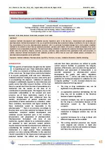
INTRODUCTION Atorvastatin calicum is ([R-(R*, R*)]-2-(4-fluorophenyl)-β, δdihydroxy-5(1-methylethyl)-3-phenyl-4-[(phenylamino)carbonyl]1H-pyrrole-1-heptanoicacid, calcium salt (2:1) trihydrate) (Fig.1) [1]. and rosuvastatin calcium is bis[(E)-7-[4-(4 fluorophenyl)-6isopropyl-2-[methyl(methylsulfonyl)amino]pyrimidin-5-yl](3R,5S)3,5 dihydroxyhept-6-enoic acid] calcium salt (Fig.1) [2] which belong to the statin class of drugs used to treat hypercholesterolemia both in patients with established cardiovascular disease as well as those who are at a high risk of developing atherosclerosis [3-5]. Statins exert their major effect – reduction of LDL level – through a mevalonic acid – the moiety that competitively inhibits HMG-Co A reductase. By reducing the conversion of HMG – Co A to mevalonate, statins inhibit an early and rate – limiting step in cholesterol biosynthesis [6-8]. The associated reduction in intracellular cholesterol concentration induces LDLreceptor expression on the hepatocyte cell surface, which results in increased extraction of LDL-C from the blood and decreased circulating LDL-C concentrations [7]. In addition statins cause reduction in total cholesterol (TC) and triglycerides (TG) and elevation in high-density lipoprotein-C (HDL-C) [3, 9]. Besides lipid lowering effects, statins also have potential roles independent of cholesterol reduction as anti-oxidative [10, 11], antitumor [12, 13], anti-inflammatory [14, 15], immune-modulator [15, 16], antimalarial [17, 18], as antifungal [19] and bone forming agents [8, 20, 21]. Thus, due to their so many beneficial effects, there is growing interest in developing analytical methods for statins monitoring. Until the approval of RST in 2003, ATV was the most efficacious drug in the statins class [22], but recent studies reported RST as a potent inhibitor of HMG-Co A reductase having a higher LDL lowering effects as compared with other statins [23,24], which demonstrates that both RST and ATV are the leading drugs in the statins class [25].
Atorvastatin calcium [1]
Rosuvastatin calcium [2] Fig. 1: It shows chemical structure of atorvastatin and rosuvastatin. To date several HPLC–UV and mass spectrophotometric methods have developed for the quantification of both ATV and RST either alone or in combination with other drugs in different matrices. ATV
Entidhar et al. Int J Pharm Pharm Sci, Vol 5, Suppl 2, 211-219 has been determined along with its metabolites using LC/MS in biological matrices [26, 27]. HPLC-UV methods have also been reported for the determination of ATV alone in human serum [28, 29], pharmaceutical preparations [30, 31], along with impurities in pharmaceutical preparations [32] and in combination with amlodipine [33, 34], nicotinic acid [35], aspirin [36], olmesartan [37], and ezetimibe [38, 39] in dosage forms. Similarly, various analytical methods have been reported for the determination of RST including, spectrophotometric and HPLC-UV [40- 43]. The simultaneous quantification of RST with gemfebrozil in human plasma [44] and with other drugs including statins in pharmaceutical preparations has been determined [45, 46], also the simultaneous analysis of RST and ATV in human serum using HPLCUV has been studied [47].
prepared in methanol. Working standard solutions of ATV and RST were prepared by serial dilution of stock solutions by their respective mobile phase, (methanol- water, 68:32 v/v for ATV and 63:37v/v for RST), at a concentration range of 20-100ng/ml of each drug, keeping IS concentration 100ng/ml and 40ng/ml, for diclofenac sodium (as IS for ATV) and naproxen sodium (as IS for RST), respectively, in each sample. Calibration curves were constructed for the standard solutions of each drug after HPLC/UV analysis. Also another calibration curve were drown at the concentration range of 20 – 200 ng/ml for ATV and RST keeping the same concentration of their respective IS, diclofenac sodium and naproxen sodium in each sample.
The aim of this new suggested method is to develop a sensitive, accurate and validated HPLC-UV method for the determination of ATV and RST in the presence of their respective internal standard, diclofenac sodium and naproxen sodium, in rat plasma at a very low concentration in ng/ml by using HPLC with UV detector, which was considered as a challenge.
Blood samples were collected from rats, in plastic tubes and centrifuged at 3000 rpm, 4◦ C for 20min. The plasma was collected and stored at −20 ◦ C until analysis. For sample preparation the plasma was first thawed at room temperature and a volume of 200μl was transferred to a glass tube and spiked with 50μl of standard solutions containing concentrations ranging from 0.2 – 1μg/ml of ATV or RST and a constant concentration of diclofenac sodium 1μg/ml or naproxen sodium 0.4μg/ml as IS for ATV and RST, respectively, in each solution. Blank plasma samples were spiked with 50μl of mobile phase. The samples were vortex- mixed for 30 sec. to be applied to the liquid–liquid extraction procedure.
The method was validated according to standard guidelines and various experimental parameters were optimized with the aim that the reported method could be applied for routine laboratory analysis of these statins and for the determination of pharmacokinetic and drug–drug interaction studies of these drugs in human and animal models.
Plasma sample preparation
MATERIALS AND METHODS
Another concentrations ranging from 1.2 – 2μg/ml of ATV and RST keeping the same concentration of their respective IS were also prepared and the plasma samples were spiked with.
Materials and reagents
Liquid-liquid extraction
Atorvastatin calcium, Rosuvastatin calcium (Biocon, India), Diclofenac sodium, Naproxen sodium (Dr. Reddy’s Co., India), Methanol, Methyl-tert-butyl ether, HPLC grade (Fisher Scientific UK, Bishop Meadow Road, Loughborough, Leics, UK), Acetic acid, glacial, biochemical grade 99.8% , Trifluroacetic acid 99% extra pure (ACROS organics, New Jersey, USA. Geel, Belgium), Ammonium acetate (Merck KGaA, Germany).
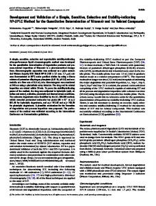
Drugs were extracted from plasma samples using liquid-liquid extraction technique. Ammonium acetate buffer (100μl of 100mM, pH 5, adjusted by glacial acetic acid) was added to each plasma sample for protein precipitation. The samples were then vortexmixed for 1min.Then methyl-tert-butyl ether 4 or 6ml was added for extraction of ATV or RST, respectively, then vortex-mixed for 2min and centrifuged at 4100 rpm, 4◦ C for 10 min. The clear supernatant layer was separated in a glass test tube and evaporated to complete dryness under the gentle stream of nitrogen at 40°C. After drying, the residue was reconstituted in 500μl of mobile phase. Samples were vortex-mixed for 1min then put in insert tubes inside the prelabeled HPLC vials and 100μl were injected into the HPLC column for analysis (Fig. 2).
Instruments Reverse-phase-performance liquid chromatography(HPLC-UV) (Thermo scientific spectra system UV 1000, Japan), Balance AG245( Mettler Toledo), Sonicater-Sonica® ultrasonic cleaner (solTec), pH/ Conductivity meter MPC227( Mettler Toledo, Taawon), ARE Heating Magnetic stirrer VELP ( Scientifica), Water bath GFL, Centrifuge 5702 R eppendorf, high speed refrigerated centrifuge, (Germany), Vortex VELP Scientifica (Europe). Preparation of standard solutions Stock solutions of ATV, RST and their respective internal standard, diclofenac sodium and naproxen sodium, each 1mg/ml were
Calibration curves were constructed for spiked plasma samples containing ATV or RST in the range of 20–100ng /ml at 20, 40, 60, 80 and 100ng/ml for each drug with diclofenac sodium 100ng/ml or naproxen sodium 40ng/ml as IS. A calibration curve for spiked plasma samples containing RST in the range of 120-200ng/ml at 120, 140, 160, 180 and 200ng/ml RST, using naproxen sodium as IS 40ng/ml, was also drawn.
Fig. 2: It shows scheme for extraction procedure of atorvastatin and rosuvastatin from rat plasma.
212
Entidhar et al. Int J Pharm Pharm Sci, Vol 5, Suppl 2, 211-219 Chromatographic conditions The HPLC analysis of the studied compounds was performed at room temperature using methanol-water (68:32 v/v for ATV and 63:37v/v for RST), as a mobile phase. Its pH was adjusted to 3.0 with trifluroacetic acid. The mobile phase was always clarified by filtration through a nylon filter paper, with pore size equal to 0.45μm, and degassed through a sonicator, then pumped at flow rate of 1 ml/min, in isocratic mode on hypersil BDS C18 column (250 mm ×4.6 mm, 5μm). The peak response was monitored at a wave length of 241nm. The sample (100μl) was injected into HPLC system and the data was acquired using Chromquest Workstation Software.
200ng/ml of ATV or RST with their respective IS, diclofenac sodium 100ng/ml or naproxen sodium 40ng/ml). Calibration curves were constructed for ATV and RST in the mobile phase and spiked plasma samples by plotting their relative area (ratio of peak area of drug to peak area of IS) against their respective concentrations using a linear least squares regression analysis. Specificity/selectivity
Optimization parameters:
The specificity/selectivity of the analytical method was investigated by confirming the complete separation and resolution of the required peaks area of the studied drugs and their respective IS in mobile phase and rat plasma samples spiked with appropriate concentration of each compound.
Extraction method optimization
LLOD and LLOQ
Parameters were used for optimization of extraction include the following:
Detection and quantification limits were determined through dilution method using S/N approach by injecting a 100μl sample. LLOD was considered as the minimum concentration with a signal to noise ratio of at least three (S/N≈3), while LLOQ was taken as a minimum concentration with a signal to noise ratio of at least ten (S/N≈10).
Change the volume of ammonium acetate buffer (100mM, pH 5), used for precipitation of plasma protein (100 or 200μl were used).
Change the volume of methyl-tert-butyl ether, used as extracted solvent (2, 3, 4, 6, or 7ml).
Change the steps of extraction (single step of extraction or two steps of extraction were studied).
All liquids used in extraction procedure should be pre-filtered to reduce the interference, also only glass equipments were used.
Chromatographic conditions optimization: Mobile phase ratio The mobile phase ratio was optimized in isocratic mode for the analysis of ATV and RST. Different ratio were studied, such as,50:50, 55:45, 60:40, and 68:32 methanol :water for ATV, while, for RST, other ratio was studied such as 63:37, 65:35, 68:32 and 70:30. The mobile phase ratio that resulted in a better resolution and higher recovery of the studied compounds was selected as the best mobile phase ratio for the analysis. Mobile phase flow rate The mobile phase flow rate was adjusted in isocratic mode for the analysis of studied compounds after applying various flow rats in the range of 0.5–1.5 ml/min. in order to get better separation. Internal standard Different compounds including diclofenac sodium, levostatin, and naproxen sodium were tried to be used as IS. The compound that showed better compatibility, best recovery and shorter analysis time was selected as IS for the suggested method. Sample size The sample loop size was evaluated in the range of 50–100 μl to adjust the sample size and minimize the problems like column loading and lack of sensitivity of the mentioned compounds. Detector’s wavelength For determination of ATV and RST using diclofenac sodium and naproxen sodium, respectively, as IS the detector’s wavelength was evaluated in the range of 239–247 nm. The wavelength that resulted in the optimal sensitivity and better resolution was chosen as the wavelength for analysis of studied compounds. Validation of the method:
Accuracy Accuracy of the suggested method was determined in term of percent recovery. Spiked rat plasma Samples were prepared and extracted to get four different concentration levels at 20, 60, 80 and(100ng/ml) of ATV, keeping the diclofenac sodium concentration constant (100ng/ml) in each sample, and (20, 40, 80 and 100ng/ml of RST), keeping the naproxen sodium concentration constant (40ng/ml) in each sample. Another set of standard mixtures of ATV and RST, at the same concentration levels, was also prepared in the mobile phase (methanol: water, 68:32, v/v for ATV and 63:37v/v for RST).The samples were injected into the HPLC system in triplicate. The percent recovery for ATV and RST was calculated using the following equation: % Recovery = [A] × 100 / [B] Where [A] is the net peak area of the drug in plasma sample, [B] is the peak area of the drug in standard mixture. Precision Method precision was determined both in terms of repeatability (injection and analysis) and intermediate precision (intra - day and inter-days reproducibility). In order to determine injection repeatability, five samples of both standard solutions (prepared in mobile phase) and extracted spiked plasma of ATV and RST were prepared individually at (20, 40, 60, 80 and100ng/ml) with their respective constant concentration of IS (diclofenac sodium at 100ng/ml for ATV and naproxen sodium 40ng/ml for RST), then each sample was injected four times into HPLC system. Repeatability of the relative area (ratio of peak area of drug to the peak area of their respect IS) was determined and expressed as mean ± standard deviation (SD) and percent relative standard deviation (%RSD) calculated from the data obtained as a precision of the suggested method. For the intermediate precision (intra-day and inter-days reproducibility), standard solutions in mobile phase and spiked plasma samples of ATV and RST at the five different concentration levels with their respective constant concentration of IS, were prepared and analyzed at three times a day and over three consecutive days. The relative peak areas were expressed as mean ±SD and %RSD calculated from data obtained. Stability
The proposed analytical method was validated according to standard guidelines [48, 49]with respect to the following parameters.
The stability studies of ATV and RST standard solutions and spiked plasma samples were carried out over a period of 24 hr at 25 ◦ C (room temperature under laboratory light) and at 2–8 ◦ C (refrigerator).
Linearity
RESULTS AND DISCUSSION
The linearity of the proposed method was established from the calibration curves constructed at several concentration levels (20-
The suggested method is new in the sense that determination of ATV and RST was carried out at ng/ml concentrations accurately
213
Entidhar et al. Int J Pharm Pharm Sci, Vol 5, Suppl 2, 211-219
0
0
-20
Volts
10.697
6.135
8.757 Atorvastatin 10.017
Name Retention Time
Volts
20
18.577
UV1000-241nm
16.257
20
accurate and quite specific and sensitive for the analysis of these compounds in mobile phase and plasma samples. Complete separation of ATV and RST was achieved at 11.35 and 6.65 min, while their respective IS, diclofenac sodium and naproxen sodium were separated at 15.73 and 10.465min, respectively by using the proposed method. 11.323
using HPLC with UV detector. The experimental parameters of the method were optimized and the method was validated according to standard guidelines [48, 49]. ATV and RST were separated applying the proposed method in standard solutions and spiked plasma samples with little interference was shown with blank plasma samples (figure 3). The suggested method was found
-20 0
2
4
6
8
10
12
14
16
18
20
Minutes
Fig. 3: It shows chromatogram of blank plasma sample. Optimization of extraction method The highest percent of recovery of ATV and RST from spiked plasma samples and the lowest interference with blank plasma samples were obtained after optimization of extraction method by using 100μl of ammonium acetate buffer (100Mm, pH5), for Protein precipitation and 4 or 6ml of methyl-tert-butylether for extraction of ATV or RST, respectively, by applying single step of extraction which increase the recovery more than 10%. Optimization of chromatographic conditions Different experimental parameters were optimized in the specified ranges to choose the optimum mobile phase ratio, mobile phase flow rate, suitable internal standard, sample size, detector’s wavelength, required to attain better resolution and higher recovery for ATV and RST. The best results were obtained by using mobile phase comprised of methanol-water in a ratio of 68: 32 (v/v) and 63:37 was selected for the analysis of ATV and RST, respectively. The selected mobile phase ratio resulted in better recoveries of ATV and RST as compared with other ratios. The retention times of the studied compounds decreased with increasing the ratio of methanol in the mobile phase. The overall analysis time decreased significantly with increasing the methanol content. This behavior may be due to change in the solubility of the analytes in the mobile phase or may be due to change in binding of the analytes to the stationary phase (column). Since both ATV (pKa = 4.46) and RST (pKa = 4.6) and are acidic compounds so their retention on the column is likely to be pH dependant. Therefore, pH 3.0 was chosen as optimum pH because of the reasonable retention times, resolution and separation of all the compounds of interest.
retention time and poor recovery with the selected solvent of RST. Therefore, naproxen was preferred as IS for RST on the basis of good resolution, compatibility and comparatively better recovery. A 100μl, sample size, gave a reasonable peak area for all analytes and was chosen as a suitable sample size for the suggested method. Detector’s wavelength has been selected after recording the sensitivities of the compounds at various wavelengths. The greater sensitivities of ATV and RST were recorded at 241nm. Method validation Linearity The linearity of the method was evaluated from the calibration curve of standard solutions and spiked plasma samples constructed at several concentration levels of ATV and RST. The relative area (ratio of peak area of the drug to the peak area of its respective IS) was yielded a linear correlation over a concentration range of 20 – 200ng/ml Calibration curves of standard solutions and spiked plasma samples with the regression equation and their respective correlation coefficient (R2) of ATV and RST are shown in figure 4 -7. Specificity/selectivity Representative chromatogram of blank plasma was confirmed the presence of very little interference from the endogenous component (Fig 3). Chromatograms of standard solutions and spiked plasma samples of ATV (Fig. 8) and RST (Fig. 9) at concentration ranging from 20–100ng/ml with their respective IS at a constant concentration confirming that ATV and its IS diclofenac sodium, also RST and its IS naproxen sodium were well resolved and completely separated at retention times of 11.35, 15.73min for ATV and diclofenac sodium and 6.65, 10.75min for RST and naproxen sod., respectively.
Similarly, the flow rate of mobile phase greatly affected the analysis of the studied compounds. Although run time decreased significantly at higher flow rates along with better resolution of the peaks, however; sensitivities of the analytes decreased. The flow rate of 1ml/min was chosen as optimal flow rate to get better separation for these compounds.
LLOD and LLOQ
Internal standard that has shown better recovery, stability and compatibility with ATV and RST was chosen among the various tested compounds. However, diclofenac sodium was used as IS for ATV, as it resulted in better recovery and good compatibility in comparison with other tested compounds. While, diclofenac sodium had a very large
Accuracy of the proposed method was determined on the basis of percent recovery at four concentration levels 20, 60, 80 and 100ng/ml for ATV and at 20, 40, 80, and 10ng/ml for RST. The average percent recoveries for ATV and RST were found to be 96.478% and 91.431%, respectively, as shown in table 1, 2.
The LLOD for ATV and RST standard solutions were found to be 1.35 and 7.2ng/ml, respectively, while LLOQ were found to be 10.3 and 8.5ng/ml, respectively. Accuracy
214
Entidhar et al. Int J Pharm Pharm Sci, Vol 5, Suppl 2, 211-219 Precision Precision data representing both repeatability (injection and analysis) and intermediate precision (intra-day and inter-days reproducibility) of standard solutions (prepared in mobile phase) and spiked plasma samples ATV and RST are summarized in tables 3 - 6. The intra-day %RSD values for the standard solutions of ATV and RST was in the range of 0.356 – 1.814 and 0.27–1.511, respectively. Similarly, their respective values for inter–day
studies were in the range of 0.867–1.883 and 0.262–1.511 for ATV and RST. Moreover, the intra-day %RSD values for spiked plasma samples of ATV and RST was in the range of 0.124–0.536 and 0.218–1.909, respectively. While, values for inter–day studies were in the range of 0.194–1.071 and 0.217–1.971, for ATV and RST, respectively. However, both intra-day and inter–days %RSD values for standard solutions and spiked plasma samples for ATV and RST were less than 2.0%, which indicates that the proposed method is precise.
Fig. 4: It shows calibration curve of atorvastatin with diclofenac sodium as internal standard in mobile phase (methanol: water 68:32).
Fig. 5: It shows calibration curve of atorvastatin with diclofenac sodium as internal standard in rat plasma.
Fig. 6: It shows calibration curve of rosuvastatin with naproxen sodium as internal standard in mobile phase (methanol: water 63:37).
215
Entidhar et al. Int J Pharm Pharm Sci, Vol 5, Suppl 2, 211-219
Fig. 7: It shows calibration curve of rosuvastatin with naproxen sodium as internal standard in rat plasma.
Fig. 8: It shows chromatogram representing complete resolution of Atorvastatin peak from its internal standard Diclofenac sodium peak at retention time 11.35 and 15.73min, respectively.
Fig. 9: It shows chromatogram representing complete resolution of rosuvastatin peak from its internal standard naproxen sodium peak at retention 6.65, 10.463 min, respectively.
216
Entidhar et al. Int J Pharm Pharm Sci, Vol 5, Suppl 2, 211-219 Table 1: It shows accuracy (% recovery) of atorvastatin from rat plasma. Concentration (ng/ml) 20 60 80 100
Area in mobile phase 4096.6 8910.3 11473.13 17416.8
area in plasma 4062.0 8046.6 11000.7 17516.0 Average
%Recovery 99.155 90.306 95.882 100.569 96.478
Table 2: It shows accuracy (% recovery) of rosuvastatin from rat plasma. Concentration (ng/ml) 20 40 80 100
Area in mobile phase 3775.136 7803.82 16122.38 23477.5
Area in plasma 3655.12 7305.5 14848.25 19532.0 Average
% Recovery 96.820 93.614 92.097 83.194 91.432
Table 3: It shows precision of atorvastatin with diclofenac sodium as internal standard in mobile phase (methanol: water 68:32). Concentration (ng/ml) 20 40 60 80 100
precision (repeatability) 0.375 ± 0.004, 1.184 0.659 ± 0.011, 1.663 0.967 ± 0.001, 0.171 1.282 ± 0.013, 0.999 1.659 ± 0.0152, 0.916
Intraday(reproducibility) 0.378 ± 0.006, 1.673 0.652 ± 0.012, 1.814 0.964 ± 0.003, 0.356 1.279 ± 0.012, 0.941 1.67 ± 1.022, 1.335
Interday (reproducibility) 0.377 ± 0.005, 1.322 0.654 ± 0.012, 1.883 0.969 ± 0.008, 0.867 1.281 ± 0.019, 1.505 1.663 ± 1.019, 1.150
Mean relative area ±SD, %RSD Table 4: It shows precision of atorvastatin with diclofenac sodium as internal standard in rat plasma. Concentration (ng/ml) 20 40 60 80 100
precision (repeatability) 0.426 ± 0.002, 0.54 0.696 ± 0.004, 0.637 1.142 ± 0.004, 0.335 1.522 ± 0.006, 0.371 1.793 ± 0.003, 0.145
Intraday(reproducibility) 0.425 ± 0.002, 0.514 0.692 ± 0.004, 0.536 1.143 ± 0.001, 0.124 1.512 ± 0.006, 0.427 1.791 ± 0.003, 0.166
Interday (reproducibility) 0.429 ± 0.005, 1.071 0.690 ± 0.004, 0.637 1.140 ± 0.002, 0.194 1.510 ± 0.008, 0.499 1.785 ± 0.009, 0.489
Mean relative area ±SD, %RSD Table 5: It shows precision of rosuvastatin with naproxen sodium as internal standard in mobile phase (methanol: water 63:37). Concentration (ng/ml) 20 40 60 80 100
precision (repeatability) 0.184 ± 0.001, 0.695 0.364 ± 0.001, 0.400 0.537 ± 0.007, 1.363 0.735 ± 0.002, 0.295 0.926 ± 0.003, 0.286
Intraday(reproducibility) 0.185 ± 0.002, 1.077 0.364 ± 0.002, 0.454 0.535 ± 0.008, 1.511 0.735 ± 0.002, 0.270 0.926 ± 0.003, 0.303
Interday (reproducibility) 0.186 ± 0.003, 1.511 0.364 ± 0.002, 0.480 0.535 ± 0.008, 1.489 0.735 ± 0.003, 0.368 0.926 ± 0.002, 0.262
Mean relative area ±SD, %RSD Table 6: It shows precision of rosuvastatin with naproxen sodium as internal standard in rat plasma. Concentration (ng/ml) 20 40 60 80 100
precision (repeatability) 0.197 ± 0.002, 0.773 0.398 ± 0.002, 0.411 0.613 ± 0.002, 0.259 0.771 ± 0.003, 0.041 0.994 ± 0.005, 0.497
Intraday(reproducibility) 0.203 ± 0.004, 1.909 0.403 ± 0.004, 0.914 0.614 ± 0.003, 0.459 0.772 ± 0.002, 0.218 0.998 ± 0.009, 0.918
Interday (reproducibility) 0.200 ± 0.004, 1.924 0.402 ± 0.004, 1.031 0.608 ± 0.012, 1.971 0.771 ± 0.002, 0.217 0.992 ± 0.014, 1.457
Mean relative area ±SD, %RSD Stability Results from the stability studies of both standard solutions and extracted spiked plasma samples of ATV and RST with their IS indicated that extracted spiked plasma samples were stable for 18 hr when stored at room temperature (25◦ C), and for 48hr when stored in refrigerator (2–8°C), after that it will be degraded. While, standard solutions were demonstrated stability for one week at 2–8 ◦ C. However, extracted spiked plasma samples, as well as all standard stock solutions and the corresponding working solutions were prepared on daily basis to ensure stability of all components. Application of the method The proposed validated HPLC/UV method will be applied, as a part of biochemical analysis, for assessing the pharmacokinetics and drugdrug interaction studies of ATV and RST in physiological fluids (plasma
and liver homogenate) using animal (rats) models later on. This method can also be applied for the quantification of these compounds in pharmaceutical preparations and in routine laboratory analysis with slight modification in the extraction procedure. CONCLUSION A new, simple, rapid and precise RP-HPLC/UV method was successfully developed for determination of ATV and RST in rat plasma. The method was validated according to standard guidelines and various experimental parameters were optimized. The extraction procedure exhibited excellent recoveries of ATV and RST. This method offers advantage for determination of two clinically important and widely prescribed statins in a HPLC/UV in ng/ml concentration which considered as a challenge.
217
Entidhar et al. Int J Pharm Pharm Sci, Vol 5, Suppl 2, 211-219 Overall, the proposed method provides high throughput for determination of ATV and RST in rat plasma with excellent accuracy, precision, selectivity and reproducibility.
19.
ACKNOWLEDGMENTS
20.
The authors express their sincere thanks to Jordanian Pharmaceutical Manufacturing Company, Naur, Jordan and College of Pharmacy, Petra University, Amman, Jordan, for providing the necessary facilities to carry out the work.
21.
REFERENCES 1.
2. 3.
4.
5.
6. 7. 8.
9.
10.
11.
12. 13.
14.
15. 16.
17.
18.
Merck index, an encyclopedia of chemicals, drugs, and biological. 4th ed., Maryadele J.O. Neil, Editor, Merck & Co. INC, USA (2006), p 864-865. Remington. The science and practice of pharmacy 21th ed., Lippincott Williams and Wilkins (2006), p1369. Law M. R., Wald N. J., Rudnicka A. R., Quantifying effect of statins on low density lipoprotein cholesterol, ischaemic heart disease, and stroke: systematic review and meta-analysis. Br.Med.J. 2003; 326: 1423-1429. Juan T., Ricardo C., Ricardo G., Lucía N., Miguel V., Eva D., Lipidlowering therapy with statins, a new approach to antiarrhythmic therapy, J. Pharmacology & Therapeutics. 2007; 114: 107–126. Lei Z., Shuning Z., Hong J., Aijun S., Yunzeng Z., Junbo G., Effects of Statin Treatment on Cardiac Function in Patients With Chronic Heart Failure: A Meta-Analysis of Randomized Controlled Trials. J. Clin. Cardiol. 2011; 34, 2: 117–123. Lennernas H., Clinical pharmacokinetics of atorvastatin. J.Clin. Pharmacokinet. 2003; 42: 1141–1160. Lippincott’s IIIustrated Reviews Pharmacology, 4th ed., Lippincott Williams and Wilkins (2009), p253-254. Pasquale M., Giustina V., Aurelio S., Francesco P., Ilenia P., Carmelna R., Salvatore D., Giovam B., Enrico C., Mevalonate pathway: Role of biphoslphonates and statins. J. Acta Medica Mediterranea. 2011; 27: 85-95. Srinivasa R., Prasad T., Mohanta G. P., Manna P. K., An Overview of Statins as Hypolipidemic Drugs. International Journal of Pharmaceutical Sciences and Drug Research. 2011; 3: 178-183. Ebru D.S., Eser Y.S., Deniz N.,Taner 0., Effect of atorvastatin therapy on oxidant-antioxidant status andatherosclerotic plaque formation. J. Vascular Health and Risk Management. 2011; 7: 333–343. Preston M., Mary F. W., Charles A. D., Robert F. J., Active Metabolite of Atorvastatin Inhibits Membrane Cholesterol Domain Formation by an Antioxidant Mechanism. Journal of Biological Chemistry. 2006; 281: 9337–9345. Carmen C., Bucharest, Romania, The statins as anticancer agents. Maedica - a Journal of Clinical Medicine. 2012; 7: 337. Sune F. N., Borge G. N., Stig E. B., Statin Use and Reduced Cancer Related Mortality. The New England Journal of Medicine. 2012; 367: 1792–1802. Anuradha G., Nancy C., Jeanette H., Jing Q., Catherine R., Julia M., Carolyn B., Jean V., Catherine F. D., Peter S., Mary B., Sybil T., Dean F., Frank M., High Dose Atorvastatin Decreases Cellular Markers of Immune Activation without Affecting HIV-1 RNA Levels: Results of a Double-blind Randomized Placebo Controlled Clinical Trial. The Journal of Infectious Diseases. 2011; 203:756–764. Aryeh M., Abeles, Michael H., Pillinger, Statins as antiinflammatory and immunomodulatory Agents. J. Arithritis & Rhumatism. 2006; 54: 393–407. Ulrike R., Franz T., Alexandra B., Helmut S., Reduction of oxidative stress and modulation of autoantibodies against modified low-density lipoprotein after rosuvastatin therapy. Br J Clin Pharmacol.2005; 61(3): 262–274. Rina P. M., Wong and Timothy M. E., Davis, Statins as Potential Antimalarial Drugs: Low Relative Potency and Lack of Synergy with Conventional Antimalarial Drugs. J. American Society for Microbiology. 2009; 53(5): 2212–2214. Veronique P., Maud H., Nathalie W., Jerome D., Sebastien B., Marine G., Eric B., Remy A., Christophe R., Bruno P., Atorvastatin as a potential anti-malarial drug: in vitro synergy
22.
23.
24.
25.
26.
27.
28.
29.
30.
31.
32.
33.
34.
in combinational therapy with quinine against Plasmodium falciparum. Malaria Journal. 2010; 9: 139–145. László G., Ildikó N., Tamás P., Csaba V., Statins as antifungal agents. World J Clin Infect Dis. 2011; 1(1): 4-10. Bernard U., Régis C., Patrick N., Michel C., Gérard-Yves P., Effects of statins on bone mineral density: A meta-analysis of clinical studies. J. Bone. 2007; 40: 1581–1587. Athanasios N. T., Charalambos D., Georgia D. K., Dimitris N. T., Antonios K., Apostolos I. H., Christos G. S., Statins, bone formation and osteoporosis: hope or hype? J. Hormones. 2012; 11(2): 126-139. Peter J., Stephanie K., Irene L., Donald H., Comparative Dose Efficacy Study of Atorvastatin Versus Simvastatin, Pravastatin, Lovastatin, and Fluvastatin in Patients With Hypercholesterolemia (The Curves Study). The American Journal of Cardiology. 1998; 81: 582–587. Peter H. J., Michael H. D., Evan A. S., Harold E. B., James M. M., Elinor M., Valerie A. C., James W. B., Comparison of the Efficacy and Safety of Rosuvastatin Versus Atorvastatin, Simvastatin, and Pravastatin Across Doses. The American Journal of Cardiology. 2003; 93: 152 –160. Michael B. C., John A., Jean-Pierre B., Hugo R., Hernández G., Sam S. M., Froukje F. S., Michael K. P., Brian S. B., Comparison of the efficacy and safety of rosuvastatin 10 mg and atorvastatin 20 mg in high-risk patients with hypercholesterolemia– Prospective study to evaluate the Use of Low doses of the StatinsAtorvastatin and Rosuvastatin. J. Trials. 2006; 7:35 Keijiro S., Bo Z., Keita N., Randomized Head-to-Head Comparison of Pitavastatin, Atorvastatin, and Rosuvastatin for Safety and Efficacy (Quantity and Quality of LDL). Circulation Journal. 2011; 10: 1281. Joyce S. M., Ileana A. I., Miroslav D., Fatemeh A., Development and validation of a sensitive, simple, and rapid method for simultaneous quantitation of atorvastatin and its acid and lactone metabolites by liquid chromatography-tandem mass spectrometry (LC-MS/MS). J. Anal Bioanal Chem. 2011; 400(2): 423–433. Ashutosh J. J., Bhavesh D., Shivprakash R., Anita A. M., Liquid Chromatographic-MS/ MS Determination of Atorvastatin and Metabolites in Human Plasma. Eurasian J. Anal. Chem. 2010; 5(1): 46-52. Gholamreza B., Bahareh M., Shahla M., Amir K., Determination of atorvastatin in human serum by reversedphase highperformance liquid chromatography with UV detection. J. Chromatography B. 2005; 826: 41–45. Hadi F., Parviz N., Abolghasem B., Hamid R. S., Rasoul D., Mohammad R. G., Quantitation of atorvastatin in human plasma using directly suspended acceptor droplet in liquid–liquid– liquid microextraction and high-performance liquid chromatography-ultraviolet detection. J. Talanta. 2009; 80: 1001–1006. Gulam M., Adnan A., Farhan J. A., Zeenat I. K., Faiyaz S., Sushma T., Stability-Indicating RP-HPLC Method for Analysis of Atorvastatin in Bulk Drug, Marketed Tablet and Nanoemulsion Formulation. J. Chil. Chem. Soc. 2010; 55(2): 184–188. Beata S., Lukasz K., Validation of HPLC Method for Determination of Atorvastatin in Tablets and for Monitoring Stability in Solid Dosage Form. J. Acta Poloniac PharmaceuticaDrug Research. 2006; 63(6): 471–476. Moynul H., Zebunnesa A., Ruhul A., Neher S. S., Development and Validation of a Spectrophotometric Method for Determination of Atorvastatin Calcium in Bulk Drug and Pharmaceutical Formulation. International Journal of Pharma Research and Development (IJPRD). 2011; 2:116–121. Manzoor A., Manohara Y.N., Ravi M.C., RP-HPLC method development and validation for simultaneous estimation of Atorvastatin calcium and Amlodipine besylate. International Journal of ChemTech Research. 2012; 4(1): 337–345. Suresh R., Manavalan R., and Valliappan K., Developing and optimizing a validated RP-HPLC method for the analysis of amlodipine and ezetimibe with atorvastatin in pharmaceutical dosage forms applying response surface methodology. Int J Pharm Pharm Sci. 2012; 4(3): 550 –558.
218
Entidhar et al. Int J Pharm Pharm Sci, Vol 5, Suppl 2, 211-219 35. Jaiprakash N. S., Zahid Z., Mohammed A., Validated Spectrophotometric Method for Simultaneous Estimation of Atorvastatin and Nicotinic acid in Combined Pharmaceutical dosage form. International Journal of PharmTech Research. 2012; 4(3): 999-1003. 36. Suma B. V., Kannan K., Madhavan V., Chandini R. N., Simultaneous Estimation and Validation of Atorvastatin Calcium and Aspirin in Combined Capsule Dosage Form by RP HPLC Method. Journal of Chemistry. 2012; 9(3):1449-1456. 37. Nikitan N. P., Parag R. P., Falguni A. T., Charmy S. K., Shailesh A. S., Ratio derivative spectrophotometric method for simultaneous estimation of olmesartan medoxomil and atorvastatin calicium in their combined tablet dosage form. Int J Pharm Pharm Sci. 2012; 4(Suppl 5): 222-226. 38. Kumar P., Ghosh A., Chaudhary M., Stability Indicating Method Development for Simultaneous Estimation of Ezetimibe and Atorvastatin in Pharmaceutical Formulations by RP-HPLC. J. Pharmaceutica Analytica Acta. 2012; 3:164-169. 39. Jayapal R. S., Rama R. K., Surya N. R., Reddanna p., Simultaneous Estimation of Atorvastatin and Ezetimibe in Pharmaceutical Formulation by RP-HPLC Method. J.Der Pharmacia Lettre. 2010; 2(1): 427-436. 40. Fazli N., Zafar I., Abad K., Lateef A., Yasar S., Amir Z. K., Jamshaid A. K., Salimullah K., Simultaneous determination of timolol maleate, rosuvastatin calcium and diclofenac sodium in pharmaceuticals and physiological fluids using HPLC-UV. Journal of Chromatography B. 2011; 879: 3434– 3443. 41. Dujuan Z., Jing Z., Xiaoyan L., Chunmin W., Rui Z., Haojing S., Han ., Guiyan ., Benjie W., Ruichen G., Validated LC-MS/MS Method for the Determination of Rosuvastatin in Human Plasma: Application to a Bioequivalence Study in Chinese Volunteers. J. Pharmacology & Pharmacy. 2011; 2: 341-346. 42. Anuradha K. G., Vishal D. S., Development and Validation of a Stability-Indicating Reversed-Phase HPLC Method for Simultaneous Estimation of Rosuvastatin and Ezetimibe from
43.
44.
45.
46.
47.
48. 49.
Their Combination Dosage Forms. Eurasian J. Anal. Chem. 2010; 5(3): 280-298. Ashwini J. P., Balap AR., Jadhav SB., Chaudhari PD., Development and Validation of UV Spectrophotometric Method for Simultaneous Determination of Rosuvastatin Calcium and Aspirin in its Pure and Pharmaceutical Dosage Forms. International Journal of Pharmaceutical and Chemical Sciences. 2012; 1(3): 659-663. Shivva V., Nikhil R.S., Thammera R. K., Menon C. A., Ramesh M., Nuggehally R. S., Simultaneous quantitation of rosuvastatin and gemfibrozil in human plasma by high-performance liquid chromatography and its application to a pharmacokinetic study. J. Biomedical Chromatography. 2006; 20: 1252–1259. Mohd M., Rahaman S.A., Ramakrishna Y. B., Ramakrishna B., Development and validation of a reverse phase HPLC method for simultaneous estimation of rosuvastatin calcium and fenofibrate in tablet dosage form. Int J Pharm Pharm Sci. 2012; 4 (Suppl 3): 150-154 Najma S., Saeed A., Shabana N. S., Nighat S., Safila N., Simultaneous Determination of Prazosin, Atorvastatin, Rosuvastatin and Simvastatin in API, Dosage Formulations and Human Serum by RP-HPLC. Journal of the Chinese Chemical Society.2010; 57: 1286-1292. Yasar S., Zafar I., Lateef A., Abad K., Muhammad I.K., Shabnam N., Fazli N., Simultaneous determination of rosuvastatin and atorvastatin in human serum using RP-HPLC/UV detection: Method development, validation and optimization of various experimental parameters. Journal of Chromatography B. 2011; 879: 557–563. Epshtein N. A., Validation of HPLC techniques for pharmaceutical analysis. Pharm. Chem. J. 2004; 38: 212-228. Guidance for Industry. Bioanalytical Method Validation. U.S. Department of Health and Human Services Food and Drug Administration, Center for Drug Evaluation and Research (CDER). May (2001), p 1 – 22.
219