f
Development and validation of implantable sensors for monitoring function of prosthetic heart valves: in vitro studies C. Lanning I
R. Shandas 1"2
1Cardiovascular Device Development Laboratory, Division of Cardiology, The Children's Hospital, Denver, USA 2Micro-electromechanical Devices in Cardiovascular Applications (MEDICA) Research Center, Department of Mechanical Engineering, University of Colorado, Boulder, USA
Abstract--The development of a "smart" heart valve prosthesis, with the intrinsic ability to monitor thrombus formation, mechanical failure and local haemodynamics and to relay this information externally, would be of significant help to clinicians. The first step towards such a valve is development of the sensors and examination of whether sensor output provides predictive information on function. Custom-made piezo-electric sensors were mounted onto the housing of mechanical valves with various layers of simulated thrombus and bioprosthetic valves with normal and stiffened leaflets. Sensor output was examined using joint time-frequency analysis. Sensors were able to detect leaflet opening and closing with high fidelity for all types of valve. The frequency content of the closing sounds for the mechanical valves contained several peaks between lOOHz and lOkHz, whereas closing sounds for the bioprosthetic valve contained energy in a lower frequency range ( < l kHz). A frequency peak of 474-15Hz was seen for the normal bioprosthetic valve; this peak increased to 1154- 12Hz for the valve with visibly stiffened leaflets. Total lowfrequency (80-3500 Hz) energy content diminished predictably with increasing levels of thrombus for the mechanical valves. Lastly, closing sound intensity correlated well with closing pressure dynamics (dp/ dt) ( y - 190x-443; r-0.90), indicating that the sensors also provide information on haemodynamics. These studies provide initial evidence regarding the use of embedded sensors to detect prosthetic valve function. Efforts to encapsulate these sensors with telemetry into a custom valve are currently underway. Keywords--Heart valve prostheses, Haemodynamics, Spectral analysis, Implantable sensors
\
Med. Biol. Eng. Comput., 2003, 41,416-424
1 Introduction
SINCE THE introduction of prosthetic heart valves, significant progress has been made in valve design with regard to durability, thrombogenicity and haemodynamic profile. However, patients still face the risk ofthrombus formation on a mechanical valve or structural stiffening and ultimate deterioration of bioprosthetic valve cusps. Current non-invasive techniques, such as cinefluoroscopy, phonocardiography, magnetic resonance imaging and echocardiography, have all been used to assess the functional state of implanted prosthetic heart valves. The spatial and temporal resolution of these techniques is such that the detection of malfunction requires a relatively large change in valve function. Therefore these methods are less reliable in detecting
Correspondence should be addressed to Dr Robin Shandas; emaih
[email protected] Paper received 20 September 2002 and in final form 6 March 2003 MBEC online number: 20033779 © IFMBE: 2003 416
J
the early onset of thrombus formation or leaflet stiffening. The development of a non-invasive method for determining prosthetic heart valve malfunction before clinical manifestation would thus be an important aid to clinical cardiologists. Numerous investigative efforts have been undertaken to address this goal. Over the past two decades, the use of spectral analysis of prosthetic heart valve sounds obtained using phonocardiography has shown promise in the early detection of complications. Spectral analysis techniques have been applied to the closing sounds of both mechanical and bioprosthetic valves, in vitro and in vivo. Using a finite element model of a porcine aortic valve, HAMID et al. (1987) showed that the primary frequency of vibration of the leaflets dunng the closing sound decreased for structural failure of the leaflets and increased based on the severity of leaflet stiffness. The study by DUtCh,S et al. (1993), which used phonocardiography to obtain closing sounds for both mitral and aortic bioprosthetic valves in vivo, also demonstrated a similar increase in the primary frequency of vibration of the closing sound owing to valve degeneration. WALKER and SCOTTEN (1991) demonstrated that pattern recognition techniques could be used to detect a broken minor Medical & Biological Engineering & Computing 2003, Vol. 41
strut from a Bjork-Shiley convexo-concave valve in vitro, and PLEMONS and HOVENGA (1995) were able to detect a single leg separation condition for a limited number of valves in vivo. Using a custom in vitro set-up, REYNOLDSand STEPHEN(1995b) demonstrated that spectral analysis could detect the addition of a small mass simulating thrombus to the structure of a mechanical valve. ERICKSON et al. (1994) demonstrated a positive trend between valve sound loudness and aortic driving pressure for several types of mechanical valve. Several investigators have modelled the closing dynamics of the mechanical valve to help understand the relationship between valve closing sounds and haemodynamics (PRABHU and HWANG, 1988; CHEON and CHANDRAN, 1994). However, these efforts are complicated by the fact that highquality phonocardiography signals cannot be obtained reliably from every patient. External acoustic recording of prosthetic heart valve sounds involves a highly complex mixed vibration and acoustic propagation problem. Vibrations are structural movements about an equilibrium, whereas acoustic propagation is initiated by the impact of valve leaflets onto the housing or each other. Both phenomena are highly interrelated, resulting in a mixed propagation of a vibro-acoustic signal. In addition, different tissues have varying propagation velocities, reflection coefficients and attenuation; the result is a complex and distorted signal at the chest-air interface (DE MOL et al., 2001). Our approach minimises some of these issues by placing the sensors directly onto the valve housing and directly telemetering the signal in digital format to an external receiver. This allows the valve signal to be obtained without attenuation, reflection or the introduction of non-linearities that occur once the signal has passed through the cardio-thoracic tissue. Thus the method builds on the above-mentioned work of analysing valve sounds and should be a step forward in non-invasively detecting early malfunction. The goal of this continuing investigation is to design, fabricate and test a custom vibration sensor for use in detecting prosthetic valve vibrations with high fidelity. This is the first step towards creating a 'smart' heart valve with the intrinsic ability to monitor valve function and relay this information externally using a telemetry link. The sensor and accompanying electronics could be incorporated into the housing of a prosthetic valve without significantly increasing the valve profile. The sensor, coupled with high-bandwidth telemetry, should allow for valve sounds to be recorded free from the above-mentioned external recording issues. The primary purpose of this study was to test whether the sensor output provided predictive information on thrombus for mechanical valves, leaflet stiffening for bioprosthetic valves and local haemodynamics for both.
2 Methods 2.1 Sensor construction
The sensor was constructed from silver-coated 28 ~tn thin-film polyvinylidene fluoride (PVDF)*. PVDF is a piezo-electric material that produces a charge across its thickness that is proportional to the change in internal strain; consequently, when PVDF is attached to a vibrating object, an electrical charge is generated that accurately characterises the amplitude and frequency of the object vibration. To create the sensor, the PVDF film was folded over to create a surface area of 3 0 m m x 15mm. The external silver coating was connected to ground to create a shield, and the internal 'active' area was connected to the lead of a shielded wire. The *Measurement Specialists, Inc., Valley Forge, PA, USA
Medical & Biological Engineering & Computing 2003, Vol. 41
entire assembly was coated with silicone and allowed to dry for 3 days. PVDF is an excellent material for sensing low-amplitude vibrations because of its excellent mechanical-to-electrical conversion ratio, low volume (30ram x 15 mm x40 ~tm), low mass ( .~
'.""... 4..
~
..., "
.~
,,,,.
.--
2., 0.~,
0.6
1
frequency, Hz
time, s
frequency, Hz
time, s
8..
>
6.4
[,
._~
r,~
frequency, Hz
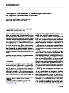
Fig. 3
420
o
time, s
Joint time-frequency domain graphs o f vibration sensor output for (a)l 17 mm HP,, (b) 19 mm HP and (c) 23 mm Biocor valves. All data were averaged over at least 39 cycles. Cardiac output was 7.91min Medical
&
Biological
Engineering
&
Computing
2003,
Vol.
41
15'
30"
% X >
>
14
25
13'
20'
12 ,..,..
O
....
,
O
"5 "5
..:.:
"5 "5
15
03
10'
O
5'
.
11'
Y_=
..... ,....
:
10'
.-... .........
O
94
5 6 7 8 9 pressure drop during closing sound, mmHg
00
10
5
10
15
20
25
30
35
40
pressure drop during closing sound, mmHg b
a
30. >
25-
.~ 20' ~
0
15-
~ lo. N N
•-
....
5.
04
5
6
7
8
9
10
11
12
pressure drop during closing sound, mmHg C
Fig. 4
Graphs of peak and mean pressure diffbrence across (a) 17mm HP valve, (b) 19mm HP valve and (c9 23 mm Biocor valve. (a) (+) Peak pressure drop: ( - ) y = 190x-443; R=0.90. (0) Mean pressure drop: ( - ) y = 190.9x- 3.7," R=0.93. (b) (+) Peak pressure drop: (-)y=O.46x + 2.96," R =0.97. (0) Mean pressure drop: ( - ) y = 1.04x + 2.82; R=0.95. (c) (+) Peak change in pressure drop: ( - ) y = O . O 3 x - 3.90; R =0.96. (0) Mean change in pressure drop: ( - ) y = O . O 4 x - 1.73; R =0.96
for the bioprosthetic valve also contains clear opening and closing signatures, although the energy content is within a lower frequency range (< 1 kHz). A frequency peak is seen at 47 -4- 15 Hz for this valve. 3.2 Haemodynamic measurements
Figs 4a and b display the relationships between the peak and mean pressure difference across the valve during the closing
sound and the total intensity of the closing sound as detected with the vibration sensor for the 17 mm HP valve (Fig. 4a) and the 23 mm Biocor valve (Fig. 4b). Both graphs show strong linear trends and indicate that the signal detected by these sensors contains information on trans-valvular haemodynamics. Fig. 4c displays a strong linear trend between the peak and mean change in the pressure difference across the valve and the total intensity of the closing sound for the 23 mm Biocor valve. Similar trends were observed for the other valves.
4" >
4-
3.:
>
=~ 2..~
3.-
=~ 2.-
03
03
E
E 1
1"-
o.~ lO:
frequency, Hz
,v
0
time, s
a
Fig. 5
frequency, Hz
,v
0
time, s
b
time-frequency domain graphs o f vibration sensor output for 2 mm HP valve ~a) without thrombus and (b) with single layer o f thrombus. All data were averaged over 35 cycles, and cardiac output was 6.61min
Joint
Medical & Biological Engineering & Computing 2003, Vol. 41
421
o.6[, .
3.3 Varying levels o f thrombus The effect of mild thrombus (one layer) on the 21 mm HP valve can be seen by examining the joint time-frequency plot of the sensor output (Fig. 5). Notice that, compared with the joint time-frequency plot for the normal condition (Fig. 5a), there is a 30% attenuation of the intensity of the closing sound between 80 Hz and 3.5kHz for the first layer of simulated thrombus (Fig. 5b). However, there is only a 12% increase in the peak trans-valvular velocity, as measured by the clinical standard of ultrasound Doppler. Table 2 lists the characteristics of the closing sound for each thrombus layer and the trans-valvular velocity as measured by Doppler echocardiography at a cardiac output of 61 min -1. Notice that the total volts between 80 Hz and 3.5kHz for the sensor located adjacent to the simulated thrombus decreased as the level of thrombus increased, whereas no such decrease was apparent for the sensor located on the opposite side of the annulus where no thrombus was present. At the same time, Doppler-measured peak velocity did not increase past 1.7 m s- 1 (~ 12 mmHg pressure drop calculated from Doppler), which clinically would still be considered mild stenosis.
3.4 S t i f f bioprosthetic valve
3.5 Sensor p l a c e d away f r o m the valve Fig. 7 contains a time domain plot of the signal obtained from the sensor placed approximately 3 cm away from the valve mounted in the aortic position of the ventricular model. The two voltage peaks above 0.03 in Fig. 7 represent the closing vibrations of the valve in the aortic position, indicating that the sensor was capable of detecting valve sounds with reasonable fidelity, even when placed away from the valve. Owing to poor repeatability of the driving pressure, averaged joint timefrequency plots could not be obtained for these conditions. Also, although numerous secondary peaks corresponding roughly to signals from the mitral valve were observed, it was difficult reproducibly to align valve signatures with mechanical
4i
3-.
,
i
..~'__
,' , ,,
=~ 2,
;,
~3~ .~
.. --,
:,.
10,
-
0
time, s
Fig. 6 Joint time-frequency domain graph o f sensor output from 24 mm Edwards" valve with visibly stiffened leaflets'. Cardiac output was 3.81min z
422
III"
f"\ ! ,
0.4
0.2
>~
~ •
.L
/
', ~.£L
l
150 100
,
",.
-50
E E
•
"
IV I
-o £
i~:O
"
/--50
I
~¢ -0.2
"1-
",J
--100
o_
-0.4 - -150
-0.6
0.4
0.6
0.8
1.0
1.2
1.4
200
time, s
Fig. 7
Time domain graph from sensor mounted near aortic valve. (- - -) Driving pressure," ( - - ) sensor output
events for each valve. This issue will require further optimisation of the sensor design and location.
4 Discussion
Fig. 6 contains the joint time-frequency plot for the bioprosthetic valve with stiffened leaflets. Whereas the primary frequency of vibration of the normal valve (Fig. 3c) was 47 -4- 15 Hz, the primary frequency of vibration for the valve with stiffened leaflets was 115 -4- 12 Hz. Student's t-test revealed that this difference was significant (p < 0.01). in addition to the increase in the primary frequency, an additional peak in the closing sound was seen at approximately 1 kHz.
frequency, Hz
- 200
The availability of prosthetic heart valves with the intrinsic ability to monitor their function automatically after implantation and provide warnings of impending failure should facilitate clinical monitoring of such devices in vivo and allow clinicians to optimise post-implantation therapies such as anti-coagulants. Such valves would have sensors incorporated into the housing and telemetry to transmit the signals to an external receiver. The first step in devising such 'smart valves' is to ensure that the sensors do provide useful information on clinically important parameters of valve function. Two important parameters for monitoring function are the presence and extent of thrombus, especially for mechanical valves, and stiffening of leaflets for bioprosthetic valves. An additional benefit would be provided if the sensors were capable of monitoring local haemodynamics. 4.1 Mechanical valve
The sensor signals from the mechanical valves were found to have excellent signal-to-noise ratios. However, even with such fidelity, very little diagnostic information is available if the signals are examined in the time domain, mainly owing to a large beat-to-beat variability in the time-domain amplitudes of the signals, in fact, the only noticeable change due to thrombus in the time domain was attenuation in the peak closing sound detected by the sensor located adjacent to the thrombus, and only for layers 4 and 5 (i.e. severe thrombus). However, when these signals were examined using a joint time-frequency representation, significant information regarding valve function could be gleaned. Each mechanical valve had its own unique frequency signature during the closing sound, i.e. the number of peaks, the amplitude of the peaks and the frequency of the peaks for the closing sound for each valve were all unique to that valve. Changes due to the addition o f simulated thrombus to the valve were most apparent in the joint time-frequency representation of the closing sound. Table 2 and Fig. 5 demonstrate the ability of the sensors to detect the level ofthrombus through changes in the energy content of frequencies below 3.5 kHz. REYNOLDS and STEPHEN(1995a) examined changes in the frequency content of the closing sounds of a mechanical valve with the addition of a small mass to the valve annulus. These investigators also observed changes in spectral content due to mass loading, although the frequency ranges (approximately 7kHz and Medical & Biological Engineering & Computing 2003, Vol. 41
above 10 kHz) were different from those found in our study. This variation in frequency range is presumably due to two differences between the studies: first, a mono-leaflet valve was used in their study; secondly, the thrombus was attached in a manner that would primarily affect the resonance frequencies of the valve structures, whereas we attached the thrombus so that it would interfere with leaflet closing. PLEMONDS and HOVENGA (1995) and DE MOL et al. (2001) have shown resonance frequencies to be located at the higher end of the frequency spectrum of the closing sound. Regardless of these differences, our results provide initial confidence that the sensors developed here can provide important and predictable information regarding the formation and extent of thrombus on mechanical valves. Further study on a wider range of valves is needed to obtain baseline information on the most common mechanical valve designs in use today. 4.2 Bioprosthetic valve
would impact the closing dynamics of a prosthetic valve in the mitral position, if a correlation between closing sound parameters and haemodynamic variables such as pressure rise could be established, the sensor would show promise as a diagnostic instrument as well. The graphs in Fig. 4 indicate that sensor output does indeed correlate to both mean and peak pressures across the valve for both mechanical and bioprosthetic valves, in this case, the total intensity of the closing sound was found to be the most useful diagnostic indicator. This makes sense, as changes in closing pressures affect the acceleration of the leaflet mass as it closes; this in turn would change the total closing sound intensity. This is in agreement with the results from ERICKSON et al. (1994), who found a positive trend between valve sound loudness and aortic drive pressure, albeit with a smaller data set (three driving pressures). Also, the trends in Fig. 4 are in agreement with the findings by EBERHARDT et al. (1995), who reported a strong linear trend between peak outlet strut load and disc closing velocity for a Bjork-Shiley valve.
Leaflet degradation is one of the primary mechanisms of valve failure for bioprosthetic valves. Although several investigators are conducting research to examine the mechanisms of such degradation, much less work is being done on methods to diagnose early failure. The pathway to leaflet failure can occur due to calcification or mechanical degradation. In both cases, leaflet stiffening can occur as a precursor to complete valve failure, due either to calcium deposition or degradation of the elastic components of the valve leaflets (LEVY et al., 1986; VESELEY et al., 2001). Current imaging and diagnostic methods (echocardiography, MRI, phonocardiography) are limited in the detection of leaflet stiffening. The sensors developed here, however, show initial promise in the detection of stiff bioprosthetic valve leaflets. As with the mechanical valves, time-domain information provided little diagnostic information. However, when multiple cycles are averaged and processed through a joint time-frequency analysis, the results become much more meaningful. An increase in the primary frequency from 47 Hz for the normal valve to 115 Hz for the stiffvalve, along with a general increase in higher-frequency content, is in agreement with the results reported by HAMIDet al. (1987) and DURAND et al. (1991). A caveat to this analysis is that, although the two valves were both porcine valves, they were from different manufacturers and differed in stent design, flexibility and construction. This may have contributed to certain differences in sensor output for the normal 23 mm Biocor and stiff23 mm Standard valve. However, high-speed video imaging of both valves (data not shown) clearly revealed structural changes in leaflet opening and closing that cannot be explained purely by differences in valve manufacture. This issue will need to be explored through a chronic in vivo experiment. Nevertheless, the fact that the sensor has the capability to distinguish changes in spectral content consistent with findings from other work is encouraging.
Valve sounds generated from within the chest are subject to attenuation, especially from the lungs, and reflections at tissue interfaces. Consequently, consistent external recordings of valve sounds are difficult to obtain. REYNOLDS and STEPHEN (1995) went so far as to submerge patients during valve sound recording to minimise the effects of the air-chest interface. Modelling acoustic sound propagation within the thorax, DE MOL et al. (2001) found that valve sound amplitude was severely position dependent and that phase reversals were possible as well. Although the model showed that submerging the patient in water did help with reflections at the chest interface, obtaining consistent, high-quality external recordings was still difficult. in our preliminary tests, we placed the sensor within an implantable package and placed the entire system approximately 3 cm away from the aortic position inside our mock left heart circulatory system, in practice, it is envisioned that this package will also contain telemetry, which will be used to transfer the valve signals to an externally located receiver. As access to such a location can be obtained through relatively minor surgery, it is reasonable to assume that the risks associated with placing this device into patients with implanted mechanical valves that may be at higher risk for failure (e.g. Bjork-Shiley valves) are exceeded by the benefits provided by the sensor, which would allow chronic on-line monitoring of valve performance with high fidelity. Our results show that the placement of such a sensor allows valve sounds to be detected with high sensitivity and without the problems associated with externally located sensors, such as signal attenuation, reflection and resonance. Further work, preferably under in vivo conditions, will be needed to develop this technique further, but these initial results are encouraging.
4.3 Use o f sensor output f o r haemodynamic diagnostics
4.5 Limitations
In addition to the detection of valve failure and thrombus formation, we wanted to determine whether these sensors could be used to provide diagnostic information regarding cardiac dynamics. For instance, the rate of ventricular pressure rise during isovolumic contraction and the rate of pressure decrease during isovolumic relaxation have been shown to provide information on systolic and diastolic function, respectively. Du6ng diastole, falling ventricular pressure is the mechanism that facilitates aortic valve closure. Thus, for a mechanical valve in the aortic position, the closing sound can be influenced by this changing pressure gradient. Similarly, systolic contraction and the associated ventricular pressure rise
As this investigation was primarily a feasibility study, we only examined the use of the proposed vibration sensor with a select number of valves. The positive results have led to the planning of chronic animal studies for a wider range of valves in the near future. The thrombus we used certainly does not match the histology of true thrombus or pannis, but did simulate massloading effects well.
Medical & Biological Engineering & Computing 2003, Vol. 41
4.4 Sensor located away from the valve
4.6 Future work These results provide initial evidence regarding the potential to create a 'smart' valve that contains sensors that monitor valve 423
function continuously after implantation. Further work is still needed in a number o f areas to realise such a valve. A similar set o f studies need to be performed for a wider range o f existing prosthetic valve designs, as our results show that each valve design and, indeed, each valve size contain unique vibration signatures that need to be documented. Likewise, further in vivo studies, both acute and chronic, are needed to assess the impact o f the physiological environment on sensor signals. Lastly, miniaturisation and integration o f the sensor, electronics and telemetry into the valve will need to be performed. All these projects are currently underway in our laboratory. Acknowledgments" This project was made possible, in part, b y grants from the Colorado Commission on Higher Education (CCHE), the American Heart Association, and The Children's Hospital Research institute, Denver.
References CHEON, G. J., and CHANDRAN, K. B. (1994): 'Transient behaviour analysis of mechanical heart valve prosthesis in the closing phase', J Biomed. Eng., 116, pp. 452-459 DE MOL, B. A., CROMHEECKE, M. E., GROEN, J. G., FABER, G., VAN DEN HEIDEN, M. S., and ONGKIEHONG,L. (2001): 'The complexity of external acoustic detection in Bjork-Shiley convexoconcave heart valves', Artif Org., 25, pp. 63-67 DURAND, L. G., Guo, Z., SABBAH, H. N., and STEIN, P. D. (1993): 'Comparison of spectral techniques for computer-assisted classification of spectra of heart sounds in patients with porcine bioprosthetic valves', Ned. Biol. Eng. Comput., 31, pp. 229-236 EBERHARDT, A. C., CHASSAING,C. E., WARD, M. A., and LEWANDOWSKI, S. J. (1995): 'Acoustic characterization of mechanical valve condition and loading', J. Heart Valve Dis'., 4, pp. 649-659 ERICKSON, R. L., THULIN,L. I., and RICHARD,G. J. (1994): 'An in vitro study of mechanical heart valve sound loudness', J. Heart Valve Dis'., 3, pp. 330-334 HAMID, M. S., SABBAH,H. N., and STEIN, R D. (1987): 'Vibrational analysis of bioprosthetic heart valve leaflets using numerical
424
models: Effects of leaflet stiffening, calcification, and perforation', Circ'. Res., 61, pp. 687-694 LEVY, e. E, SCHOEN, E J., and GOLOMB, G. (1986): 'Bioprosthetic heart valve calcification: Clinical features, pathobiology and prospects for prevention', CRC Crit. Rev. Biocompatibility, 2, pp. 147-187 PLEMONS, T. D., and HOVENGA,M. (1995): 'Acoustic classification of the state of artificial heart valves', J. Acoust. Soc. Am., 97, pp. 2326-2333 PRABHU, A. A., and HWANG, N. H. C. (1988): 'Dynamic analysis of flutter in disk type mechanical heart valve prostheses', J. Biomech., 21, pp. 585-590 REYNOLDS, K. J., and STEPHEN, R. O. (1995a): 'Acoustic analysis of the closing sounds of implanted prosthetic heart valves', J. Acoust. Soc. Am., 98, pp. 69-77 REYNOLDS, K. J., and STEPHEN, R. O. (1995b): 'Detection of mechanical changes to prosthetic heart valves by spectral analysis of valve closing sounds', J. Acoust. Soc. Am., 98, pp. 60-68 VESELEY, I., BARBER, J. E., and RATLIFF,N. B. (2001): 'Tissue damage and calcification may be independent means of bioprosthetic heart valve failure', J Heart Valve Dis'., 10, pp. 471-477 WALKER, D. K., and SCOTTEN, L. N. (1991): 'Discrimination in vitro between the acoustic emission from Bjork-Shiley convexo-concave valves with and without a broken minor strut', Ned. Biol. Eng.'Comput., 29, pp. 457-464
Author's biography Robin Shandas received a Bachelor's degree in Electrical Engineering from the University of California, Santa Barbara in 1987 and his Master's and Ph.D. degrees in Bioengineering from the University of California, San Diego in 1990 and ~ 1993 respectively. He is currently Joint Associate /~'~ Professor in the Departments of Mechanical Engio neering and Pediatric Cardiology at the University of Colorado. He also directs the Cardiovascular Device Development Laboratory at The Children's Hospital, Denver. His research interests revolve around cardiovascular imaging, bio/ fluid mechanics and biomedical devices.
Medical & Biological Engineering & Computing 2003, Vol. 41