AVIAN DISEASES 57:544–554, 2013
Development, Application, and Results of Routine Monitoring of Marek’s Disease Virus in Broiler House Dust Using Real-Time Quantitative PCR Stephen W. Walkden-Brown,AF Aminul Islam,AB Peter J. Groves,C Ambrosio Rubite,D Sue M. Sharpe,E and Susan K. BurgessA A
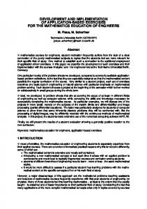
Animal Science, School of Environmental and Rural Science, University of New England, Armidale, NSW 2351, Australia C Faculty of Veterinary Science, University of Sydney, Camden, NSW 2570, Australia D Baiada Poultry, Laverton, VIC 3026, Australia E Birling Avian Laboratories, Bringelly, NSW 2556, Australia Received 25 September 2012; Accepted 19 November 2012; Published ahead of print 8 January 2013
SUMMARY. Results are presented from four studies between 2002 and 2011 into the feasibility of routinely monitoring Marek’s disease virus serotype 1 (MDV-1) in broiler house dust using real-time quantitative PCR (qPCR) measurement. Study 1 on two farms showed that detection of MDV-1 occurred earlier on average in dust samples tested using qPCR than standard PCR and in spleen samples from five birds per shed assayed for MDV-1 by qPCR or standard PCR. DNA quality following extraction from dust had no effect on detection of MDV-1. Study 2 demonstrated that herpesvirus of turkeys (HVT) and MDV serotype 2 (MDV-2) in addition to MDV-1 could be readily amplified from commercial farm dust samples, often in mixtures. MDV-2 was detected in 11 of 20 samples despite the absence of vaccination with this serotype. Study 3 investigated the reproducibility and sensitivity of the qPCR test and the presence of inhibitors in the samples. Samples extracted and amplified in triplicate showed a high level of reproducibility except at very low levels of virus near the limit of detection. Mixing of samples prior to extraction provided results consistent with the proportions in the mixture. Tests for inhibition showed that if the template contained DNA in the range 0.5–20 ng/ml no inhibition of the reaction was detectable. The sensitivity of the tests in terms of viral copy number (VCN) per milligram of dust was calculated to be in the range 24–600 VCN/mg for MDV-1, 48–1200 VCN/mg for MDV-2, and 182–4560 VCN/mg for HVT. In study 4 the results of 1976 commercial tests carried out for one company were analyzed. Overall 23.1% of samples were positive for MDV-1, 26.1% in unvaccinated and 16.4% in vaccinated chickens. There was marked regional and temporal variation in the proportion of positive samples and the MDV-1 load. The tests were useful in formulating Marek’s disease vaccination strategies. The number of samples submitted has increased recently, as has the incidence of positive samples. These studies provide strong evidence that detection and quantitation of MDV-1, HVT, and MDV-2 in poultry house dust using qPCR is robust, sensitive, reproducible, and meaningful, both biologically and commercially. Tactical vaccination based on monitoring of MDV-1 rather than routine vaccination may reduce selection pressure for increased virulence in MDV-1. RESUMEN. Desarrollo, aplicacio´n y resultados de un muestreo rutinario del virus de la enfermedad de Marek en polvo de casetas avı´colas mediante PCR cuantitativo en tiempo real. Se presentan los resultados de cuatro estudios entre los an˜os 2002 y 2011 sobre la viabilidad de un muestreo para la enfermedad de Marek serotipo 1 (MDV-1) llevado a cabo en polvo de casetas avı´colas de pollo de engorde mediante PCR cuantitativo en tiempo real (qPCR). El estudio 1 llevado a cabo en dos granjas mostro´ que la deteccio´n de MDV-1 en promedio ocurrı´a antes en las muestras de polvo con el me´todo cuantitativo de PCR en comparacio´n con el convencional y en las muestras de bazo de cinco aves por caseta analizadas para detectar al virus de Marek 1 por PCR en tiempo real o por PCR convencional. La calidad del ADN despue´s de la extraccio´n a partir de polvo no tuvo ningu´n efecto sobre la deteccio´n del virus de Marek 1. El estudio 2 demostro´ que el herpesvirus de pavos (HVT) y el serotipo 2 del virus de Marek (MDV-2), adema´s del serotipo1 podrı´an ser fa´cilmente amplificados a partir de muestras de polvo de granjas comerciales, a menudo en mezclas. El serotipo 2 se detecto´ en 11 de 20 muestras a pesar de la ausencia de la vacunacio´n con este serotipo. El estudio 3 investigo´ la reproducibilidad y la sensibilidad de la prueba de PCR cuantitativo y la presencia de factores inhibidores en las muestras. Las muestras extraı´das y amplificadas por triplicado mostraron un alto nivel de reproducibilidad, excepto a niveles muy bajos del virus que estaban cerca del lı´mite de deteccio´n. El mezclado de muestras antes de la extraccio´n proporciono´ resultados acordes con las proporciones de la mezcla. Las pruebas de inhibicio´n mostraron que si el ADN extraı´do estaba en un rango de 0.5–20 ng/ml no se detecto´ inhibicio´n de la reaccio´n. Se calculo´ que la sensibilidad de las pruebas en te´rminos de nu´mero de copias virales (VCN) por miligramo de polvo esta´ en el rango de 24 a 600 copias virales por mg para el serotipo 1, de 48 a 1200 copias virales por mg para el serotipo 2, y de 182 a 4560 copias virales por mg para el herpesvirus de los pavos. En el estudio 4 se analizaron los resultados de 1976 pruebas llevadas a cabo por una empresa. En general el 23.1% de las muestras fueron positivas para el serotipo 1, el 26.1% en aves no vacunadas y el 16.4% en los pollos vacunados. Hubo una marcada variacio´n regional y temporal de la proporcio´n de muestras positivas y en la carga del virus de Marek 1. Las pruebas fueron u´tiles en la formulacio´n de estrategias de vacunacio´n contra la enfermedad de Marek. El nu´mero de muestras presentadas ha aumentado recientemente, de igual manera que la incidencia de muestras positivas. Estos estudios proporcionan una fuerte evidencia de que la deteccio´n y cuantificacio´n del virus de Marek 1, del Herpesvirus de los pavos, y del serotipo 2, en el polvo de casetas avı´colas mediante PCR cuantitativo es robusto, sensible, reproducible y significativo, tanto biolo´gica como comercialmente. La ta´ctica de vacunacio´n basada en el seguimiento del serotipo 1en lugar de la vacunacio´n de rutina puede reducir la presio´n de seleccio´n para una mayor virulencia en el serotipo 1. Key words: dander, poultry dust, qPCR, Marek’s disease virus, surveillance, epidemiology, diagnosis Abbreviations: HVT 5 herpesvirus of turkeys; MD 5 Marek’s disease; MDV-1 5 Marek’s disease virus serotype 1; MDV2 5 Marek’s disease virus serotype 2; PCR 5 polymerase chain reaction; qPCR 5 real-time quantitative PCR; UNE 5 University of New England; VCN 5 viral copy number
B F
Present address: Department of Microbiology, John Hunter Hospital, Newcastle, NSW 2310, Australia. Corresponding author. E-mail:
[email protected]
544
Monitoring of Marek’s disease virus in poultry dust
Marek’s disease (MD) is an important disease of chickens with worldwide occurrence, particularly where there are large concentrations of commercial poultry. The disease has a number of interesting features that have enabled it to persist widely and prove difficult to eliminate or control permanently. It is caused by Gallid herpesvirus 2, an alphaherpesvirus in the genus Mardivirus, which is commonly referred to as Marek’s disease virus serotype 1 (MDV-1) (25). MDV1 is a lymphotrophic oncogenic virus, inducing a range of pathological syndromes including immunosuppression, central and peripheral nervous system dysfunction, and most notably, lymphomas in a wide range of visceral and other organ systems (36). While the primary target cell for MDV-1 pathology is the lymphocyte, replication of fully infectious virus occurs in the epithelial cells of the keratinizing layer of the feather follicle epithelium, which then slough off and are shed as highly infective dander (4). MDV-1 is contagious and is transmitted by the inhalation of this infective dander (2). The virus in dander is persistent in the environment and retains infectivity for many months at room temperature (6). Infection is characterized by periods of latency and appears to persist indefinitely in the host if not terminated by host mortality (39). Other related MDV-1s in the genus Mardivirus include Gallid herpesvirus 3, an avirulent chicken herpesvirus commonly referred to as MDV serotype 2 (MDV-2); and Meleagrid herpesvirus 1, a naturally occurring herpesvirus of turkeys commonly referred to as herpesvirus of turkeys (HVT) or MDV serotype 3 (MDV-3) (25). The latter viruses are essentially nonpathogenic in chickens and are used as live vaccines to control MD, as are selected attenuated MDV-1 strains. The vaccine strains may be used alone or in any combination of the three types and were first introduced to control the disease in 1970. The vaccines induce protection against MD, but do not prevent infection with wild-type oncogenic MDV-1, so multiple serotypes of MDV-1 may co-exist in the host (37). The vaccine viruses also shed freely in dander (15,19) and transmit efficiently between chickens in the case of MDV-2 (38) and the Rispens CVI998 MDV-1 vaccine (19) but not HVT (7). MD has largely been controlled in areas of high chicken population density by routine vaccination. This is mandatory in long-lived layer and breeder chickens in most parts of the world, but it is less uniformly applied to the much larger population of shorter-lived broiler (meat) chickens. Nevertheless in countries such as the United States and Australia, in ovo vaccination of broiler embryos for MD control is widespread. One of the key features of MD has been a well-documented increase in virulence over time, which has seen it evolve from the classical form characterized mainly by peripheral nerve lesions causing paresis and paralysis in older chickens first described by Josef Marek (23), to modern acute forms in which chickens are affected at much younger ages, with lymphomas predominating together with central nervous system lesions in very young birds (26,35). This increase in virulence has been marked by sequential vaccine failure in the United States (34,35). Vaccination has been implicated in the increase in virulence of MDV (24,35). MD vaccines are ‘‘imperfect’’ and such imperfect vaccines that do not prevent co-infection of the host have been postulated to favor evolution towards greater virulence in the target pathogen (10). In the past, monitoring and diagnosis of MDV infection has been difficult. The virus is strongly cell associated, so isolation from the host requires inoculation of intact infective cells into cultured cells or embryos. Differentiation between the different MDV serotypes or species is also problematic given the widespread use of vaccination, co-infection with wild-type MDV-1, and persistence of infections. The virus does induce characteristic pathology but this requires the presence of disease and skilled interpretation. The complete
545
sequencing of the genome of the three members of the Mardivirus genus (20,21,22,30) and subsequent development of numerous standard and then fully quantitative PCR tests that differentiate them have thus been of great benefit for the diagnosis of MDV infection. We recognized the potential of such tests to provide quantitative information on the replication and shedding of MDV in various tissues and materials, including feather dander (13,29), and commenced a program of research with the ultimate goal of improving the management of MDV by replacing routine or ‘‘blanket’’ vaccination of broilers by a more tactical deployment of vaccination based on environmental monitoring of MDV levels in poultry house dust. Such an approach potentially reduces the cost of MD control and selection pressure for increased virulence in MDV. This paper reviews our experiences in developing and validating the use of real-time quantitative PCR to quantify MDVs in poultry house dust, and the results of several years of commercial application of the method. Specific objectives were 1) to determine whether MDV-1, HVT, and MDV-2 could be reliably amplified from environmental dust samples collected from commercial poultry farms; 2) to evaluate the sensitivity of such tests in comparison with tests based on samples from individual chickens; and 3) to review the findings of the application of the MDV-1 test for 7 yr within a single commercial company.
MATERIALS AND METHODS The objectives were met in four separate experiments or studies as follows: Study 1, longitudinal farm study with unvaccinated broilers on two commercial farms (2002–2003); Study 2, cross-sectional farm study of dusts from 17 farms (2003–2004); Study 3, tests of repeatability, PCR inhibition, and assay sensitivity (2004–2006); and Study 4, analysis of results of commercial use of the dust test (2005–2011). Longitudinal farm study with unvaccinated broilers (Study 1). Study 1 involved two commercial broiler operations in Victoria (Farms A and B). Each farm had three sheds and operated on an all-in all-out basis and both were placed with mixed-sex Cobb broilers from parent flocks vaccinated for MD using the Rispens CVI988 vaccine. Both were placed on new litter. Farm A was placed with 44,000 birds in early October 2002 while Farm B was placed with 117,000 birds in late October 2002. Flocks were killed out at final ages of 55 and 57 days, respectively. Chicks were spray-vaccinated against infectious bronchitis at hatch but were not vaccinated against MD. MD had been noted as a problem in unvaccinated flocks in Victoria at this time. On Farm A, five birds per shed were sacrificed for spleen collection on days 8, 15, 27, 34, 41, and 48 of age, and shed dust was sampled on days 27, 34, 41, and 48. On Farm B, five spleens per shed were sampled on days 12, 19, 25, 32, 41, and 47, and 53 shed dust was sampled on days 19, 25, 41, 47, and 53. Dust samples were collected from various surfaces in the shed (mainly wall studs and other protuberances or indentations, curtains, feeder lines) and were frozen at 220 C until despatch to University of New England (UNE) for DNA extraction and real-time quantitative PCR (qPCR) analysis. Cross-sectional farm study to detect different MDV serotypes in dust (Study 2). Study 2 tested the ability to use qPCR to amplify MDV-1, MDV-2, and HVT in field dust samples from commercial broiler farms. Twenty dust samples from five Australian states were used for this study. Samples were collected between 12 and 56 days of age from various surfaces in the shed (curtains, feeder lines, wall studs, fan housings, etc.) and frozen at 220 C until despatch to UNE for DNA extraction and qPCR analyses for MDV-1. Tests of reproducibility, PCR inhibition and assay sensitivity (Study 3). To test the reproducibility of the qPCR tests in dust, dust samples from 11 commercial farms varying in MDV-1 load were
546
S. W. Walkden-Brown et al.
Table 1. Number of dust samples included in the analysis, by region and year. Region
Code
2005
2006
2007
2008
2009
2010
2011
Total
South Australia NSW non-Riverina Victoria NSW Riverina Western Australia Queensland Total
A B C D E F —
99 63 11 — — — 173
131 145 111 — — — 387
136 48 53 — — — 237
140 76 16 — — — 232
147 51 18 — — — 216
152 22 32 67 16 27 316
164 66 49 61 24 51 415
969 471 290 128 40 78 1976
subjected to DNA extraction, quantification, and qPCR for MDV-1 three times, with the qPCR being done in duplicate each time. As a further test, dust samples from 16 farms were paired and mixed in ratios of 100:0, 75:25, 50:50, 25:75, and 0:100 by mass and subjected to DNA extraction, quantification, and qPCR for MDV-1 in duplicate. For each sample containing a mixture of dusts the predicted DNA concentration, viral copy number (VCN) per reaction, and VCN per milligram of dust were calculated based on the values obtained for the pure samples for each mixture component, and the predicted values compared with the actual values obtained. To test for inhibition in the PCR reaction, three separate tests were carried out. Firstly a 5 3 10-fold serial dilution series was carried out on DNA extracted from seven farm dust samples covering a range of MDV1 load values and samples assayed at each dilution to detect interference at high concentrations and identify the optimum DNA concentration to use in the test. Subsequently two separate spiking tests were performed, with DNA extracted from known positive and negative farm dust samples, plus a water sample being spiked with a known small amount of plasmid DNA containing the target region. Samples were then subjected to qPCR for MDV-1, and the increase in detected MDV-1 was analyzed. In test 1, five water samples, 11 negative dust samples, and nine positive dust samples were spiked, while in the test 2 the respective numbers were five, six, and six. Commercial use of the dust qPCR test for MDV-1 (Study 4). Study 4 utilized records of DNA samples from 1976 individual chicken broiler house dust samples submitted to UNE by a single commercial poultry company for analysis of MDV-1 load by qPCR, between 2005 and 2011. Samples from 2004 and 2012 were excluded because they did not comprise complete years. The number of samples used in the study, by region and year is shown in Table 1. DNA extraction and quantitation. DNA was extracted from 10 mg of spleen or 5 mg of dust using QIAamp DNA mini kits according to manufacturer’s instructions (Qiagen, Clifton Hill, Australia) and stored at 220 C. DNA was eluted in a final volume of 100 ml. Prior to use in the qPCR assay, extracted DNA was quantified using spectrophotometric analysis (Bio-Rad, Hercules, CA; SmartSpec TM 3000 or NanoDropH ND-1000 UV-Vis) for DNA yield, purity, and further dilution for qPCR assays. The purity of DNA samples was determined by the A260/A280 ratio and for later samples, also the A260/230 ratio. An A260/A280 ratio of ,1.8 is generally accepted as ‘‘pure’’ for DNA with lower values suggestive of protein contamination. Expected 260/230 values for pure DNA are commonly in the range of 2.0–2.2, with a lower ratio suggestive of the presence of nonprotein contaminants. Measurement of MDV-1 load. Standard PCR. Serotype-1 MDV-1– specific PCR amplifying a 199-bp fragment of the glycoprotein A gene using 1 ml of extracted DNA template was performed using a modification of the method of Reddy et al. (18). The 50-ml PCR amplification contained 40.75 ml of milliQ water, 5 ml of 103 PCR buffer (QIAGEN, Pty. Ltd, Australia), 1 ml of dNTPs (2 mM), 1 ml each of the two primers (10 pmol/ml), 0.25 ml Taq polymerase (Qiagen, Hilden, Germany), and 1 ml of template (undiluted eluate and 1:10 and 1:100 dilutions). The reaction mixture was overlaid with 30 ml of mineral oil; started at 94 C for 4 min, followed by 40 amplification cycles of 1 min at 94 C, 1 min at 55 C, and 1 min at 72 C; followed by a final extension at 15 C for 10 min; and held at 4 C. The PCR products were analyzed by electrophoresis on a 1.2% agarose gel with TBE buffer
containing GelStar Nucleic Acid Gel Stain (FMC BioProducts, Philadelphia, PA) and bands were visualized under ultraviolet light. Real-time quantitative PCR. MDV-1 and HVT genome copy numbers were determined in the extracted DNA by qPCR as described by Islam et al. (13). For MDV-2 the genome copy numbers were determined by the absolute quantitation method of Renz et al. (29). Amplification and data acquisition were carried out using Rotor Gene 3000 real-time PCR machine (Qiagen, Australia) and each 72-well rotor contained four- or five-point standard curves, two quality controls, and unknown samples, all in duplicate. Viral loads in dust DNA were expressed as VCN per milligram of dust. Samples were randomized across assays to minimize individual assay effects and amplified in duplicate. The lower limit detection (mean 6 SEM) based on the mean calculated VCN of the lowest standard in each of the assays in Studies 1 and 2 were 4 6 0.3, 9 6 7, and 129 6 3.5 VCN per reaction, for the MDV-1, HVT, and MDV-2 assays, respectively. Neither the standard nor qPCR assays differentiate wild-type MDV from the Rispens CVI988 vaccine strain. However, all of the work reported in this paper was in broiler chickens, and these are not vaccinated with the Rispens or other serotype-1 vaccines in Australia. Thus any MDV-1 detected is unlikely to be the Rispens vaccine strain. Measurement of chicken DNA load in dust. Chicken DNA was quantified using the chicken a 2 (VI) collagen gene-specific qPCR assay described by Islam et al. (14). Statistical analysis. Much of the data are presented descriptively, but some were subject to more formal analysis. Analysis of count data was by contingency table analysis with the chi square test. The effect of design factors on continuous variables was investigated using analysis of variance following examination of distributions and residuals to determine whether transformation was required prior to analysis. Relationships between measured variables were explored using linear regression analysis or in some cases logistic regression. All data were analyzed using JMPTM 9 (SAS Institute Inc., Cary, NC) statistical software. A significance level of P , 0.05 is used throughout. Means are presented 6SEM unless otherwise specified.
RESULTS
Longitudinal farm study with unvaccinated broilers (Study 1). The DNA content of the eluate from 5-mg dust samples (mean 6 SE) from Farms A and B were 58.4 6 8.3 and 53.1 6 6.2 ng/ml respectively. The mean A260/280 ratios for the dust samples were 1.22 6 0.28 and 0.99 6 0.33, respectively, indicating significant protein contamination, while the mean content of chicken DNA was 0.027 6 07 and 0.013 6 03 ng/ml, respectively. Thus, the chicken DNA content of the extracted dust DNA was extremely low, being 0.046% and 0.024% of total DNA, respectively. Despite this, the MDV-1–specific target was amplified reliably from dust samples, with all submitted samples being positive for MDV-1 by qPCR and 18 of 27 samples (68%) being positive for MDV-1 by standard PCR (Table 2). There was no significant association between A260/280 ratio and log10 VCN per reaction or per milligram of dust (R2 5 0.003, P 5 0.06). On the other hand, there was a significant negative linear relationship between the chicken DNA content in the
547
Monitoring of Marek’s disease virus in poultry dust
Table 2. Summary of mean DNA concentration and quality (A260/280 ratio) and MDV-1–specific PCR results from chicken spleen or poultry house dust samples collected from three sheds on unvaccinated broiler Farms A and B. A single dust sample was collected at each sampling for each shed, and spleens from five chickens. Shaded values indicate the test which first detected MDV-1 in the shed. DNA conc. (ng/ml)
Standard PCRA
A260/280 ratio
qPCR Spleen
qPCR DustA
Farm
Shed
Day
Spleen
Dust
Dust
%Pos spleen
Dust MDV +/2
Log10 VCN/rxB
%Pos
Log10 VCN/rx
MDV +/2
A
1
27 34 41 48 27 34 41 48 27 34 41 48 12 19 25 32 41 47 53 12 19 25 32 41 47 53 12 19 25 32 41 47 53
319 371 349 335 373 371 417 274 313 386 387 326 318 339 330 291 220 300 294 376 309 325 229 245 326 317 367 368 307 258 278 314 295
86 59 124 76 66 28 36 32 40 73 29 52 NSC 75 29 NS 50 63 44 NS 40 47 NS 32 52 31 NS 93 42 NS 58 110 29
1.20 1.41 0.98 1.60 1.31 1.17 1.18 1.44 1.40 1.09 0.88 1.01 NS 0.84 0.97 NS 0.53 0.96 0.87 NS 0.90 0.96 NS 1.36 0.96 0.80 NS 1.11 1.19 NS 0.98 1.15 1.21
20 0 60 80 0 20 80 100 0 0 100 100 0 0 0 60 100 100 100 0 0 20 100 100 60 80 0 0 0 100 100 80 100
2 + + + 2 + + + 2 + + 2 NS 2 2 NS + + + NS + 2 NS + 2 + NS 2 + NS + + +
0.39 0 2.38 2.60 0 0.52 1.77 3.13 0 0 1.77 3.02 0 0 0 2.34 3.26 3.33 3.21 0 0 0 3.47 2.70 3.05 2.96 0 0 0 2.30 2.98 3.04 3.07
20 0 80 100 0 20 80 100 0 0 60 100 0 0 0 80 100 100 100 0 0 0 100 100 100 100 0 0 0 80 100 100 100
2.09 1.85 2.82 3.88 2.17 2.29 3.37 3.63 2.09 1.80 2.59 3.73 NS 0.59 1.82 NS 3.75 4.36 4.91 NS 2.35 1.55 NS 4.57 4.30 4.78 NS 1.01 2.34 NS 3.75 4.49 4.60
+ + + + + + + + + + + + NS + + NS + + + NS + + NS + + + NS + + NS + + +
2
3
B
1
2
3
A
(2) 5 negative; (+) 5 positive. Log10 viral copy number detected per reaction. C NS 5 not sampled. B
sample and log10 VCN per reaction (R2 5 0.31, P 5 0.003) or log10 VCN per milligram of dust (R250.25, P 5 0.007). This is likely due to a significant negative linear relationship between the chicken DNA content in the sample and the age of chickens (R2 5 0.24, P 5 0.09). In terms of sensitivity of detection of MDV-1, there was a clear advantage in qPCR of dust over standard PCR of dust or spleen from five birds, or qPCR from spleen of five birds. On Farm B, MDV-1 was first detected in dust at day 19 in all three sheds and by qPCR of dust alone in two of those sheds (Table 2). On Farm A the picture was less clear because sampling commenced later, but in all cases MDV-1 was first detected by qPCR of dust, sometimes with other samples also being positive, but less uniformly so. The MDV-1 load data made biological sense with the level of MDV-1 in dust increasing exponentially over time on both farms, with some variation between sheds (Fig. 1). This was associated with concomitant increases in the proportion of spleen samples becoming positive from sampled chickens (Table 2). Farm B tended to have higher levels of MDV-1 in dust at equivalent time points to Farm A. No gross MD was observed on either farm but performance was poor on both farms and tended to be worse on Farm B than on Farm
A (Table 3). There was a marked difference in late mortality after day 28 between the farms with values of 1.96% and 4.03% for Farms A and B, respectively. Cross-sectional farm study to detect different MDV serotypes in dust (Study 2). Table 4 shows the MDV-1, MDV-2, and HVT load in broiler house dust sampled from 17 broiler flocks across Australia. HVT was detected in dust from all farms claiming to have HVT vaccination, but also for two farms for which vaccination was not reported. MDV-1 was detected in samples from vaccinated and unvaccinated flocks in New South Wales (NSW) and South Australia (SA) but was not detected in samples from Victoria (VIC) or the few samples from Queensland (QLD) and Western Australia (WA). Interestingly MDV-2 was detected in 11 of 20 samples despite no history of vaccination with serotype 2 vaccines. Tests of reproducibility, PCR inhibition, and assay sensitivity (Study 3). Reproducibility. The repeatability of measurement of DNA and MDV-1 content in 11 dust samples is summarized in Table 5. There was a high level of agreement between triplicate extractions from the same dust samples, apart from those with viral loads around the limit of assay sensitivity, for which some replicates amplified and some did not. The mean CVs of 10.8% for DNA
548
S. W. Walkden-Brown et al.
Fig. 1. Mean MDV-1 load in poultry house dust (VCN/mg dust) dust samples collected at various times from three sheds containing broiler chickens unvaccinated against MD on Farms A and B.
concentration and 2.4% for MDV-1 load per milligram of dust (excluding borderline samples with uneven amplification) are indicative of good reproducibility of results. This was confirmed by analysis of variance, which revealed that for DNA concentration, VCN per reaction and VCN per milligram of dust, respectively, 96.5%, 99.0%, and 93.3% of the total variation for these variables was due to between-sample variation with only 3.5%, 1.0%, and 6.7% due to within-sample variation. Mixing of dust samples. Eight different combinations of dusts from two different farms were combined in ratios of 100:0, 75:25, 50:50, 25:50, and 0:100 by weight. Four of the 16 dust samples involved were negative and 12 were positive for MDV. Predicted values for extracted DNA concentration and MDV-1 load in mixtures (based on the values obtained for the pure dusts involved in each mixture) were compared against the actual measured values using linear regression. There was poor agreement between predicted and actual extracted DNA concentrations (R2 5 0.06, P 5 0.25, N 524) with significant underestimation of actual DNA concentrations at lower predicted values (Fig. 2A), but close agreement between predicted and actual Log10 MDV load in dust (R2 5 0.97, P , 0.0001, N 524) (Fig. 2B). PCR inhibition. Effect of DNA concentration on qPCR performance. Amplification of some samples was reduced at DNA concentrations above of 30 ng/ml and totally suppressed at concentrations above 100 ng/ml (Fig. 3). At very low concentrations (,0.5 ng/ml) in samples containing little MDV-1, the virus became undetectable. MDV-1 load was best determined at the DNA concentration range of 0.5–20 ng/ml where all seven positive samples amplified (Fig. 3). Nonspecific qPCR inhibition as detected by spiking samples with plasmid DNA. Addition of a fixed amount of plasmid DNA in two separate tests produced a similar increase in MDV-1 values in water, DNA extracted from MDV-1 negative dust samples, or DNA extracted from MDV-1 positive dust samples (test 1, P 5 0.76; test
2, P 5 0.72) (Fig. 4). In test 1 the MDV-1 load in positive samples ranged from 2.4 to 6014 VCN per reaction, while in test 2 the range was 251 to 5399 VCN per reaction. Sensitivity of detection of MDV-1, HVT, and MDV-2 in dust. The lower limits of detection of MDV-1, MDV-2, and HVT copy number per milligram of dust in the assay system depend on the limits of detection per reaction and the amount of DNA extracted from the dust sample. The calculated limits range from 24 to 600 VCN/mg dust for MDV-1, 48–1200 VCN/mg dust for MDV-2, and 182–4560 for HVT (Table 6). Commercial use of the dust qPCR test for MDV-1 (Study 4). DNA concentration and quality. The distributions of extracted DNA concentration A260/280 ratio and A260/230 ratios are shown in Fig. 5. DNA concentration had an approximately normal distribution with a mean of 518 ng/ml, median of 525 ng/ml, and standard deviation of 281 ng/ml. The A260/280 ratio had a highly skewed distribution with a median value of 0.44, and 80% of the samples had values between 0.35 and 0.62. The A260/230 ratio was also highly skewed with a median value of 0.47, and 80% of the samples had values between 0.34 and 0.86. There was little variation in the DNA concentration or quality between regions, but there was significant (P , 0.001) trend towards lower DNA yields and higher quality over time. For the period 2005–2007 the mean (6 SEM) DNA concentration was 670 6 10 ng/ml, log10 A260/280 was 20.396 6 006, and log10 A260/230 was 20.410 6 06. For the period 2008–2011 the respective values were 431 6 8 ng/ml, 20.310 6 05, and 20.258 6 06. Incidence of MDV-1–positive samples. Of the 1976 dust qPCR tests for MDV-1, 456 (23.1%) were positive for MDV-1. Vaccination with HVT significantly reduced the ratio of positive samples. For Regions A, B, and C, which have a reasonable numbers of samples from known vaccinated and unvaccinated flocks over a long time frame, vaccination reduced the percentage of positive flocks from 26.1% (65/249) to 16.4% (137/835) (P , 0.01).
Table 3. Summary of performance data on Farms A and B. Cumulative mortality (%) Farm
Day 7
Day 28
Final
Average adj. age (d)
Average weight (kg)
Adj. FCRA (feed:gain)
A B
2.8 1.17
5.13 3.04
7.09 7.07
47.04 48.63
2.489 2.332
2.097 2.171
A
FCR 5 feed conversion ratio (feed:live weight) adjusted to a fixed weight of 2.45 kg.
549
Monitoring of Marek’s disease virus in poultry dust
Table 4.
Viral load of three serotypes of MD viruses in poultry house dust collected from commercial broiler farms across Australia. Viral load (Log10 VCN/mg dust)
State
Reported MD vaccination
Day of age
MDV-1
MDV-2
HVT
NSW NSW NSW NSW NSW NSW QLD QLD VIC VIC VIC VIC SA SA SA SA SA SA SA WA
HVT HVT HVT HVT Nil Nil HVT Nil HVT Nil Nil Nil Nil Nil Nil Nil Nil Nil Nil Nil
28 35 49 56 33 33 31 12 49 46 49 54 35 49 52 53 56 56 58 52
0 4.91 0 0 3.98 3.88 0 0 0 0 0 0 3.03 5.81 0 5.09 6.19 5.04 6.36 0
0 4.61 5.24 4.45 3.74 3.73 0 0 0 5.29 5.04 0 0 5.46 0 5.29 5.17 0 4.72 0
3.59 3.85 3.27 4.18 0 2.93 2.85 1.64 4.94 0 0 0 0 0 0 0 0 0 0 0
There was also significant variation between regions and years as shown in Fig. 6. Region A, which supplied the most samples, showed a gradual increase in positive samples to a peak of 26% in 2009, followed by a marked reduction to a level of only 3% in 2011. A somewhat similar pattern was seen in Region B, but in Region C the proportion of positive samples has increased irregularly over time reaching 55% in 2011. Region D supplied samples only from 2010 but had exceptionally high percentages of positive samples, 85 and 84% in 2010 and 2011, respectively. Region E, which also commenced sampling in 2010, also had a comparatively high percentage of positive samples, 50% and 37.5% in 2010 and 2011, respectively. Region F had a moderate incidence of positive samples, below the overall average. The percentage of MDV-1–positive samples also varied significantly with month of the year (P , 0.001). Higher percentages were observed in the cooler months of April to September, but also in the warm month of December (Fig. 6). With regard to the effect of chicken age, the earliest samples were submitted from the third week of life (days 15–21), and from samples in that week of life 3 of 23 samples (13.0%) were positive for MDV-1. For the fourth, fifth, sixth, seventh, and eighth weeks of life, the proportions varied little,
being 6 of 21 (28.6%), 19 of 77 (24.7%), 228 of 951 (24.0%), 155 of 754 (20.6%), and 31 of 114 (27.2%), respectively. MDV-1 viral load in dust samples. Mean MDV-1 viral load differed significantly between regions and years in a very similar fashion to the percentage of MDV-1–positive samples (Fig. 7A), reflecting the large influence of the negative samples. When the negative samples were excluded, the magnitude of the differences due to year or region was greatly reduced (Fig. 7 B) but differences of up to 2.5 logs still remained. The overall mean log10 VCN per milligram of dust (6 SEM) of 1976 samples was 0.91 6 0.04; whereas, that from the 456 positive samples was 3.95 6 0.53 VCN/ mg. Among these positive samples, there was a higher MDV-1 load in dust from known unvaccinated flocks (4.04 6 0.13, n 5 75) than from known HVT-vaccinated flocks (3.53 6 0.06, n 5 139). Increasing chicken age had little influence on the MDV-1 load in dust in positive samples with mean (6 SEM) values for the third, fourth, fifth, sixth, seventh, and eighth weeks of life being 3.50 6 0.60, 4.30 6 0.37, 4.28 6 0.24, 3.93 6 0.08, 3.95 6 0.09, and 4.03 6 0.22, respectively. Association between DNA concentration and quality and MDV1 detection. There was a significant overall negative association
Table 5. DNA concentration and MDV-1 viral load in dust samples from 11 farms, extracted in triplicate, and subjected to MDV-1– specific qPCR. Extracted DNA concentration (ng/ul)
MDV-1 viral load (log10 VCN/mg dust)
Farm
n
Mean
SD
CV%
Mean
SD
CV%
Comment
A B C D E F G H I J K Mean
3 3 3 3 3 3 3 3 3 3 3
494 691 784 753 755 645 124 219 157 400 495 501
76 33 60 21 46 43 13 16 26 123 52 —
15.4 4.8 7.7 2.8 6.1 6.6 10.2 7.4 16.5 30.8 10.5 10.8
5.34 5.05 0 4.31 3.27 2.07 6.83 6.99 0.70 0 0.94 3.2
0.10 0.05 0 0.02 0.29 1.80 0.10 0.04 1.22 0 1.63 —
1.85 0.95 — 0.41 8.96 86.6 1.42 0.55 173.2 — 173.2 49.7 (2.36)
— — — — — 2 of 3 replicates amplified — — 1 of 3 replicates amplified — 1 of 3 replicates amplified (excluding samples with nonamplifying reps)
550
S. W. Walkden-Brown et al.
Fig. 2. Predicted DNA concentration (A) and MDV-1 load (B) regressed against actual values measured in 24 dust samples comprising eight mixtures of two different farm dust samples mixed in rations of 75:25, 50:50, and 25:75.
between DNA concentration and log10 MDV-1 VCN per milligram of dust (P , 0.0001, R2 5 0.013). However, this overall effect masked a significant positive association in MDV-1–positive samples (P , 0.001, R2 5 0.03). This positive association was present in all years. The overall negative association was due to negative samples having significantly higher DNA concentrations on average (540 6 8 ng/ml) than positive samples (456 6 14 ng/ml). For the log10 transformed A260/280 ratio there was an inverse situation with a trend towards a significant overall positive association with log10 MDV-1 VCN per milligram of dust (P 5 0.06, R2 5 0.003) but a significant negative association in positive samples (P 5 0.005, R2 5 0.027). The negative association in MDV-1 positive samples was evident in each year’s data but only achieved statistical significance in 2011. MDV-1–negative samples had slightly but significantly higher log10 A260/280 ratios (20.336 6 05) than positive samples (20.306 6 08). For the log10 transformed A260/230 ratio, there was a weak positive overall association with log10 MDV-1 VCN per milligram of dust (P 5 0.001, R2 5 0.009) and no significant association in positive samples (P 5 0.51), although this tended towards negative.
Fig. 3. MDV-1 loads in dust from seven farms at different concentrations of DNA in template. Each samples was diluted 5 3 10-fold commencing with undiluted sample as eluted from the extraction kit. Results take into account dilution, so no effect of dilution would manifest as different dilutions all having the same value. The shaded area demarcates the range of concentrations at which all of the samples amplified. The dotted line indicates the DNA template concentration (5 ng/ml) samples are diluted to prior to qPCR assay in the laboratory.
MDV-1–negative samples had slightly but significantly higher log10 A260/230 ratios (20.302 6 06) than positive samples (20.257 6 0.011). DISCUSSION
All three serotypes of MDV-1 were successfully quantified in commercial broiler farm dust from around Australia using qPCR. The overall data show that the DNA extracted from crude poultry house dust is suitable material for amplification of all three serotypes of MDV-1, alone or in combination, despite the apparently poor quality of the extracted DNA material. At the DNA concentration used in the MDV-1 qPCR test (5 ng/ml) no inhibitory effect on the reaction was observed, and addition of small amounts of plasmid DNA to a range of samples extracted from dust revealed no inhibition of the PCR. The test results made biological sense on two Victorian farms where exponential increases in viral load over the life of a broiler placement were associated with poor performance and MDV-1 was ultimately isolated from the dust on both properties (data not presented). Deployment of the test over 7 yr in a commercial environment showed that the majority of dust samples from broiler flocks were negative for the presence of MDV-1 but that there were marked differences in prevalence and viral load due to region, year, time of year and chicken vaccination status. Regarding the first objective, DNA extraction from poultry dust using commercial kits was technically straightforward and yielded DNA concentrations covering a wide range from 20 to 1200 ng/ml. However, the A260/A280 ratio was always less than the 1.8 seen with pure DNA (median value in field samples of 0.44), suggesting that all DNA samples were contaminated with protein (3). The A260/A280 ratio was also always less than the 2–2.2 seen with pure DNA (median value in field samples of 0.47), suggesting that the DNA samples also contained other nonprotein contaminants. The high level of contaminants is not surprising given the heterogenous nature of poultry house dust, which may contain feed particles, litter material, feces, and microbes in addition to the targeted feather dander. Indeed, analysis of chicken DNA content in the samples showed that a very low proportion (,0.1%) of the extracted DNA was of chicken origin, and this proportion declined as the birds aged. The quality, but not amount of DNA extracted from field dust samples, was lower than that extracted from dander collected from isolator exhaust ducts. Analysis of DNA extracted from 100 samples of such dander from an isolator experiment using layer chickens between the ages of 1 and 8 wk revealed a mean DNA concentration
551
Monitoring of Marek’s disease virus in poultry dust
Fig. 4. Effect of adding a fixed amount of plasmid DNA containing the qPCR MDV-1 target sequence on the MDV-1 viral copy number per reaction (means 6 SEM of change in VCN per reaction). Two separate tests were carried out in different years by different operators. Total n 5 25 in test 1 and n 5 17 in test 2. Differences between groups were nonsignificant in both tests (P . 0.7). Numbers of samples represented by each column from left to right are 5, 11, 9, 5, 6, and 6, respectively.
of 107 6 8.3 ng/ml, A260/280 of 1.57 6 0.04, and A260/230 of 1.13 6 0.03 (Renz et al., unpubl. data, 2005). The sample dilution in Study 3 showed that at very high concentration of DNA and/or contaminants there was partial or total inhibition of the PCR, while at low concentrations test sensitivity was reduced. This work showed that within a working range of 0.5–20 ng/ml of DNA in the template there was no evidence of inhibition. This was further confirmed by a series of tests, using a concentration of 5 ng/ml of DNA template and involving addition of small amounts of plasmid DNA to samples extracted from dust or to water controls, that revealed no inhibition of the PCR. Therefore, samples that tested negative were likely to be true negatives, not false negatives. Nevertheless, some inhibition in some samples cannot be ruled out. Chicken melanin is an inhibitory factor in PCR (1,12). This inhibitor is found in colored chickens such as brown egg layers. However, dust samples used in this study were obtained from broiler chickens that are white. Extension of this work to colored layers would require sample treatment to overcome this inhibition. It has also been reported from a large epidemiological study of broiler farms that wood-based litter materials provide a significant ‘‘protective effect’’ against detection of MDV-1 in dust (11). We have subsequently observed significant inhibition in detection of MDV-1 from litter material of hardwood origin (32) but have yet to demonstrate this in dust samples. Other components of Study 3 showed that amount and quality of DNA extracted from dust and the amount of MDV-1 detected in it were highly reproducible and that mixtures of different samples gave predictable results based on the proportions of the different samples combined. The one exception to the high level of reproducibility is that of samples around the limit of detection, for which only some of the replicates may amplify. This poses some challenges in interpretation of results in this area and raises the issue of whether samples need to be tested in duplicate. To date all samples have been assayed in duplicate.
A significant finding of Study 2 was that MDV-1, MDV-2, and HVT were all detectable in broiler house dust, often in combination. This was not surprising in the case of flocks vaccinated with HVT because earlier reports have confirmed significant shedding of HVT from vaccinated chickens (15,16). More surprising is that HVT was detected on two farms for which HVT vaccination was not recorded. Lateral transmission of HVT is relatively ineffective in chickens (7) so this is a possible explanation. Errors in laboratory submission forms or farm records are an alternative explanation. The possibility of false positives is considered to be unlikely due to the specificity of the primers and probes used in the qPCR assay (14). Detection of MDV-2 in 50% of samples was an interesting finding. An MDV-2 vaccine was extensively used in layer and breeder chickens in the past in Australia but was never widely used in broilers. In 2009 (after Study 2) a bivalent HVT-SB1 vaccine was introduced, but it is also rarely used in broiler flocks. In the late 1970s, avirulent MDV-1 was isolated from commercial chickens in Australia but not classified (27). As with MDV-1, MDV-2 spreads laterally between chickens. This study suggests that MDV-2 may be circulating naturally in Australian chicken flocks, providing protection against MD. However, further studies are required to ascertain the relationship between the circulating MDV-2 viruses and existing vaccinal strains. Regarding the second objective, the longitudinal Study 1 in six sheds of unvaccinated broiler chickens clearly demonstrated that qPCR of dust samples was a very sensitive means of detecting MDV1 in a flock. In all six sheds the earliest detection of MDV-1 was with qPCR of dust samples. In four sheds this was the only test to detect MDV-1 at this time, while in the other two sheds, MDV-1 was also detected by other tests at the same sampling. These results clearly show the greater sensitivity of the qPCR dust test for detecting MDV-1 compared with standard PCR of the same sample, or standard PCR or qPCR of spleen tissue collected from five birds per shed. The greater sensitivity of the dust qPCR test adds to its other considerable advantages of requiring only a single sample per shed,
Table 6. Lower limits of detection of MDV-1, MDV-2, and HVT in the qPCR assays of dust over a range of DNA extraction yields. Limits are based upon lower limits of detection of MDV-1, MDV-2, and HVT of 5, 10, and 38 VCN per reaction. Detection limit/mg dust (VCN)
Dust weight (mg)
DNA content (ng/ml in 100 ml eluate)
Total DNA in 5 mg dust (ng)
DNA per reaction (ng)
MDV-1
MDV-2
HVT
5 5 5
30 150 750
600 3000 15,000
25 25 25
24 120 600
48 240 1200
182 912 4560
552
S. W. Walkden-Brown et al.
Fig. 5. Distribution histograms and outlier box plots for DNA concentration (ng/ml) following extraction from dust samples (left, n 5 1752), A260/280 ratio (middle, n 5 1118), and A260/230 ratio (right, n 5 1111). In the outlier box plots the horizontal line is the median, the triangle represents the mean and standard error, the box ends represent the 25th and 75th quantiles (first and third quartiles) and the whiskers represent 1.5 times the interquartile range. The bracket alongside the box plot indicates the most concentrated 50% of samples.
no bird sacrifice, no freezer storage of sample or cold chain to the laboratory required, no post-PCR steps required to interpret the results, and absolute quantification of the genome copies of the target organism. The actual sensitivities of the qPCR tests as defined by their lower detection limits in dust could be calculated and were shown to range from 24 to 600 VCN/mg dust for MDV-1, from 48 to 1200 VCN/mg dust for MDV-2, and from 182 to 4560 for HVT depending on the amount of DNA extracted from the dust sample. Study 1 also showed the biological relevance of the results with MDV-1 load in dust and spleen increasing as birds increased in age. This was associated with inferior chicken performance on these farms, although no frank MD lesions were reported from either farm. However, MDV-1 was successfully isolated from both properties, and the isolate 02LAR from Farm B was adapted to cell culture and extensively characterized in pathotyping experiments in specific-pathogen-free chickens (28), commercial broiler chickens (33), and commercial layers. These studies have classified it as a very virulent isolate under the classification system proposed by Witter (34). In commercial unvaccinated broilers challenged with 500 plaque-forming units of isolate 02LAR, lymphomas were detectable from day 27 of age and by day 56, 70.4% of chickens had lymphomas (33). However, in that experiment MDV-1 load in dander was greater than 106 VCN/mg dust after day 21 (15) and 107 thereafter; whereas, in farm B dust from study 1, viral load in dust
never exceeded 105 VCN/mg dust. This may explain the lack of MD tumors observed in Study 1. The third objective was to review 7 yr of experience of using the qPCR dust test for MDV-1 in broiler flock dust samples within a single company. Sample numbers submitted by the company have increased sharply since 2009 and this continued into 2012, suggesting that the test is proving useful for the management of MD. The increase has been due in part to extension of the testing to new regions, some of which show a very high level of MDV-1 in their dust samples. The company uses the information provided by these tests, together with their own standard PCR test of dust for HVT, to monitor the need for vaccination against MD in their broiler flocks and to assess the efficacy of vaccination where it is used. The lack of major association between measures of DNA quality and the level of MDV-1 detected in dust samples confirms the findings of Studies 1 and 3 that the qPCR tests are robust and largely uninfluenced by the quality of the extracted DNA. Although MDV-1 is considered to be ubiquitous wherever poultry are kept intensively, we were only able to detect its presence in 26% of samples from unvaccinated flocks and 16% of HVTvaccinated flocks. This is consistent with previous reports of a moderate reduction in shedding of MDV-1 in dander in HVTvaccinated broilers challenged with MDV-1 (15,16), and a reduction in the efficacy of transmission of MDV-1 to HVT-vaccinated
Fig. 6. Percentage of MDV-1–positive dust samples by region and year (left) and month (right). Refer to Table 1 for numbers of samples in each region by year category. The numbers of samples in each month ranged from 99 in December to 225 in August.
553
Monitoring of Marek’s disease virus in poultry dust
Fig. 7. Mean MDV-1 viral load (VCN/mg dust) in broiler house dust samples by region and year with negative (zero) samples included (A) or excluded (B). Refer to Table 1 for numbers of samples in each region by year category.
broilers (8,17). In some regions (e.g., Region A) the monitoring program appears to have reduced the mean load of MDV-1 in dust samples, while in others (e.g., Region C) it clearly has not. Because the tests are used tactically and submission is strongly influenced by regional managers within the company, year-to-year changes in viral load within a region should not be over-interpreted. For example, increases in load may reflect an increased emphasis on sampling problem farms or a reduction in vaccination in an area in response to monitoring. The prevalence of MDV-1 of 26% in unvaccinated flocks is lower than might be expected and is certainly lower than the 59% (27/52) of broiler dust samples positive for MDV-1 in a recent survey in Iraq (30). The reasons may include good biosecurity and between batch cleanout procedures in the Australian broiler industry (9), including good spatial separation of farms in some regions. It is also possible that the results include a significant number of false negatives, due to the detection limit of the method. However, as shown in Study 1, this method of detecting MDV-1 presence if a flock appears to be the most sensitive method currently available. As shown in Study 2, it is also possible that avirulent MDV-2 viruses are circulating within the industry, and these could be causing a reduction in shedding of MDV-1. A somewhat unexpected finding was that the proportion of MDV-1–positive samples and the viral load in dust did not increase systematically between samples collected between the fourth and eighth week of age. This appears inconsistent with the expected picture of MDV-1 spreading within a broiler flocks from a low initial infection base, with disease progression terminated by chicken slaughter. In reality, however, it may simply reflect a policy of sampling at risk flocks at an earlier age, without resampling. Given that most broiler batches are only sampled once in the commercial tests, no equivalent of the longitudinal profile available from Study 2 is available. The higher prevalence of positive dust samples during the cooler months is consistent with similar seasonal variation in broiler carcass condemnations due to MDV-1 in the United States (5) and most likely reflects reduced ventilation of poultry houses during this time of year and resultant increased exposure to infective dander. In summary, the data presented in this paper provide strong evidence that detection and quantitation of MDV-1, HVT, and MDV-2 in poultry house dust using serotype-specific qPCR is robust, sensitive, reproducible, and biologically meaningful. The value of the test for MDV-1 has been confirmed in 7 yr of application in a commercial environment. It makes possible a move from managing MD in broilers by blanket vaccination to a more tactical approach based on the measured level of virus in chicken
populations. This should reduce the selection pressure for increased virulence in MDV-1 which widespread vaccination with ‘‘imperfect’’ vaccines apply (10). The ability to monitor the MDV-1 status of a flock of chickens using a single environmental sample that is stable at room temperature represents a major advance in our ability to diagnose and manage MD. While there are reasons why MDV-1 is a particularly suitable candidate for this approach, we believe that it is likely that this principle and approach will be extended to other diseases of poultry in the future.
REFERENCES 1. Baigent, S. J., L. J. Petherbridge, K. Howes, L. P. Smith, R. J. Currie, and V. K. Nair. Absolute quantitation of Marek’s disease virus genome copy number in chicken feather and lymphocyte samples using real-time PCR. J. Virol. Methods 123:53–64. 2005. 2. Biggs, P. M. Spread of Marek’s disease. In: Current topics in microbiology. L. N. Payne, ed. Martinus Nijhoff, Boston, MA. pp. 329–340. 1985. 3. Bustin, S., and T. Nolan. Template handling, preparation and quantification. In: A-Z of quantitative PCR. S. A. Bustin, ed. International University Line, La Jolla, CA. pp. 141–191. 2004. 4. Calnek, B. W., H. K. Adldinger, and D. E. Kahn. Feather follicle epithelium: a source of enveloped and infectious cell-free herpesvirus from Marek’s disease. Avian Dis. 14:219–233. 1970. 5. Calnek, B. W., and R. L. Witter. Marek’s disease. In: Diseases of poultry, 10th ed. B. W. Calnek, ed. Iowa State University, Ames, IA. pp. 369–413. 1997. 6. Carrozza, J. H., T. N. Fredrickson, R. P. Prince, and R. E. Luginbuhl. Role of desquamated epithelial cells in transmission of Marek’s disease. Avian Dis. 17:767–781. 1973. 7. Cho, B. R., and S. G. Kenzy. Horizontal transmission of turkey herpesvirus to chickens. 3. Transmission in three different lines of chickens. Poult. Sci. 54:109–115. 1975. 8. Crabb, J. R., S. W. Walkden-Brown, S. J. Baigent, L. P. Smith, and V. K. Nair. Rate of natural transmission of Marek’s disease virus (MDV) to susceptible chickens in the post-challenge period: effect of vaccination with HVT and association with MDV genome count in air. Proc. Aust. Poult. Sci. Symp. 20:184–187. 2009. 9. East, I. J. Adoption of biosecurity practices in the Australian poultry industries. Aust. Vet. J. 85:107–112. 2007. 10. Gandon, S., M. J. Mackinnon, S. Nee, and A. F. Read. Imperfect vaccines and the evolution of pathogen virulence. Nature 414:751–755. 2001.
554
S. W. Walkden-Brown et al.
11. Groves, P. J., S. W. Walkden-Brown, A. F. M. F. Islam, P. S. Reynolds, M. L. King, and S. M. Sharpe. An epidemiological survey of MDV in Australian broiler flocks. In: Proc. 8th International Marek’s Disease Symposium, Townsville, Australia. p. 26. 2008. 12. Handberg, K. J., O. L. Nielsen, and P. H. Jørgenson. Use of serotype1and serotype 3-specific polymerase chain reaction for the detection of Marek’s disease virus in chickens. Avian Pathol. 30:243–249. 2001. 13. Islam, A., B. F. Cheetham, T. J. Mahony, P. L. Young, and S. W. Walkden-Brown. Absolute quantification of Marek’s disease virus and herpesvirus of turkeys in PBL, feather tip and dust samples using real-time PCR. J. Virol. Methods 132:127–134. 2006. 14. Islam, A., B. Harrision, B. F. Cheetham, T. J. Mahony, P. L. Young, and S. W. Walkden-Brown. Differential amplification and quantitation of Marek’s Disease viruses using real-time polymerase chain reaction. J.Virol. Methods 119:103–113. 2004. 15. Islam, A., and S. W. Walkden-Brown. Quantitative profiling of the shedding rate of the three Marek’s disease virus (MDV) serotypes reveals that challenge with virulent MDV markedly increases shedding of vaccinal viruses. J. Gen. Virol. 88:2121–2128. 2007. 16. Islam, A. F. M. F., S. W. Walkden-Brown, P. J. Groves, and G. J. Underwood. Kinetics of Marek’s disease virus (MDV) infection in broiler chickens 1: effect of varying vaccination to challenge interval on vaccinal protection and load of MDV and herpesvirus of turkey in the spleen and feather dander over time. Avian Pathol. 37:225–235. 2008. 17. Islam, A. F. M. F., S. W. Walkden-Brown, P. S. Reynolds, and P. J. Groves. Rate of transmission of MDV in vaccinated and unvaccinated chickens. In: Proc. 8th International Marek’s Disease Symposium, Townsville, Australia. p. 27. 2008. 18. Islam, A. F. M. F., S. W. Walkden-Brown, C. W. Wong, P. J. Groves, S. K. Burgess, K. E. Arzey, and P. Young. Influence of vaccine deposition site on post-vaccinal viraemia and vaccine efficacy in broiler chickens following in ovo vaccination against Marek’s disease. Avian Pathol. 30:525–533. 2001. 19. Islam, T., K. G. Renz, S. W. Walkden-Brown, and S. Ralapanawe. Viral kinetics, shedding profile and transmission of serotype 1 Marek’s disease vaccine Rispens/CVI988 in maternal antibody-free chickens. Avian Dis. 57(Suppl. 1):454–463. 2013. 20. Izumiya, Y., H. K. Jang, M. Ono, and T. Mikami. A complete genomic DNA sequence of Marek’s disease virus type 2, strain HPRS24. Curr. Top. Microbiol. Immunol. 255:191–221. 2001. 21. Kingham, B. F., V. Zelnik, J. Kopacek, V. Majerciak, E. Ney, and C. J. Schmidt. The genome of herpesvirus of turkeys: comparative analysis with Marek’s disease viruses. J. Gen. Virol. 82:1123–1135. 2001. 22. Lee, L. F., P. Wu, D. X. Sui, D. L. Ren, J. Kamil, H. J. Kung, and R. L. Witter. The complete unique long sequence and the overall genomic organization of the GA strain of Marek’s disease virus. Proc. Natl. Acad. Sci. U. S. A. 97:6091–6096. 2000. 23. Marek, J. Multiple Nervenentzuendung (Polyneuritis) bei Huehnern. Dtsch. Tierarztl. Wochenschr. 15:417–421. 1907. 24. Nair, V. Evolution of Marek’s disease—a paradigm for incessant race between the pathogen and the host. Vet. J. 170:175–183. 2005. 25. Osterrieder, K., and J.-F. Vautherot. The genome content of Marek’s disease-like viruses. In: Marek’s disease. An evolving problem. F. Davison, and V. Nair, eds. Elsevier, Amsterdam. pp. 17–31. 2004.
26. Osterrieder, N., J. P. Kamil, D. Schumacher, B. Karsten Tischer, and S. Trapp. Marek’s disease virus: from miasma to model. Nat. Rev. Microbiol. 4:283–294. 2006. 27. Prasad, L. B. M., J. Scott, and P. B. Spradbrow. Isolation of Marek’s disease herpesvirus of low pathogenicity from commercial chickens. Aust. Vet. J. 53(8):405–406. 1977. 28. Renz, K. G., J. Cooke, N. Clarke, B. F. Cheetham, Z. Hussain, A. F. M. Fakhrul Islam, G. A. Tannock, and S. W. Walkden-Brown. Pathotyping of Australian isolates of Marek’s disease virus and association of pathogenicity with meq gene polymorphism. Avian Pathol. 41:161–176. 2012. 29. Renz, K. G., A. Islam, B. F. Cheetham, and S. W. Walkden-Brown. Absolute quantification of Marek’s disease virus serotype 2 using real-time polymerase chain reaction in field dust samples, spleens, and feather tips. J. Virol. Methods 135:186–191. 2006. 30. Tulman, E. R., C. L. Afonso, Z. Lu, L. Zsak, D. L. Rock, and G. F. Kutish. The genome of a very virulent Marek’s disease virus. J. Virol. 74:7980–7988. 2000. 31. Wajid, S., M. E. Katz, K. G. Renz, and S. W. Walkden-Brown. Prevalence of Marek’s disease virus in different chicken populations in Iraq and indicative virulence based on sequence variation in the EcoRI-Q (meq) gene. Avian Dis. 57(Suppl 1):562–568. 2013. 32. Walkden-Brown, S. W., P. W. Hunt, J. McNally, M. D. Cressman, S. K. Burgess, and A. F. M. F. Islam. Development of methods for recovery and quantitation of viral nucleic acids from broiler litter. Proc. Aust. Poult. Sci. Symp. 24:171–174. 2013. 33. Walkden-Brown, S. W., J. Cooke, A. Islam, K. Renz, Z. Hussain, F. Islam, G. A. Tannock, and P. J. Groves. Pathotyping of Australian isolates of Marek’s disease virus. Proc. Aust. Vet. Poult. Alliance 2007:32–37. 2007. 34. Witter, R. L. Increased virulence of Mareks disease virus field isolates. Avian Dis. 41:149–163. 1997. 35. Witter, R. L. The changing landscape of Marek’s disease. Avian Pathol. 27:S46–S53. 1998. 36. Witter, R. L., and K. A. Schat. Marek’s disease. In: Diseases of poultry, 11th ed. Y. M. Saif, ed. Iowa State Press, Ames, IA. 2003. 37. Witter, R. L., J. M. Sharma, L. F. Lee, H. M. Opitz, and C. W. Henry. Field trials to test the efficacy of polyvalent Marek’s disease vaccines in broilers. Avian Dis. 28:44–60. 1984. 38. Witter, R. L., R. F. Silva, and L. F. Lee. New serotype 2 and attenuated serotype 1 Marek’s disease vaccine viruses: selected biological and molecular characteristics. Avian Dis. 31:829–840. 1987. 39. Witter, R. L., J. J. Solomon, L. R. Champion, and K. Nazerian. Long-term studies of Marek’s disease infection in individual chickens. Avian Dis. 15:346–365. 1971.
ACKNOWLEDGMENTS We thank Peter Young, Tim Mahony and Greg Underwood for their insights and contributions to the development of this concept. Funding for the work came from Australian Research Council grants LP 0211607 and LE0347223.