JOURNAL OF CLINICAL MICROBIOLOGY, Sept. 2005, p. 4744–4750 0095-1137/05/$08.00⫹0 doi:10.1128/JCM.43.9.4744–4750.2005
Vol. 43, No. 9
Development of a Nested PCR Method Targeting a Unique Multicopy Element, ISMap02, for Detection of Mycobacterium avium subsp. paratuberculosis in Fecal Samples J. R. Stabel* and J. P. Bannantine USDA-ARS, National Animal Disease Center, Bacterial Diseases of Livestock Research Unit, 2300 Dayton Rd., Ames, Iowa 50010 Received 22 December 2004/Returned for modification 20 March 2005/Accepted 13 May 2005
This study describes the development of a nested PCR assay that uses a unique element (ISMap02) for Mycobacterium avium subsp. paratuberculosis that is present at six copies within the genome. In addition, the sensitivity of the assay with this element was compared to the sensitivity of detection of the IS900 element in both conventional and real-time PCR assays. The specificity of the ISMap02 element was evaluated by PCR of the DNA extracted from isolates of M. avium subsp. paratuberculosis and M. avium subsp. avium, as well as DNA from M. fortuitum, M. scofulaceum, M. phlei, M. smegmatis, and M. gordonae. Only M. avium subsp. paratuberculosis DNA was detectable after amplification with the ISMap02 primers. The sensitivity of detection for the ISMap02 element in either a conventional or a real-time PCR format was less than 100 fg DNA or 102 CFU/ml in serial titration curves with pure bacteria. These results were comparable to those obtained for the IS900 element. Experimental spiking of a negative fecal sample followed by M. avium subsp. paratuberculosis DNA extraction resulted in detection thresholds of 102 CFU/g for the IS900 element and 103 CFU/g for the ISMap02 element by using a real-time PCR format, but this sensitivity dropped 10-fold for both elements in a conventional PCR format. Analyses of fecal samples obtained from naturally infected animals demonstrated a sensitivity for the detection of M. avium subsp. paratuberculosis DNA by use of the ISMap02 element similar to that achieved by use of the IS900 element when it was used in a conventional PCR format. The real-time PCR format improved the levels of detection of both elements, but not to a significant degree. In conclusion, the ISMap02 element provides a very sensitive and specific alternative as a diagnostic reagent for use in PCR assays for the detection of paratuberculosis. of disease has not yet been identified, but the gamma interferon test has shown promise for the early detection of infection in cattle and sheep (11, 22, 25). Fecal detection of M. avium subsp. paratuberculosis in infected animals is the only methodology that can traverse both the subclinical and the clinical stages of disease in a somewhat reliable manner. However, culture methods for M. avium subsp. paratuberculosis require long incubation times, creating management problems for producers who need to make quick decisions for the purchase of replacement animals or to cull infected animals from their herd. Current methods for the primary culture of M. avium subsp. paratuberculosis from fecal samples require 8 to 16 weeks on solid medium and up to 8 weeks in liquid medium systems. Nucleic acid detection methods by the PCR have made rapid detection of M. avium subsp. paratuberculosis in fecal samples possible, reducing the time to detection to 2 to 3 days. In recent years, advances have been made in the improvement of methods for the detection of M. avium subsp. paratuberculosis DNA. Application of real-time, nested, and TaqMan PCR methods, along with immunomagnetic bead separation of the microorganism and dot blot hybridization of PCR products, to assays for the detection of M. avium subsp. paratuberculosis have improved the sensitivity of detection and have made it possible to perform semiquantitative analysis (3, 8, 14, 15, 18, 28). However, the majority of these assays have been based upon the amplification of the M. avium subsp. paratuberculosis IS900 element. The IS900 element is present at approximately 17 copies in the genome, and
Paratuberculosis has become more widespread in both domestic and wild ruminants in the United States and other parts of the world in recent years. The escalation of disease incidence in some areas of the world has prompted the implementation of national control programs (10, 13, 16). However, a successful control program is dependent upon an accurate and sensitive diagnostic tool for detection of infected animals. Antemortem diagnosis of paratuberculosis has been characterized as being both inaccurate and frustrating due to the lack of sensitivity of some of the current diagnostic tools. Unfortunately, the host immune response to infection with Mycobacterium avium subsp. paratuberculosis, the causative agent of paratuberculosis, complicates the use of a single tool to detect infection on an individual-animal basis. During the early stages of infection the host response is primarily cell mediated, with little to no humoral immunity noted (24). A reciprocal shift in host immunity is generally noted during the clinical stage of infection, with reduced cell-mediated immunity and strong humoral responses. Therefore, serologic tools such as enzymelinked immunosorbent assay and agar gel immunodiffusion are more useful during the clinical stages of disease, when the titers of antibodies to M. avium subsp. paratuberculosis are measurable. A single diagnostic tool for use in the early stages
* Corresponding author. Mailing address: USDA-ARS, National Animal Disease Center, Bacterial Diseases of Livestock Research Unit, 2300 Dayton Rd., Ames, IA 50010. Phone: (515) 663-7304. Fax: (515) 663-7458. E-mail:
[email protected]. 4744
PCR METHOD FOR DETECTION OF M. PARATUBERCULOSIS
VOL. 43, 2005
tests that detect this element thereby have a higher level of sensitivity of detection than tests that detect other single-copy genes (9). The only other multicopy gene that has been identified is the ISMav2 element (27). This gene is present at at least three copies in the genome and has the potential for use as a diagnostic target for the detection of M. avium subsp. paratuberculosis but has not yet been adequately evaluated with fecal samples. The recent completion of the M. avium subsp. paratuberculosis genome sequence has provided some alternative genes that may be useful for diagnostic tests (2, 19). Although the majority of the specific genes that have been identified thus far have been present at a single copy within the genome, one element has been found to be present at six copies. This element has been named ISMap02 and has no readily identifiable function at this time. However, due to its multiplicity within the genome, it may be useful as a target gene for diagnostic assays. The current study is an evaluation of the sensitivity and specificity of the newly identified ISMap02 element in both conventional and real-time PCR tests for the detection of M. avium subsp. paratuberculosis in bovine fecal samples in comparison to the sensitivity and specificity of the IS900 element target. MATERIALS AND METHODS Fecal culture of M. avium subsp. paratuberculosis. Culture of M. avium subsp. paratuberculosis was performed with fecal samples obtained from naturally infected cows by using the double-centrifugation, double-decontamination procedure, as described previously (23). Each fecal sample was inoculated onto four tubes of Herrold’s egg yolk medium containing mycobactin J (2 mg/liter) and antibiotics (naladixic acid, 50 g/ml; vancomycin, 50 g/ml; amphotericin B, 50 g/ml). Samples were read after 8 weeks of incubation at 39°C. Positive samples were scored as ⫹4 (⬎75 CFU/g feces), ⫹3 (31 to 75 CFU/g feces), ⫹2 (8 to 30 CFU/g feces), ⫹1 (1 to 7 CFU/g feces), and ⬍⫹1 for any other samples with positive results. The identities of the organisms in the samples were verified as M. avium subsp. paratuberculosis by growth rate, colony morphology, and ZiehlNeelsen staining for acid fastness. Negative control samples were also obtained from noninfected cows with no history of paratuberculosis in the herd of origin and with negative results by multiple fecal culture tests and no detectable serum antibody to M. avium subsp. paratuberculosis. Preparation of experimental samples. The sensitivities of the PCR tests were evaluated by using naturally and experimentally infected fecal samples. Fecal samples from naturally infected cows were cultured and categorized according to the average number of CFU per g of feces. In addition, titration curves were generated by using pure cultures of M. avium subsp. paratuberculosis and negative fecal samples spiked with known quantities of M. avium subsp. paratuberculosis. Briefly, a pure culture of M. avium subsp. paratuberculosis (strain K-10) was harvested in the log phase of growth (A540 ⫽ 0.4). The bacteria were pelleted by centrifugation at 7,500 ⫻ g, washed twice with phosphate-buffered saline (pH 7.4), and resuspended in phosphate-buffered saline to a concentration of 109 CFU/ml. The pure culture was then serially diluted to yield 10-fold dilutions ranging from 108 to 1 CFU/ml in a volume of 5 ml of 1⫻ Tris-EDTA buffer (TE; 10 mM Tris-HCl, 1 mM EDTA; pH 7.6). Tenfold dilutions of strain K-10 of M. avium subsp. paratuberculosis were used to spike feces from a known negative cow to yield final concentrations ranging from 108 to ⬍1 CFU/g of feces. The concentrations of stock bacterial preparations were determined by extrapolation by using a previously constructed titration curve for comparison of the A540 of 10-fold dilutions of bacteria to the numbers of CFU/ml obtained after culture on Herrold’s egg yolk medium for 8 weeks. This method of bacterial enumeration has proven to be consistent within ⫾1 log10. Samples consisting of pure bacteria in buffer as well as spiked and naturally infected fecal samples were then processed by the National Animal Disease Center DNA extraction protocol (26). M. avium subsp. paratuberculosis DNA extraction. Extraction of DNA from fecal samples obtained from naturally infected cows, fecal samples experimentally spiked with M. avium subsp. paratuberculosis, and pure bacteria samples in buffer was performed by a recently described protocol (26). Briefly, samples were diluted in 1⫻ TE, vortexed for 5 s, and allowed to settle for 2 min. The samples
4745
were centrifuged at 200 ⫻ g for 30 s, and the supernatants from each sample were transferred to new tubes. The supernatants were diluted 1:100 in 1⫻ TE buffer. One milliliter of each dilution was placed into 1.5-ml sterile, DNase-free and RNase-free Eppendorf tubes and centrifuged at 13,000 ⫻ g for 2 min. The supernatants were discarded, and the pellets were washed two times with 1 ml of 1⫻ TE buffer. The pellets were resuspended in 500 l of 1⫻ TE buffer and placed in a heating block at 100°C for 10 min. During each sample run a negative control containing 1⫻ TE buffer was included in the heating block as a sentinel for DNA cross-contamination during sample processing. After the sample was cooled to room temperature, 4 l of RNase (500 g/ml; Roche, Indianapolis, IN) was added to each sample. The samples were stored at ⫺20°C until PCR analyses were performed. Genomic DNA extraction was performed by resuspension of the bacterial pellets (⬃500 mg) from cultures of M. avium subsp. paratuberculosis (bovine, ovine, and mink isolates and laboratory strains 19698 and K-10), M. avium subsp. avium (n ⫽ 21; bovine, deer, swine, gazelle, elk, human, and avian isolates), M. fortuitum, M. scrofulaceum, M. smegmatis, M. phlei, and M. gordonae in Buffer B1 (QIAGEN, Inc., Valencia, CA) and incubation with 100 l of lipase (4 ⫻ 106 U/0.9 ml; Sigma Chemical Co., St. Louis, MO) at 37°C for 2 h. Lysozyme (100 l; 100 mg/ml; Sigma) was added, and the samples were incubated at 37°C for 3 h. After the addition of 500 l of proteinase K (20 mg/ml; QIAGEN), the samples were incubated at 37°C for 1.5 h. Four milliliters of Buffer B2 (QIAGEN) was added, and the samples were mixed gently and incubated overnight at 50°C. After centrifugation at 10,000 rpm for 25 min, the sample supernatants were column purified by using a QIAGEN kit. DNA was precipitated from the eluate by the addition of 100% isopropanol. Following centrifugation at 18,000 rpm for 30 min, the DNA pellets were washed in 70% ethanol, air dried, and resuspended in 1 ml of ultrapure distilled water (GIBCO-BRL, Grand Island, NY). The samples were stored frozen at ⫺20°C until analyses were performed. Genomic preparations of M. avium subsp. paratuberculosis strains 19698 and K-10 were serially diluted (10-fold dilutions) in 1⫻ TE from 10 ng/l to 1 fg/l for sensitivity curves. PCR analyses. (i) Conventional PCR. The primer sequences used for the detection of M. avium subsp. paratuberculosis-specific genetic element IS900 by PCR were 5⬘-CCGCTAATTGAGAGATGCGATTGG-3⬘ (forward primer) and 5⬘-AATCAACTCCAGCAGCGCGGCCTCG-3⬘ (reverse primer) and amplified a 229-bp gene sequence, as described previously (21). Primers specific for the ISMap02 genetic element were selected for use in a nested PCR format. The primer sequences for the initial amplification were 5⬘-GCACGGTTTTTCGGA TAACGAG-3⬘ (forward primer) and 5⬘-TCAACTGCGTCACGGTGTCCT G-3⬘ (reverse primer) and resulted in a 278-bp product. The primers nested within the first set, 5⬘-GGATAACGAGACCGTGGATGC-3⬘ (forward primer) and 5⬘-AACCGACGCCGCCAATACG-3⬘ (reverse primer), were used for a second amplification reaction and yielded a 117-bp product. The reaction mixture consisted of ultrapure distilled water (DNase and RNase free), GeneAmp 10⫻ PCR buffer II, 2.5 mM MgCl2, 0.25 mM deoxynucleoside triphosphates, 0.3 M primers, and 2 U AmpliTaq Gold. Controls consisted of reaction mixture alone (negative control) and a positive control containing 1 l of genomic DNA from M. avium subsp. paratuberculosis (strain 19698 or K-10). Sample tubes contained 1 l of M. avium subsp. paratuberculosis DNA extracted from fecal samples or diluted bacteria. Samples were run in duplicate according to the following protocol: 1 cycle at 94°C for 5 min and 20 cycles at 94°C for 45 s, 58°C for 1 min, and 72°C for 2 min, followed by a final extension cycle at 72°C for 7 min. For the nested PCR, the following protocol was used with 1 l of the amplicon from the first PCR used as the template for the second amplification: 1 cycle at 94°C for 5 min and 30 cycles at 94°C for 45 s, 60°C for 1 min, and 72°C for 2 min, followed by a final extension cycle at 72°C for 7 min. PCR amplicons and a X174 molecular weight marker were then electrophoresed in a 4% NuSieve 3:1 Plus agarose gel in 1⫻ Tris-borate-EDTA (1 M Tris-HCl, 0.9 M boric acid, 0.01 M EDTA) buffer at 65 V for 1 h. The gels were then stained with ethidium bromide and captured on a Gel-Doc imager by using Multi-analyst software (Bio-Rad). (ii) Real-time PCR. Samples were also run by using a real-time PCR format with SYBR Green as the fluorochrome (MJ Research, Waltham, MA). The reaction mixture consisted of 2⫻ QuantiTect master mix (QIAGEN) containing proprietary concentrations of HotStar Taq polymerase, deoxynucleoside triphosphates, including dUTP, 5 mM MgCl2, SYBR Green I, and ROX (passive reference dye). To the reaction mixture, 0.625 M of each primer, RNase-free distilled water, and either 1 l of genomic DNA or 1 l of fecal extract DNA (IS900) or the amplicons from the first conventional PCRs (ISMap02) were added, and the PCRs were run in duplicate under the same cycling conditions described above for the conventional PCRs.
4746
STABEL AND BANNANTINE
J. CLIN. MICROBIOL.
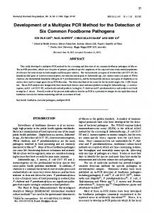
FIG. 1. Specificity analyses of the ISMap02 element by a conventional nested PCR protocol for amplification of a 117-bp product from DNA isolated from bacterial cultures of various species of mycobacteria. (A) Top of gel, lane 1, 100-bp molecular mass marker; lane 2, blank; lanes 3 and 4, M. fortuitum; lanes 5 and 6, M. scrofulaceum; lanes 7 and 8, M. smegmatis; lanes 9 and 10, M. phlei; lanes 11 and 12, M. gordonae. Bottom of gel, lane 1, 100-bp molecular mass marker; lane 2, blank; lanes 3 and 4, M. avium subsp. paratuberculosis DNA (strain K-10); lanes 5 and 6, M. avium subsp. paratuberculosis DNA (strain 19698). (B) Top of gel, lane 1, 100-bp molecular mass marker; lane 2, blank; lanes 3 and 4, M. avium subsp. avium (strain 1030); lanes 5 and 6, M. avium (strain 1115); lanes 7 and 8, M. avium (strain 1167); lanes 9 and 10, M. avium (strain 1156); lanes 11 and 12, M. avium (strain 1355). Bottom of gel, lane 1, 100-bp molecular mass marker; lane 2, blank; lanes 3 and 4, M. avium (strain 1177); lanes 5 and 6, M. avium (strain 1247); lanes 7 and 8, M. avium (strain 1025); lanes 9 and 10, M. avium (strain 1015); lane 11, negative control (master mix only); lane 12, positive control (strain 19698 DNA).
Statistical analyses. In order to compare the sensitivity of detection by realtime PCR versus that by conventional PCR and also to compare the efficacy of the IS900 element target versus that of the ISMap02 element target, the data were collapsed into the two variables of interest for each analysis (Stata Statistics/ Data Analysis; StataCorp, College Station, TX). McNemar’s chi-square test was used to derive an exact P value, and then a Bonferroni correction was done to compensate for the running of two analyses with the same data set. Due to the Bonferroni correction, the results would be considered significantly different if P values were ⱕ0.025. A 95% confidence interval was calculated for the significant comparison by using a risk ratio. A ratio of 1.0 would mean that there was no difference between the two tests, and a ratio of 1.5 would mean that a test would correctly identify 50% more samples. The confidence interval was calculated around that risk ratio.
RESULTS Results from the specificity evaluation of the nested PCR format for detection of the ISMap02 element are presented in Fig. 1. No products were observed when the primers were tested with DNA from laboratory cultures of M. fortuitum, M. scrofulaceum, M. smegmatis, M. phlei, or M. gordonae (Fig. 1A). In addition, all M. avium isolates evaluated were negative (Fig. 1B). The sensitivities of detection were compared between the ISMap02 nested protocol and the IS900 standard protocol for conventional PCR in our laboratory. Sample analyses were performed with DNA extracted from the K-10 strain of M. avium subsp. paratuberculosis (Fig. 2). Tenfold dilutions of bacterial DNA demonstrated a detection threshold of less than
FIG. 2. Comparison of the sensitivities of detection of the IS900 element and the ISMap02 element targets in a conventional PCR format. A single amplification of a 229-bp sequence was performed for IS900, and a two-step nested amplification of a 117-bp sequence was performed for ISMap02. DNA samples consisted of Mycobacterium avium subsp. paratuberculosis DNA (strain K-10) ranging in concentration from 10 ng/l to 1 fg/l (10-fold dilutions in buffer prior to amplification). Top gel, IS900, lane 1, 100-bp molecular mass marker; lane 2, negative control (master mix only); lane 3, 1 fg/l; lane 4, 10 fg/l; lane 5, 100 fg/l; lane 6, 1 pg/l; lane 7, 10 pg/l; lane 8, 100 pg/l; lane 9, 1 ng/l; lane 10, 10 ng/l; lane 11, positive control (strain 19698 DNA). Bottom gel, ISMap02, lane 1, 100-bp molecular mass marker; lane 2, negative control (master mix only); lane 3, 1 fg/l; lane 4, 10 fg/l; lane 5, 100 fg/l; lane 6, 1 pg/l; lane 7, 10 pg/l; lane 8, 100 pg/l; lane 9, 1 ng/l; lane 10, 10 ng/l; lane 11, positive control (strain 19698 DNA).
VOL. 43, 2005
PCR METHOD FOR DETECTION OF M. PARATUBERCULOSIS
FIG. 3. Sensitivity of detection of the 117-bp target after amplification of the ISMap02 element in a conventional nested PCR format. Samples represented are DNA extracted from a pure culture of M. avium subsp. paratuberculosis (strain 19698) at a stock concentration of 109 CFU/ml and DNA extracted from 10-fold serial dilutions of the bacterial stock. Top, lane 1, 100-bp molecular mass marker; lane 2, blank; lanes 3 and 4, 0 CFU/ml; lanes 5 and 6, 10 CFU/ml; lanes 7 and 8, 102 CFU/ml; lanes 9 and 10, 103 CFU/ml; lanes 11 and 12, 104 CFU/ml. Bottom, lane 1, 100-bp molecular mass marker; lane 2, blank; lanes 3 and 4, 105 CFU/ml; lanes 5 and 6, 106 CFU/ml; lanes 7 and 8, 107 CFU/ml; lanes 9 and 10, 108 CFU/ml; lanes 11 and 12, negative control (master mix only).
approximately 100 fg of DNA for the ISMap02-specific nested PCR protocol and 10 fg of DNA for the IS900-specific PCR protocol. A serial titration of pure bacterial culture (strain 19698) demonstrated a sensitivity of detection of 102 CFU/ml for the ISMap02 element by conventional PCR (Fig. 3). Significant differences in the sensitivity of detection were not observed by real-time PCR analyses for either the IS900 element or the ISMap02 element, based upon the area under the curve and melting curve plots (Fig. 4). The IS900 element was detected in all dilutions of DNA within about 35 cycles of PCR in the real-time system, whereas the second amplification of the ISMap02 element was detected more rapidly at less than 20 cycles of the 30-cycle run. The sensitivity of detection of M. avium subsp. paratuberculosis DNA extracted from negative fecal material spiked with dilutions of K-10 bacteria resulted in a detection limit between 103 and 104 CFU/g for the ISMap02 element and a detection limit of 103 CFU/g for the IS900 element by conventional PCR (data not shown). The sensitivity of detection of M. avium subsp. paratuberculosis in spiked fecal samples improved up to 1 log10 by real-time PCR for either the IS900 or the ISMap02 element. Further comparison of the sensitivities of these two proto-
4747
FIG. 4. Histogram of real-time PCR results for comparison of the sensitivities of detection of the IS900 element (top) and the ISMap02 element (bottom) for DNA samples extracted from M. avium subsp. paratuberculosis (strain K-10). A single amplification of a 229-bp sequence was performed for IS900, and a two-step nested amplification of a 117-bp sequence was performed for ISMap02. DNA samples consisted of M. avium subsp. paratuberculosis DNA (strain K-10) ranging in concentration from 10 ng/l to 1 fg/l (10-fold dilutions in buffer prior to amplification). The red lines in the histograms represent the sensitivity detection threshold for each target sequence. Red line in the histogram at the top, 10 fg DNA/l; red line in the histogram at the bottom, 100 fg DNA/l.
cols was performed with fecal samples from infected cows and known negative cows (Table 1). All the samples from infected cows had previously been positive by the IS900-specific conventional PCR in our laboratory (21) and, therefore, provided an excellent platform for comparison of the sensitivities of detection of the IS900 and ISMap02 elements. As expected, all samples were positive by IS900-specific conventional and realtime PCRs. One sample from the group with a positive fecal sample score of ⫹3 did not result in a positive band on the gel after conventional PCR was performed but was positive after the real-time method was performed. The sensitivity of detection of the ISMap02 element by the nested protocol by either conventional or real-time PCR was similar to that achieved for the IS900 element. One fecal sample from a cow that had clinical disease and high numbers of M. avium subsp. paratuberculosis organisms was negative for the ISMap02 element by both PCR tests. Amplification of either the IS900 element or the ISMap02 element was not demonstrated for the known negative fecal samples by either conventional or real-time PCR. The results of the amplification of the ISMap02 element by the nested protocol with fecal samples that were positive for the IS900 element are shown in Fig. 5. Further comparison of the results of the PCR methods for the detection of M. avium subsp. paratuberculosis in a crosssection of fecal samples from infected cows with various rates of shedding is shown in Table 2. Although fecal culture data were available for these cows, there was no correlation with historical PCR data. These samples were chosen because of stratification of the numbers of viable M. avium subsp. paratu-
4748
STABEL AND BANNANTINE
J. CLIN. MICROBIOL.
TABLE 1. Comparison of conventional and real-time PCR methods for detection of M. avium subsp. paratuberculosis-specific IS900 and ISMap02 elements in fecal DNA extracts No. of samples positive/no. of samples tested (%) Culture status
⫹4 (⬎75 CFU/g) ⫹3 (31–75 CFU/g) ⫹2 (8–30 CFU/g) ⫹1 (1–7 CFU/g) Negative
Conventional PCR
Real-time PCR
IS900
ISMap02
IS900
ISMap02
16/16 (100) 9/10 (90) 18/18 (100) 16/16 (100) 0/21
15/16 (94) 10/10 (100) 18/18 (100) 16/16 (100) 0/21
16/16 (100) 10/10 (100) 18/18 (100) 16/16 (100) 0/21
15/16 (94) 10/10 (100) 18/18 (100) 16/16 (100) 0/21
berculosis organisms recovered by primary isolation from fecal samples, which represented a cross-section of an infected herd. Of the results presented here, amplification of either the ISMap02 element or the IS900 element by conventional PCR resulted in positive identification of cows that were shedding low, moderate, or high levels of M. avium subsp. paratuberculosis in their feces. Amplification of the IS900 element was more sensitive than amplification of the ISMap02 element, although it missed one low-level shedder and one moderatelevel shedder in this group. The real-time PCR assay for the ISMap02 element was able to detect two animals that were negative by the conventional method. Real-time PCR with the IS900 element also detected one additional positive animal
that was negative by the conventional method. However, when we compared more samples from infected cows with a score of ⫹1, there was little improvement in the sensitivity of detection by the real-time method compared to that by the conventional method. Interestingly, the conventional PCR did not detect all samples that had bacterial loads of ⬎75 CFU/g (too numerous to count), regardless of the gene amplified. One high-level shedder was not detected by real-time PCR with the ISMap02 element; however, the sample from this animal was highly contaminated, and this may have contributed to the inaccuracy. Overall, there were no significant differences in the sensitivities of detection when the results obtained with the two gene targets within an assay (i.e., the conventional or the real-time PCR) were compared. In addition, when all data were grouped together by assay, regardless of the gene target, no differences were observed between the conventional and the real-time methods. However, grouping of the data by gene targets demonstrated a significant increase (P ⬍ 0.02) in the sensitivity of
TABLE 2. Comparison of conventional and real-time PCR assays for IS900 and ISMap02 elements for the detection of Mycobacterium avium subsp. paratuberculosis DNA in fecal samples of infected cows with various shedding levelsb Result by:
FIG. 5. Representative results of amplification of a 117-bp sequence of ISMap02 by the conventional nested PCR protocol in DNA extracted from fecal samples of known infected cows. Top, lane 1, 100-bp molecular mass marker; lane 2, blank; lanes 3 and 4, 3⫹ shedder; lanes 5 and 6, 2⫹ shedder; lanes 7 and 8, 1⫹ shedder; lanes 9 and 10, 2⫹ shedder; lanes 11 and 12, 2⫹ shedder. Bottom, lanes 1 and 2, 2⫹ shedder; lanes 3 and 4, 1⫹ shedder; lanes 5 and 6, 2⫹ shedder; lanes 7 and 8, 2⫹ shedder; lanes 9 and 10, negative control (master mix only); lanes 11 and 12, positive control (strain 19698 DNA). Shedders were defined as ⫹4 (⬎75 CFU/g feces), ⫹3 (31 to 75 CFU/g feces), ⫹2 (8 to 30 CFU/g feces), ⫹1 (1 to 7 CFU/g feces), and ⬍⫹1.
Sample no.
Fecal culture
1 2 3 4 5 6 7 8 9 10 11 12 12 14 15 16 17 18
1, 0, 0, 0 1, 0, 0, 0 1, 0, 2, 0 1, 5, 0, 0 1, 3, 0, 1 2, 1, 2, 3 0, 3, 3, 3 3, 2, 1, 3 4, 6, 5, 4 14, 11, 29, 19 30, 26, 38, 26 26, 27, 25, 28 T, T, T, T T, T, T, T T, T, T, T T, T, T, T T, T, T, T T, T, T, T
a
IS900 conventional PCR
ISMap02 conventional PCR
IS900 real-time PCR
ISMap02 real-time PCR
P N P P P P P P P P N P P P P P P P
P N P N P P P N P P N N P P P P N P
P N P P P P P P P P P P P P P P P P
P N P N P P P P P P P N P P P P N P
a Colony forming units on each of four slants of Herrold’s egg yolk medium; T, 150 CFU/slant. b P, positive; N, negative.
PCR METHOD FOR DETECTION OF M. PARATUBERCULOSIS
VOL. 43, 2005
detection of the IS900 element compared to that of the ISMap02 element. The risk ratio for comparison of the detection of a positive sample by both PCR methods with the IS900specific primers than that with the ISMap02-specific primers was 1.27 (95% confidence interval, 1.06 to 1.52), indicating that in this data set, amplification of the IS900 element by both conventional and real-time PCR resulted in the detection of approximately 27% more positive fecal samples than amplification of the ISMap02 element. DISCUSSION As methods for the detection of bacterial DNA in fecal samples become more sensitive and laboratories become more adept, it is likely that the PCR methodology will usurp other diagnostic methods in the field. The use of direct PCR with fecal DNA extracts has several advantages, including a more rapid time to the end point compared to that for culture of the bacterium (3 days versus 8 weeks). In addition, the procedure for the extraction of fecal DNA in preparation for PCR has become more simple and inexpensive in recent years, circumventing the need for expensive reagents or kits (26). Most importantly, the sensitivities of the PCR methods appear to continually improve, with the sensitivity approaching or surpassing that of fecal culture. Historically, diagnostic assays for the direct PCR of fecal extracts for the detection of M. avium subsp. paratuberculosis have utilized the IS900 element sequence as the target. The IS900 element is a 1,451-bp sequence that is present at 15 to 20 copies within the M. avium subsp. paratuberculosis genome (31). The multicopy nature of the sequence makes it ideal as a target sequence for the diagnosis of paratuberculosis, with a higher level of sensitivity achieved with these sequences as targets than that achieved with single-copy genes as targets. Although other insertion elements, such as IS1311, ISMav2, and ISMpa1, have been observed within the genome, they have not been demonstrated to be specific to M. avium subsp. paratuberculosis, precluding their use as diagnostic reagents (17, 27, 32). Other elements that have demonstrated high specificities for the detection of M. avium subsp. paratuberculosis in PCR assays are F57, HspX, and, more recently, genomic target 251 (5, 20, 30). There is some dissent as to whether the IS900 element is indeed unique to M. avium subsp. paratuberculosis. Two studies have demonstrated the presence of IS900 in species of environmental mycobacteria, M. cookii and M. scrofulaceum (4, 7). The IS900 regions of these two species that were sequenced shared 94% and 79% homologies with the IS900 region of M. avium subsp. paratuberculosis that was sequenced, respectively, suggesting that use of the IS900 sequence as a diagnostic tool in the field may yield false-positive results in rare instances. More recently, in silico analysis of the published IS900-specific primer sequences frequently used by diagnostic laboratories demonstrated that these primer pairs would amplify IS900-like sequences (30). Stringent selection of new IS900-specific primers indicated that the primer set would amplify M. avium subsp. paratuberculosis strains but not non-M. avium subsp. paratuberculosis strains or IS900-like sequences. Previously, in our laboratory, a PCR analysis of over 100 DNA preparations isolated from M. avium subsp. paratuberculosis, M. avium, M. intracellulare, and other Mycobacterium spp. such
4749
as M. celatum, M. leprae, M. scofulaceum, and M. tuberculosis demonstrated 99.2% specificity for the IS900 sequence (6). Therefore, the use of IS900-specific primers must be viewed with some caution unless adequate controls are included within the assay. In the present study, the specificity of the ISMap02 element compared well to that of the IS900 element. The ISMap02 element did not amplify DNA from any of the closely related species of mycobacteria, in particular, the M. avium isolates and M. scrofulaceum. The ISMap02 genetic element is 1,674 bp, and six copies are present in the M. avium subsp. paratuberculosis K-10 genome (designated Map0338c, Map2416c, Map2502, Map2566, Map3357c, and Map3467c). Prior to this study, the entire 1,674-bp sequence of ISMap02 was used to search the nonredundant sequence databases by BLAST analysis (1). No matches were identified by this search other than an identity to self. Further, comparison of the ISMap02 sequence to the 381 complete and incomplete genomes available in GenBank resulted in the return of only the self sequence from the K-10 genome. Finally, PCR analyses of ISMap02 demonstrated that the sequence was present in all 39 isolates of M. avium subsp. paratuberculosis examined but was absent from all other mycobacterial species and subspecies tested (19). These analyses further substantiate the specificity of this sequence. In addition, using a nested format and real-time PCR technology, we were able to demonstrate a difference in sensitivity of detection of only 1 log10 between the two genes. Due to the additional copies present within the M. avium subsp. paratuberculosis genome, it is not surprising that the sensitivity should be somewhat higher for the IS900 element than for the ISMap02 element. Yet, the evaluation of fecal samples obtained from naturally infected animals suggests that the ISMap02 element could be useful as a target sequence for use as a diagnostic tool for the detection of M. avium subsp. paratuberculosis. In general, the sensitivity of detection of M. avium subsp. paratuberculosis DNA is highly correlated with the level of shedding of the bacterium in the feces. However, fecal samples that are positive by culture methods and even samples with high numbers of bacteria are sometimes falsely identified as negative by PCR. PCR-based diagnostic tests with fecal samples are often hampered by the presence of inhibitors such as phytic acid and other complex polysaccharides (12, 29). It is also highly probable that feces from cows with clinical paratuberculosis, which are typically high-level shedders, may contain heme and epithelial cells, a result of sloughing of the thickened mucosa associated with end-stage disease. These components have been found to be inhibitory to PCRs as well (12). The comparison of conventional and real-time PCR techniques was informative and yielded significant information about the sensitivities of the two methods. The real-time technology improved detection of the M. avium subsp. paratuberculosis genes negligibly, as evaluated within this study. However, identification of positive samples circumvents the labor associated with the gel documentation protocol required for conventional PCR. In addition, one can control the stringency for the designation of positive samples by defining the cycle threshold or the area under the melting curve (Fig. 6). This precludes much of the speculation of false-positive results that has been a factor with gel analyses in the past due to the use of
4750
STABEL AND BANNANTINE
FIG. 6. Histogram depicting the melting curve results for a realtime nested PCR assay for detection of the ISMap02 element in DNA extracted from fecal samples from a known infected cow (red line) and a known noninfected cow (green line).
a poor loading technique, the presence of faint banding patterns, or the presence of bands of the incorrect size. Therefore, this methodology would be the most appropriate for laboratories that must make critical judgments about the status of an unknown sample. Although real-time PCR is a bit more expensive to perform due to the initial outlay for the equipment, this study suggests that it is worth the additional cost. In conclusion, a PCR test developed with a newly identified gene unique to M. avium subsp. paratuberculosis, ISMap02, was comparable in sensitivity to a nested conventional or real-time PCR with the IS900 element. This offers an alternative or an additional method for the rapid detection of M. avium subsp. paratuberculosis in biological samples from suspect animals. ACKNOWLEDGMENTS We express our appreciation to Tonia McNunn and Trudy Bosworth for excellent technical assistance and tremendous effort on this project. REFERENCES 1. Altschul, S. F., W. Gish, W. Miller, E. W. Myers, and D. J. Lipman. 1990. Basic local alignment search tool. J. Mol. Biol. 215:403–410. 2. Bannantine, J. P., J. K. Hansen, M. L. Paustian, A. Amonsin, L. L. Li, J. R. Stabel, and V. Kapur. 2004. Expression and immunogenicity of proteins encoded by sequences specific to Mycobacterium avium subsp. paratuberculosis. J. Clin. Microbiol. 42:106–114. 3. Christopher-Hennings, J., M. A. Dammen, S. R. Weeks, W. B. Epperson, S. N. Singh, G. L. Steinlicht, Y. Fang, J. L. Skaare, J. L. Larsen, J. B. Payeur, and E. A. Nelson. 2003. Comparison of two DNA extractions and nested PCR, real-time PCR, a new commercial PCR assay, and bacterial culture for detection of Mycobacterium avium subsp. paratuberculosis in bovine feces. J. Vet. Diagn. Investig. 15:87–93. 4. Cousins, D. V., R. Whittington, I. Marsh, A. Masters, R. J. Evans, and P. Kluver. 1999. Mycobacteria distinct from Mycobacterium avium subsp. paratuberculosis isolated from the faeces of ruminants possess IS900-like sequences detectable by IS900 polymerase chain reaction: implications for diagnosis. Mol. Cell. Probes 13:431–442. 5. Ellingson, J. L., C. A. Bolin, and J. R. Stabel. 1998. Identification of a gene unique to Mycobacterium avium subspecies paratuberculosis and application to diagnosis of paratuberculosis. Mol. Cell. Probes 12:133–142. 6. Ellingson, J. L. E., J. R. Stabel, W. R. Bishai, R. Frothingham, and J. M. Miller. 2000. Evaluation of the accuracy and reproducibility of a practical PCR panel assay for rapid detection and differentiation of Mycobacterium avium subspecies. Mol. Cell. Probes 14:153–161. 7. Englund, S., G. Bolske, and K. E. Johansson. 2002. An IS900-like sequence found in a Mycobacterium sp. other than Mycobacterium avium subsp. paratuberculosis. FEMS Microbiol. Lett. 209:267–271. 8. Fang, Y., W. Wu, J. L. Pepper, J. L. Larsen, S. A. E. Marras, E. A. Nelson, W. B. Epperson, and J. Christopher-Hennings. 2002. Comparison of realtime, quantitative PCR with molecular beacons to nested PCR and culture methods for detection of Mycobacterium avium subsp. paratuberculosis in bovine fecal samples. J. Clin. Microbiol. 40:287–291. 9. Green, E. P., M. L. Tizard, M. T. Moss, J. Thompson, D. J. Winterbourne, J. J. McFadden, and J. Hermon-Taylor. 1989. Sequence and characteristics of IS900, an insertion element identified in a human Crohn’s disease isolate of Mycobacterium paratuberculosis. Nucleic Acids Res. 17:9063–9073.
J. CLIN. MICROBIOL. 10. Groenendaal, H., and D. T. Galligan. 2003. Economic consequences of control programs for paratuberculosis in midsize dairy farms in the United States. J. Am. Vet. Med. Assoc. 223:1757–1763. 11. Huda, A., G. Jungersen, A. B. Christoffersen, and P. Lind. 2003. Diagnosis of bovine paratuberculosis by interferon-gamma (IFN-gamma) test. Acta Vet. Scand. 44:281. 12. Inglis, G. D., and L. D. Kalischuk. 2003. Use of PCR for direct detectin of Campylobacter species in bovine feces. Appl. Environ. Microbiol. 69:3435– 3447. 13. Kennedy, D. J., and M. B. Allworth. 2000. Progress in national control and assurance programs for bovine Johne’s disease in Australia. Vet. Microbiol. 77:443–451. 14. Khare, S., T. A. Ficht, R. L. Santos, J. Romano, A. R. Ficht, S. Zhang, I. R. Grant, M. Libal, D. Hunter, and L. G. Adams. 2004. Rapid and sensitive detection of Mycobacterium avium subsp. paratuberculosis in bovine milk and feces by a combination of immunomagnetic bead separation-conventional PCR and real-time PCR. J. Clin. Microbiol. 42:1075–1081. 15. Kim, S. G., S. J. Shin, R. H. Jacobson, L. J. Miller, P. R. Harpending, S. M. Stehman, C. A. Rossiter, and D. A. Lein. 2002. Development and application of quantitative polymerase chain reaction assay based on the ABI 7700 system (TaqMan) for detection and quantification of Mycobacterium avium subsp. paratuberculosis. J. Vet. Diagn. Investig. 14:126–131. 16. Luyven, G., A. Vom Schloss, and M. Sasserath. 2002. Paratuberculosis eradication programs in Northrhine-Westfalia. Dtsch. Tierarztl. Wochenschr. 109:524–527. 17. Olsen, I., T. B. Johansen, H. Billman-Jacobe, S. F. Nilsen, and B. Djonne. 2004. A novel IS element, ISMpa1, in Mycobacterium avium subsp. paratuberculosis. Vet. Microbiol. 98:297–306. 18. O’Mahony, J., and C. Hill. 2002. A real-time PCR assay for the detection and quantitation of Mycobacterium avium subsp. paratuberculosis using SYBR Green and the Light Cycler. J. Microbiol. Methods 51:283–293. 19. Paustian, M. L., A. Amonsin, V. Kapur, and J. P. Bannantine. 2004. Characterization of novel coding sequences specific to Mycobacterium avium subsp. paratuberculosis: implications for diagnosis of Johne’s disease. J. Clin. Microbiol. 42:2675–2681. 20. Rajeev, S., Y. Zhang, S. Sreevatsan, A. S. Motiwala, and B. Byrum. 2005. Evaluation of multiple genomic targets for identification and confirmation of Mycobacterium avium subsp. paratuberculosis isolates using real-time PCR. Vet. Microbiol. 105:215–221. 21. Shin, S. J., Y. F. Chang, C. Huang, J. Zhu, H. S. Yoo, K. S. Shin, S. Stehman, S. J. Shin, and A. Torres. 2004. Development of a polymerase chain reaction test to confirm Mycobacterium avium subsp. paratuberculosis in culture. J. Vet. Diagn. Investig. 16:116–120. 22. Stabel, J. R. 1996. Production of gamma-interferon by peripheral blood mononuclear cells: an important diagnostic tool for detection of subclinical paratuberculosis. J. Vet. Diagn. Investig. 8:345–350. 23. Stabel, J. R. 1997. An improved method for cultivation of Mycobacterium paratuberculosis from bovine fecal samples and comparison to three other methods. J. Vet. Diagn. Investig. 9:375–380. 24. Stabel, J. R. 2000. Cytokine secretion by peripheral blood mononuclear cells from cows infected with Mycobacterium paratuberculosis. Am. J. Vet. Res. 61:754–760. 25. Stabel, J. R., and R. H. Whitlock. 2001. An evaluation of a modified interferon-gamma assay for the detection of paratuberculosis in dairy herds. Vet. Immunol. Immunopathol. 79:69–81. 26. Stabel, J. R., T. L. Bosworth, T. A. Kirkbride, R. L. Forde, and R. H. Whitlock. 2004. A simple, rapid, and effective method for the extraction of Mycobacterium paratuberculosis DNA from fecal samples for polymerase chain reaction. J. Vet. Diagn. Investig. 16:22–30. 27. Strommenger, B., K. Stevenson, and G. F. Gerlach. 2001. Isolation and diagnostic potential of ISMav2, a novel insertion sequence-like element from Mycobacterium avium subspecies paratuberculosis. FEMS Microbiol. Lett. 196:31–37. 28. Taddei, S., C. Robbi, C. Cesena, I. Rossi, E. Schiano, N. Arrigoni, G. Vicenzoni, and S. Cavirani. 2004. Detection of Mycobacterium avium subsp. paratuberculosis in bovine fecal samples: comparison of three polymerase chain reaction-based diagnostic tests with a conventional culture method. J. Vet. Diagn. Investig. 16:503–508. 29. Thornton, C. G., and S. Passen. 2004. Inhibition of PCR amplification by phytic acid, and treatment of bovine fecal specimens with phytase to reduce inhibition. J. Microbiol. Methods 59:43–52. 30. Vansnick, E., P. De Rijk, F. Vercammen, D. Geysen, L. Rigouts, and F. Portaels. 2004. Newly developed primers for the detection of Mycobacterium avium subspecies paratuberculosis. Vet. Microbiol. 100:197–204. 31. Whipple, D. L., P. Kapke, and C. Vary. 1990. Identification of restriction fragment length polymorphisms in DNA from Mycobacterium paratuberculosis. J. Clin. Microbiol. 28:2561–2564. 32. Whittington, R., I. Marsh, E. Choy, and D. Cousins. 1998. Polymorphisms in IS1311, an insertion sequence common to Mycobacterium avium and M. avium subsp. paratuberculosis, can be used to distinguish between and within these species. Mol. Cell. Probes 12:349–358.