Hindawi Publishing Corporation International Journal of Analytical Chemistry Volume 2015, Article ID 748056, 5 pages http://dx.doi.org/10.1155/2015/748056
Research Article Development of a Rapid and Simple Method for Detection of Protein Contaminants in Carmine Norihisa Nakayama,1 Yutaka Ohtsu,2 Daisuke Maezawa-Kase,2 and Ken-Ichi Sano1 1
Graduate School of Environmental Symbiotic System and Department of Innovative Systems Engineering, Nippon Institute of Technology, 4-1 Gakuendai, Miyashiro-machi, Saitama 345-8501, Japan 2 Kishi Kasei Company, 1-10-8 Fukuura, Kanazawa-ku, Yokohama 234-0004, Japan Correspondence should be addressed to Ken-Ichi Sano;
[email protected] Received 1 December 2014; Revised 16 March 2015; Accepted 19 March 2015 Academic Editor: Shaoping Li Copyright © 2015 Norihisa Nakayama et al. This is an open access article distributed under the Creative Commons Attribution License, which permits unrestricted use, distribution, and reproduction in any medium, provided the original work is properly cited. Protein contaminants in carmine can cause dyspnea and anaphylactic reactions in users and consumers of products containing this pigment. The method generally used for detection of proteins in carmine has low reproducibility and is time-consuming. In this study, a rapid, simple, and highly reproducible method was developed for the detection of protein contaminants in carmine. This method incorporates acidic protein denaturation conditions and ultrafiltration. To prevent protein aggregation, sodium dodecyl sulfate containing gel electrophoresis running buffer was used for dispersing the carmine before filtration. An ultrafiltration device was used to separate the protein contaminants from carminic acid in the carmine solution. Two ultrafiltration devices were compared, and a cylindrical device containing a modified polyethersulfone membrane gave the best results. The method had high reproducibility.
1. Introduction Carmine is a natural red pigment extracted from dried scale insects (cochineal, Dactylopius coccus) [1]. The main chemical component of this pigment is carminic acid (7-𝛼-d-glucopyranosyl-9,10-dihydro-3,5,6,8-tetrahydroxy1-methyl-9,10-dioxoanthracene carboxylic acid, Figure 1). Carmine is widely used for coloring food products, cosmetics, and medicines [2, 3]. In 2012, the Japanese Consumer Affairs Agency published a report on carmine detailing its potential links to dyspnea and anaphylactic reactions [4]. This report detailed approximately 20 articles from 1960s about anaphylaxis resulting from the use of the cosmetics or consumption of food containing carmine. Carminic acid is not the cause of these allergic reactions; they are rather caused by protein contaminants in the carmine [5, 6]. Some of the allergenic protein contaminants have been identified, and one of them has high homology with phospholipase A, which is a well-known allergen in vespidae venom [7]. Hence, the use of carmine in foods is regulated both in Japan and European Union.
For consumer safety, allergenic protein contaminants in carmine need to be monitored. The general method for detection of protein contaminants in carmine involves suspension of the pigment in an acidic solution (1% aqueous solution of phosphoric acid) and then separation of any protein contaminants from the solution by HPLC [8]. The collected proteins are identified by SDS-PAGE [9]. This method is time-consuming because about ten HPLC analyses are required to obtain a sufficient quantity of protein for SDSPAGE analysis. Furthermore, the reproducibility of the HPLC separation is low. Therefore, an alternative method that is rapid, simple, and has high reproducibility is needed. In this study, we developed a rapid and simple method for detection of protein contaminants in carmine with high reproducibility.
2. Materials and Methods Three samples of dry carmine were used. Sample A was purchased from Biocon (cochineal extract, lot number M008363). Sample B was manufactured by Kishi Kasei Company (carminic acid, lot number 174302), and we carried
2
International Journal of Analytical Chemistry OH H
H A
OH
O
O
OH
B
C
H
O
OH HO
H HO
OH
H
OH O
OH
Figure 1: Carminic acid. 1% phosphoric acid
out further purification of Sample B as follows. Firstly, Sample B was dissolved in 5% of phosphoric acid and loaded on a spherical polymerized divinylbenzene resin. Then bound carminic acid was eluted by 50% ethanol and dried (Sample C). To separate the protein fraction from the carminic acid, we used an ultrafiltration device with a molecular weight cut-off of 3,000 Da. Two ultrafiltration devices were tested; one was v-shaped, with a regenerated cellulose membrane (Amicon Ultra-0.5 mL 3 K, Millipore, Billerica, MA), and the other was cylindrical, with a modified polyethersulfone membrane (Nanosep 3 K Omega, Pall Corporation, Port Washington, NY). The initial sample volume was 500 𝜇L, and the filtration was done by centrifugation until the samples were concentrated to about 0.4–0.7 of their initial volume. The time for centrifugation depended on each samples. The protocol was repeated more than 14 times. After these filtration steps, the concentration of carminic acid in each sample would be diluted by at least 6 × 10⁴ times mathematically. Electrophoresis was carried out using precast gels (15% Choju Gel, Oriental Instruments, Sagamihara, Japan) and stained using Silver Stain MS kit (Wako Chemicals, Osaka, Japan) [10].
3. Results and Discussion 3.1. Removal of Carmine Acid from Cochineal Dispersed in Protein Denaturant Solution. Initially, we investigated the separation of proteins from the carmine solution. Three types of carmine were used, as detailed in Section 2. Each carmine sample was dispersed in phosphoric acid (1% (mass fraction) aqueous solution) at a final concentration of 50 mg/mL using ultrasonication. A white precipitate of aggregated protein contaminants was observed when Sample A (cochineal extract, lot number M008363, Biocon) was dispersed in phosphoric acid (Figure 2). We had confirmed low reproducibility of HPLC separations, such as disappearance and shift of a peak from contaminant proteins (data not shown). The aggregation of protein contaminants in the phosphoric acid would reduce the reproducibility of protein separation. As a logical approach to prevention of aggregation of protein contaminants, we made use of protein denaturant first. Urea and guanidine hydrochloric acid are commonly used for
+8 M urea
+6 M guanidine HCl
Figure 2: Carmine (Sample A, cochineal extract, Biocon) suspensions. (A) is suspended in 1% phosphoric acid. (B) is dispersed in 1% phosphoric acid and 8 mol/L urea. (C) is dispersed in 1% phosphoric acid and 6 mol/L guanidine hydrochloride.
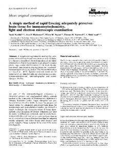
dissolution of protein aggregates. Therefore, in the present method, urea (8 mol/L) and guanidine hydrochloric acid (6 mol/L) were added to dissolve the precipitate (Figure 2). Detection of proteins is usually performed by colorimetric methods such as biuret method and ninhydrin reaction. Free carminic acid interferes with the detection of allergenic proteins in the carmine by colorimetric analysis, and it needs to be removed before analysis. In addition, to detecting the allergenic proteins, protein fractions of >6 kDa need to be collected, and determining the molecular weight of protein contaminants is also important. Both SDS-PAGE and mass spectrometric analysis are generally used methods for determination of molecular weight of proteins. Bound carminic acid on protein shift in molecular weight is found in mass spectrometric analysis, and a smear band pattern is also acquired from SDS-PAGE. Consequently, in the present study, two ultrafiltration devices were compared for separating the protein fraction from the carmine solution. Both of these had molecular weight cut-offs of 3,000 Da and are described in Section 2. The initial sample volume was 500 𝜇L, and the filtration was done by centrifugation until the samples were concentrated to about 0.4–0.7 of their initial volume. The protocol was repeated more than 14 times. The optimum initial concentration of carmine was investigated. 50, 100, and 200 mg/mL concentrations of carmine were suspended in each solution. At 100 and 200 mg/mL of carmine, carminic acid suspensions were difficult to remove by both ultrafiltration devices, and a concentration of 50 mg/mL was used in subsequent experiments. After filtration, the concentration of carminic acid in each sample was diluted by at least 6 × 10⁴ times. The Amicon Ultra-0.5 v-shaped ultrafiltration device could not quantitatively remove the carminic acid, and thus the SDS-PAGE results were obscured by remaining pigment (Figure 3(a)). However, the cylindrical ultrafiltration device did remove sufficient carminic acid to allow the protein
1% phosphoric acid + 6 M guanidine HCl
1% phosphoric acid + 8 M urea
Molecular weight marker 120 90
64 48 36
64 (kDa)
(kDa)
120 90
1% phosphoric acid
3 1% phosphoric acid + 6 M guanidine HCl
1% phosphoric acid + 8 M urea
1% phosphoric acid
Molecular weight marker
International Journal of Analytical Chemistry
28
48 36 28
20 20
9
9
V-shaped filter device (a)
Cylindrical filter device (b)
Figure 3: SDS-PAGE of protein contaminants in carmine (Sample A, cochineal extract, Biocon) after silver staining. (a) Amicon Ultra-0.5 ultrafiltration device. (b) Nanosep 3 K Omega ultrafiltration device.
contaminant band to be clearly observed at around 60 kDa. Although the cylindrical ultrafiltration device provided more effective removal of carminic acid than the v-shaped device, the detection of low molecular weight proteins (