1979; Bellows et al. 1981, \982a,b) or outside (Grinnell & Lamke,. 1984) pre-made collagen gels. Behaviour of connective tissue cells is influenced greatly by ...
J. Cell Set. 84, 183-200 (1986)
183
Printed in Great Britain © The Company of Biologists Limited 1986
DEVELOPMENT OF A THREE-DIMENSIONAL EXTRACELLULAR MATRIX SYNTHESIZED BY HUMAN DIPLOID FIBROBLASTS IN VITRO EVA E. QWARNSTROM AND ROY C. PAGE Department of Pathology, SM-30, School of Medicine, Department of Periodontics, School of Dentistry, and Center for Research in Oral Biology, University of Washington, Seattle, WA 98195, USA
SUMMARY Development and maturation of an extracellular matrix, synthesized by human gingival fibroblasts, have been studied microscopically. Pairs of demineralized, fibronectin-coated slices of human tooth root, 300/im thick, were placed on confluent cell layers, defining a 0-5 mm wide space. The cultures were grown under standard conditions with ascorbic acid (SO/igmP 1 ) added daily. At various times up to 13 weeks, the cultures were fixed and the samples prepared for light and electron microscopy. Cells from the monolayer became attached to, and migrated up, the vertical root surface and, during the time studied, completely filled the space between the root slices with an extracellular matrix. A close association was seen between the cell membrane and collagen fibres in the demineralized surface initially. A thin layer of fibrillar material was deposited between the cell and the vertical surface, and eventually an extracellular matrix surrounding the cells and attaching to the root surface was present. Samples fixed in the presence of Ruthenium Red showed intense staining of the fibrillar material, indicating the presence of anionic molecules. Additional cells migrated onto the newly synthesized matrix and up the root surface. Growth of the fibrillar networks on either side, horizontally and vertically, continued and, eventually, an extracellular matrix attaching to the vertical surfaces completely filled the previously empty space. Immunocytochemical staining showed that the matrix contained hyaluronic acid, chondroitin sulphate, dermatan sulphate and fibronectin at this time. Collagen fibres were observed at 6 weeks, and at later times collagen types I, III and V were the primary matrix components. The fibroblasts attaching to the root slice and those present at the edge of the matrix had an elongated, polar form. The cells within the matrix frequently showed a stellate appearance with numerous extended processes, in contact with fibrillar material or collagen fibres. Fibroblast processes were at later times seen to enclose bundles of collagen fibres and to mediate cell-to-cell contact, occasionally via desmosome-like structures. The structure and composition of the matrix and the appearance and apparent behaviour of the cells were similar to that observed in the healing wound. This system thus could provide a model for studying various aspects of regeneration of extracellular matrix.
INTRODUCTION
Wound healing and tissue regeneration consist of complex events, many of which are known from in vivo studies. Formation of a blood clot is followed by infiltration of polymorphonuclear leucocytes and macrophages. Fibroblasts in the periphery proliferate and migrate into the wound during formation of an extracellular matrix Key words: collagen, extracellular matrix, fibroblasts, glycosaminoglycans, immunocytochemistry, wound healing.
184
E. E. Qwarnstrom and R. C. Page
and angiogenesis (Ross, 1968; Castor, 1981; Marks, 1981). Once structurally completed, maturation and reorganization of the collagenous matrix occurs until functional integrity is reconstituted. The process is regulated by hormones, such as platelet-derived growth factor (Kohler & Lipton, 1974; Ross et al. 1974), fibroblast growth factor (Gospodarowicz, 1974) and interleukin-1 (Schmidt et al. 1982) and complement (Sobel et al. 1983; Cooper, 1985). Some aspects of wound healing, such as the ability of fibroblasts to attach and spread, have been studied in detail in vitro (Culp et al. 1979; Singer, 1979; Yamada, 1983). Synthesis by these cells of various types of extracellular matrix components has been investigated (Narayanan et al. 1978; Savage & Swann, 1985). Contraction and reorganization of extracellular matrix have been studied after plating fibroblasts within (Belle* a/. 1979; Bellows et al. 1981, \982a,b) or outside (Grinnell & Lamke, 1984) pre-made collagen gels. Behaviour of connective tissue cells is influenced greatly by their environment (Gospodarowicz et al. 1978; Hay, 1981). So, in studies on the role of defined factors on connective tissue regeneration, it would be valuable to use a model in which the matrix surrounding the cells more closely resembles that
in vivo. This paper describes an in vitro model in which a space is filled successively with an extracellular matrix, which is synthesized by the fibroblasts and which they inhabit. The overall events were examined by light microscopy and scanning electron microscopy. The interaction between the fibroblasts and the space-limiting surfaces, and the synthesized matrix, as well as morphological characteristics of the cells, were studied using transmission electron microscopy. The types of matrix components present in the fibrillar network were detected by the use of specific stains for light microscopy and transmission electron microscopy. Further determination of the composition of the extracellular matrix was done by immunocytochemical staining for various types of glycosaminoglycans and fibronectin. In addition, staining for various types of collagens was done. MATERIALS AND METHODS
Cell lines Human gingival fibroblast strains 24 and 42 (HGF24, HGF42) (Narayanan & Page, 1976) derived from specimens of human gingiva were used at 5th, 6th, 9th or 10th transfer. The cells were plated subconfluently (250 000 cells/dish) on 35 mm tissue culture dishes and maintained until confluent in Dulbecco—Vogt (DV) medium containing 10 % heat-inactivated foetal calf serum (FCS, Gibco Laboratories) in a humidified atmosphere of 95 % air and 5 % CO 2 at 37°C.
Preparation of root pieces Extracted human teeth obtained from the Oral Surgery Clinic at the University of Washington School of Dentistry were kept frozen (—20cC) until used. Following thawing, the roots were planed to create a smooth dentin surface with occasional patches of cementum, the organic component primarily composed of collagen. The roots were cut transversely into 200— 300 Jim thick sections using a rotary saw. Root pieces were extensively decalcified in formic acid (4M, 1 week) or left in phosphate-buffered saline (PBS). Decalcified and non-decalcified root pieces were washed repeatedly in PBS, and DV medium containing 10% heat-inactivated FCS and added antibiotics (ampicillin 1-25/igml""1; garamycin
Fibroblast extracellular
matrix in vitro
185
2 / i g m P 1 ; mycostatin lOunitsml" 1 ; penicillin 100 units ml" 1 ; streptomycin lOO/igml"1) and then incubated in this medium for 4 days at 37°C. Following rinses in PBS, the root pieces that were non-decalcified were subjected to citric acid (pH 1-5) treatment for 3 min to decalcify the outer 3 /im (Register & Burdick, 1975). For consistency, the already decalcified root pieces were similarly treated. The root pieces were again rinsed in PBS until neutralized, and subsequently coated with plasma fibronectin (100^gml" 1 in serum-free DV medium) for 15 min. The prepared root pieces, totally decalcified or surface-decalcified, were carefully laid on top of the confluent cell layer in pairs with a gap of 0-5-1 mm. The cultures were maintained in DV medium containing 10% FCS as above, and were given ascorbic acid (50//gml~ ) daily.
Microscopy At various times (5, 7, 10 days, and 2, 3, 6, 9, 12, 13 weeks) specimens were rinsed twice in PBS and fixed using a modified Karnovsky's (1965) fixative containing 2 % formaldehyde (Polysciences) and 2 % glutaraldehyde (Ted Pella) in 0-1 M-sodium cacodylate buffer (pH 7-4) with 0-05 % CaCl2 overnight at room temperature, followed by rinsing in sucrose buffer (0-1 M-sodium cacodylate, 6 % sucrose and 1 mM-CaClz). After postfbtation in 1 % osmium tetroxide in sucrose buffer (1 • 5 h at room temperature), the samples were rinsed and stained en bloc with 3 % uranyl acetate in 70% ethanol during dehydration through a graded series of ethanols (35% to 100%). Some samples were processed with Ruthenium Red added to fixative (0-2%), buffer ( 0 1 % ) and osmium tetroxide (0-05 %) (Wight, 1980) for staining anionic molecules. Four to eighteen specimens were fixed at each time point and prepared for light microscopy, or scanning or transmission electron microscopy. Specimens for scanning electron microscopy (SEM) containing extensively or surface-decalcified root slices were critical point dried, coated with gold/palladium and examined in a JEOL 35C microscope. Specimens for transmission electron microscopy (TEM) contained mostly extensively decalcified root slices. Surface-decalcified root pieces prepared for TEM were demineralized using EDTA (0'2 M) for about 10 days following fixation, and were then put in sucrose buffer with 0-5 M-CaCl2 added. Data shown are from samples containing totally decalcified root slices unless stated otherwise. All samples for TEM were embedded in the dish, in Polybed 812 (Polysciences). Thick (1 fim) cross-sections through the space between the two root pieces, were cut perpendicular to the bottom of the dish, stained with Toluidine Blue and examined by light microscopy. Thin sections of selected areas were cut with a diamond knife and mounted on Formvar-coated copper grids. The sections were stained with uranyl acetate and lead citrate, and examined in a JEOL 100B electron microscope. Samples for light microscopy only were dehydrated following fixation and embedded in JB-4 plastic, sectioned (3 jim) and stained with haematoxylin-eosin, periodic acid-Schiff-haematoxylin (PAS), or Alcian Blue. In addition, some samples were embedded in paraffin, sectioned (5 /jxn), and stained with van Gieson (Mollory, 1961) or Trichrome (Gomori, 1950). Samples for immunocytochemical staining were fixed in Carnoy's fixative (30% chloroform/ 10 % acetic acid/60 % ethanol, by vol.) at 3 and 13 weeks and embedded in paraffin. Deparaf finized sections (5 ^m) were incubated with primary antibodies diluted in PBS overnight in the cold (4°C). Hyaluronic acid was detected using monoclonal antibody l-B-5 (1/50) (Caterson et al. 1985) following digestion with Streptomyces hyaluronidase (6TRUml~', 1 h, 37°C) (Miles Scientific Laboratories, Naperville, IL). Monoclonal antibodies 9-A-2 (1/40) (Couchman et al. 1984) or 2-13-6 ( l / l 0 ) (B. Caterson, personal communication) were used to localize dermatan sulphate by comparing staining after digestion with chondroitinase ABC (O^unitsmP 1 , Miles Lab.) and chondroitinase AC II (0-67 units ml~'; Miles Lab.; 1 h, 37 °C; the three antibodies were a gift from Dr Bruce Caterson, University of West Virginia, Morgantown). Localization of chondroitin sulphate was done using a monoclonal antibody, 941 (O-Smgml" 1 ; Lark et al. 1985a, and unpublished data; gift from Dr Michael Lark, University of Washington, Seattle). A polyclonal rabbit antibody (1/100; Cappel, Cooper Biomedical Inc., Malvern, PA) was used to determine the presence of fibronectin. In addition, sections were incubated with antisera against collagen types I and V (rat sera; 1/200), and III (sheep serum; 1/200) (Narayanan et al. 1985; gift from Dr A. S. Narayanan, University of Washington, Seattle). Before incubation with the primary antibody, endogenous peroxidase was blocked (0-3 % H2O2 in methanol; 20 min, RT), and specific
186
E. E. Qwarnstmm and R. C. Page
blocking sera (l/lOO in PBS) were applied ( l h , RT). Secondary antibodies diluted in PBS (hyaluronic acid and dermatan sulphate: goat anti-mouse, l/lOO, Cappel; chondroitin sulphate: rabbit anti-mouse, l/lOO, Cappel; fibronectin: sheep anti-rabbit, 1/100, Cappel; collagen types I and V: goat anti-rat, 1/100, Cappel; and collagen type III: rabbit anti-sheep, 1/400, Vector Laboratories, Inc., Burlingame, CA; and goat anti-rabbit, l/lOO, Cappel) were applied for 1 h at room temperature. Following application of peroxidase-antiperoxidase (mouse, 1/500, Miles Lab.; rat, 1/200, Pel-Freez Biologicals, Rogers, AR; rabbit, 1/200, Cappel), the sections were incubated in 0-03 % DAB (3,3'-diaminoben2idine tetrahydrochloride dihydrate) with 0-03 % HZC>2 in Tris buffer ( 0 - 0 5 M ) for 2-6min. Negative controls included omission of the first antibody; additional controls for hyaluronic acid and dermatan sulphate localization included omission of enzyme treatment.
RESULTS
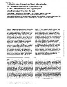
The initial event in development of the three-dimensional matrix was a reorientation of the fibroblasts so that one end became attached to the vertical root surface while the opposite part of the cell remained in contact with the culture dish. After 5-7 days, a faint fibrillar network was present at the lower part of each of the vertical surfaces, close to the bottom of the dish (Fig. 1A). The elongated fibroblasts present on top of the matrix were oriented with their long axis from the floor of the dish to the vertical root surface forming a continuous layer (Fig. IB). At 10 days, cells were observed along the entire vertical surface and a monolayer of cells was covering the top of the root pieces. Continuous movement of fibroblasts up along the root surface was apparent, paralleling a successive vertical and horizontal growth of the extracellular matrix. Eventually, the two fibrillar networks from the opposing vertical surfaces merged and, at 3 weeks, a continuous sparse cell-containing matrix filled about half the depth of the space (Fig. 1C). The majority of cells in the centre of the matrix had an elongated shape, and were oriented along fibres spanning the space, whereas the growth pattern on top of the root pieces was less organized (Fig."ID). Successive growth of the matrix continued in a vertical direction and, at about 9 weeks, a fibrillar network containing numerous fibroblasts filled the initially empty space (Fig. IE). The cells present within the matrix at later times had numerous extended processes, frequently resulting in a stellate appearance. Elongated cells at the top of the matrix maintained the orientation across the space; the superficial layers were continuous with multilayers on top of the root pieces. The fibrillar matrix between the cells progressively grew more dense throughout the period studied (Fig. IF). The general development of the matrix was the same whether the root slices were totally demineralized or demineralized only on the surface, and were independent of the angle of the root surface with the bottom of the dish. The cells initially attaching to, and migrating up, the vertical surfaces of the root slice were elongated with an elliptical nucleus (Fig. 2A). Some cells showed pronounced polarity and were characterized by a flattened leading edge with the nucleus located in the broader, trailing part of the cytoplasm. The cell membrane of the attaching fibroblasts was in close contact with collagen bundles, irregularly
Fibroblast extracellular matrix in vitro
•^L_.._-,v Fig. 1. Development of the matrix. A. A faint fibrillar network is present at the lower part of the vertical surface after 7 days in culture (double arrows). Some cells are attached to the demineralized root slice (arrows), whereas others are present within the network (arrowheads). Monolayer on the bottom of the dish (ml). Slices of tooth root (r). Light micrographs, Toluidine Blue stain. X120. Inset, X160. B. Cells aligned from the floor of the dish (d) onto the bottom part of the vertical root surface (r) after 7 days in culture. Scanning electron micrograph. X130. C. At 3 weeks, a continuous matrix between the two vertical surfaces has developed. Note the horizontal spindle-shaped cells with parallel fibrils (arrows). Light micrograph, Toluidine Blue stain. XllO. D. Cells lined up across the space (s) after 3 weeks in culture. Cells at the top of the space have detached during critical point drying, making it possible to see several cell layers (arrows). Top of root slices (r). Scanning electron micrograph, surface-decalcified root slices. X40. E. At 9 weeks a dense network has filled the entire space between the vertical surfaces of the root slices. Numerous cells with a stellate appearance are present within the matrix (double arrows). A multilayer is seen at the top of the matrix and on the top horizontal surface of the root slices (arrows). Light micrograph, Toluidine Blue stain. X 110. F. Dense fibrous material is seen between cells present on the top of the matrix. Top surface of cells (/); fibrous matrix (arrow). SEM, 6 weeks. X700.
187
E. E. Qwarnstrom and R. C. Page
2A Fig. 2. A. An elongated cell present on the vertical surface at an early stage of development of the matrix. Close association is seen between the fibroblast and the demineralized root surface (r). Nucleus (n). TEM, 7 days. X9000. B. Higher magnification shows a close association between collagen bundles on the demineralized surface (c) and the cell membrane (arrows). TEM, 10 days. X53 500. C. Cell processes (p) are extended into concavities in the surface of the root slice (r). Nucleus (n). TEM, 3 weeks. X14000.
Fibroblast extracellular matrix in vitro
189
oriented within the demineralized dentin or cementum (Fig. 2B). The close relationship of the fibroblasts to the demineralized surface resulted in the projection of cell processes into deep concavities in the vertical surface (Fig. 2C). At a later stage, a space-containing fibrillar material was seen between the cells and the vertical surface (Fig. 3). The fibrillar material stained intensely with Ruthenium Red (Fig. 4), indicating the presence of anionic moieties. Initially, a sparse matrix was extended between the vertical surface and the cells at the bottom of the dish. Elongated fibroblasts in association with the matrix were parallel to and in close contact with the fibrils. Additional cells were seen to attach following deposition of fibrillar material and, eventually, several layers of elongated spindle-shaped cells, separated by fibrils, were present. Eventually, cells totally surrounded by fibrils frequently showed a more rounded appearance, whereas fibroblasts present at the outer surface of the network remained elongated and spindle-shaped (Fig. 5). Thin fibrils developed in close association with fibroblast processes and appeared to mediate attachment between the cell membrane and the demineralized surface (Fig. 5, inset). During continued growth and maturation of the matrix, the irregular orientation of the fibrils became more pronounced, and the cells within the network showed many extended processes, resulting in a stellate appearance (Figs 6, 11). These cells contained a fairly lobulated nucleus and were characterized by an extensive rough endoplasmic reticulum and a pronounced Golgi apparatus, and contained bundles of microfilaments, frequently located at the periphery of the cytoplasm. Fibrillar material of the type described was the most prominent matrix component during the first 3 weeks in culture. Immunocytochemical staining showed the presence of various types of glycosaminoglycans at early time points. Thus, the matrix stained for chondroitin sulphate (Fig. 7A). The presence of dermatan sulphate was indicated by a much stronger staining of the matrix following digestion with chondroitinase ABC (Fig. 7B) than after treatment with chondroitinase AC II (data not shown). In addition, hyaluronic acid (Fig. 7C) and an apparently large amount of fibronectin (Fig. 7D) were present in the early fibrillar network. Bundles of fibrillar material with a faint banding pattern were observed in proximity to the fibroblast cell surface at 3 weeks (Fig. 8). Pronounced banding pattern, characteristic of collagen fibres, was seen initially after 6 weeks in culture and became successively more prominent (Fig. 9). Intense staining of the matrix by van Gieson and Trichrome indicated that it was highly collagenous. Immunocytochemical staining showed the presence of collagen types I (Fig. 10A), III (Fig. 10B) and V (Fig. IOC) in the extracellular matrix at later times. Antisera for collagen types III and V stained in addition the cell surface of fibroblasts present in the matrix. Some Alcian Blue and PAS staining at later times suggested the continued presence of carbohydrate moieties (data not shown). At the electron microscopic level, Ruthenium Red staining was found along collagen fibres. Attachment between the cells and the surrounding matrix appeared to be localized primarily at the distal end of the extended processes (Fig. 11). Single contact points (Fig. 12A) and, less commonly, a series of contact points (Fig. 12B) were seen
190
E. E. Qwarnstwm and R. C. Page
Figs 3-5. For legends see p. 192
Fibroblast extracellular matri nx in vitro
?••• '-v, s/A M
Figs 6-7. For legends see p. 192
192
E. E. Qwarnstwm and R. C. Page
between the cell membrane at these sites and the extracellular fibrils. Close association between the extended cellular processes and collagen fibres was frequently observed at later times (Fig. 12C). As the collagen matrix grew denser, cell processes enclosing bundles of collagen fibres were occasionally seen (Fig. 13). Contact between cells within the matrix was seen most frequently between extended processes (Fig. 14), and occurred occasionally, via desmosome-like structures (Fig. 14, inset).
DISCUSSION An in vitro model consisting of the development of a three-dimensional matrix has been characterized. Slices of human tooth root placed in pairs on top of a cell layer provided vertical collagen surfaces with which the cells could interact. During the 13 weeks of the experimental period, gingival fibroblasts, originally constituting a monolayer, migrated up the vertical surface and successively filled the space above the cell layer with extracellular matrix, which the cells also inhabited. The attachment of the cells on the vertical surface and their continued migration upwards were apparently prerequisites for the development of the matrix. Initially, cells were attached to the root surface while still adhering to the bottom of the dish, part of the cell surface apparently not in contact with either substratum. Substrate contact at the front and the rear only has been noted in moving nbroblasts (Ingram, 1969). The pronounced polarity with a flattened leading edge, shown by some cells present on the vertical root surface, was probably also a consequence of migration Fig. 3. At a later stage, a space containing fibrillar material (arrows) is present between the fibroblasts (/) and the demineralized surface (r). TEM, 10 days, x44 500. Fig. 4. The fibrillar material (arrows) present between the root slice (r) and the fibroblast (/) stained intensely with Ruthenium Red. TEM, 10 days. X30500. Fig. 5. A fibrillar network has developed (arrows) within which several cell layers are present. The fibroblasts {/) within the extracellular matrix close to the demineralized surface (r) show a rounded, somewhat stellate, appearance while cells at the outer surface of the network maintain a more elongated form ( / / ) . TEM, 3 weeks. X9500. Inset: fibrillar material (arrow) is connecting the demineralized root surface (r) with the cell process (/>). TEM, 10 days, x62000. Fig. 6. Fibroblasts (/) present within the matrix frequently had a rounded cell body with extended processes (/>), giving the cells a stellate appearance. TEM, 9 weeks. X9000. Fig. 7. Immunocytochemical staining using peroxidase-antiperoxidase. At early time points, the matrix contains: A. Chondroitin sulphate; B, dermatan sulphate; C, hyaluronic acid; D, fibronectin. Light micrographs at 3 weeks. X600. Fig. 8. Bundles of filaments manifesting a faint periodicity present in a 3-week sample (arrows). Fibroblast (/). TEM, X30700. Fig. 9. After 6—9 weeks, collagen fibres with the characteristic banding pattern were the major matrix component. TEM, 12 weeks. X61 500. Fig. 10. Immunocytochemical staining using peroxidase-antiperoxidase. After 6 weeks in culture and at later time points, collagen was the most prominent matrix component. A. Collagen type I, X600; B, collagen type III, X600; C, collagen type V, X560. Light micrographs at 13 weeks. D. Negative control, X400.
Fibroblast extracellular matrix in vitro
IV *
193
194
E. E. Qwarnsttvm and R. C. Page
(Ingram, 1969; Abercrombie et al. 1970). Deposition of an extracellular matrix apparently made possible migration of additional cells in a vertical direction, probably by creating an increasing number of sites for cell attachment. Synthesis
12A Fig. 11. A cell located near the centre of the matrix showing a stellate appearance, an irregularly shaped nucleus (n), and microfilament bundles (mf). Extracellular fibrillar material (arrows) is seen close to the cell surface, particularly in areas of extended processes. TEM, 6 weeks. X8300. Fig. 12. Cell matrix attachment sites. A. Attachment site of fibrillar material (arrow) located on a cell process (p). TEM, 6 weeks. x33 300. B. Several attachment sites present along the cell membrane (arrows). Fibroblast (/), fibrillar matrix (fin). TEM, 6 weeks. X14900. C. Cell process (p) in close contact with a collagen fibre (c) in a 12-week sample. TEM. X36600.
Fibroblast extracellular matrix in vitro
mf
Fig. 13. Fibroblasts cell processes (arrows) partially encompass bundles of collagen fibres (c). Microfilament bundles (mf) are seen in the cell periphery'. TEM, 12 weeks. X23 200. Fig. 14. Cell-to-cell contact is seen between extended processes (p) from neighbouring cells in the collagen matrix in a 12-week sample. TEM. X7600. Inset: desmosome-like structures (d) were observed in regions of cell-to-cell contact. TEM, 12 weeks. X41 000.
195
196
E. E. Qwarnstrom and R. C. Page
of a fibrillar material, and a continuous upward movement of cells, resulted in the successive growth of the triangular matrix. In addition, an increasing number of cells following division of cells present within or at the edge of the newly synthesized matrix (Gospodarowicz et al. 1978) probably increased the rate of matrix formation. The initial reorientation and attachment of the cells could be due to fibroblast substratum preference (Postlethwaite et al. 1978; Bellows et al. 1980), resulting in a cell membrane—collagen fibre interaction. Enhanced cell attachment to root surfaces following demineralization (Register & Burdick, 1975; Boyko et al. 1980; Fernyhough & Page, 1983; Pitaru et al. 1984) has been suggested to be due to liberation of collagen fibres (Register, 1973; Garrett et al. 1978). The cell reorientation, mobility and attachment were probably affected by the presence of fibronectin on the root slice, serving as a chemoattractant (Gauss-Muller et al. 1980; Postlethwaite et al. 1981) and enhancing cell attachment (Klebe, 1974; Pearlstein, 1976). The fibrillar material constituting the initial matrix stained with Ruthenium Red, indicating that it contained anionic moieties. The staining was probably partly due to the presence of hyaluronic acid, chondroitin sulphate and dermatan sulphate, as shown by the immunocytochemical staining. Gingival fibroblasts have been shown to synthesize these types of glycosaminoglycans in vitro (Bartold & Page, 1985). The Ruthenium Red staining could, however, partially result from the presence of an apparently high amount of fibronectin in the matrix at early times. Glycosaminoglycans are the initial extracellular material synthesized by fibroblasts during tissue regeneration (Ross, 1968; Castor, 1981), and fibronectin is present at early times during matrix formation in vivo (Grinnell et al. 1981; Forrest, 1983). The late onset of collagen formation compared to that occurring in the healing wound (Castor, 1981) may be a result of a slow conversion of procollagen to collagen in vitro. The fibrillar aggregation, initially observed at 3 weeks, was similar in appearance to fibrils seen in close association with myofibroblasts in granulation tissue (Baur & Parks, 1983). The faint periodicity observed in some of these bundles could indicate that they constitute sites for collagen fibril assembly. The presence of collagen types I, III and V in the matrix correlates well with biochemical data on synthesis by gingival fibroblasts in vitro (Narayanan & Page, 1976, 1983). Peripheral Ruthenium Red staining of the mature collagen fibres could be due to codistribution with fibronectin (Furcht et al. 1980). Attachment of fibroblasts to matrix components may be mediated in several ways (Culp et al. 1979; Rollins et al. 1982). Fibronectin on the cell surface could bind hyaluronic acid (Yamada et al. 1980), present in the extracellular matrix. Alternatively, cell surface proteoglyeans, in particular heparan sulphate, could mediate close contact with extracellular fibronectin (Lark et al. 19856). In addition, a more specific interaction via a fibronectin receptor at the cell surface is possible (Yamada et al. 1985). The attachment of cells to the collagen matrix could be partly a direct interaction since this occurs in three-dimensional gels of native hydrated collagen (Schor & Court, 1979; Schor et al. 1981). Enhancement of the collagen
Fibroblast extracellular matrix in vitro
197
attachment with serum or synthesized fibronectin is, however, likely (Kleinman etal. 1981). The spindle shape of the cells, observed on the vertical surface of the root slices, was probably related to the close contact to, and spreading on, the surface (Rosen & Culp, 1977; Grinell & Hays, 1978). The shape of the cells in contact with the synthesized material was apparently influenced by the geometry of the matrix and the number and distribution of the attachment sites. The elongated shape seen initially was probably due to attachment of the cells to a sparse number of fibrils oriented primarily across the space. The stellate shape may be caused by the irregular orientation of the fibrils, resulting in a random distribution of the cells within the matrix and of contact sites throughout the cell surface. Thus, matrix contact points, primarily present at extended processes, if connected to actin filaments in the cytoskeleton (Singer et al. 1984; Woods et al. 1984) could result in the stellate shape of the fibroblasts observed at later times. In addition, the differences in appearance of the cells could be caused by an altered tension in the matrix with time. Tension in the thin matrix initially in part may be responsible for the elongated cell shape (Bellows et al. 1982a; Haston et al. 1983). This may also explain why the top layers of cells maintained this appearance throughout matrix development. The generally more stellate appearance of the cells in the centre of the matrix at later times could be a result of less tension within the thicker and more stable network. This indicates that the overall geometry of the space also may have an effect on the cell shape by influencing matrix organization and tension. The increased density of the matrix observed at later times could be partly a result of contraction by the cells. It has been suggested that cell processes enclosing bundles of collagen, noted at later times, result in changes in the shape of pre-made collagen gels (Grinnell & Lamke, 1984). The presence of microfilaments in the cytoplasm of these cells supports this view (Gabbiani et al. 1978; Gabbiani, 1981). In addition, development of the desmosome-like intercellular attachment sites, observed at later times, could also be related to contraction by the cells and have an effect on reorganization of the matrix (Grinnell & Lamke, 1984). Using this model, the fibroblasts were allowed to migrate into and occupy a space, which successively was filled with a matrix, both synthesized and inhabited by the cells. The composition of the matrix, an early deposition of glycosaminoglycans and fibronectin, followed by procollagen synthesis and formation of collagen fibres, was similar to that in a healing wound. The resident fibroblasts interacting with the various types of components of the three-dimensional matrix showed appearance and behaviour resembling those seen during tissue regeneration. The longer time needed for development of the matrix, compared to that occurring in vivo, probably reflects that it is a simplified system. With this in mind, it could provide a well-defined model for studying the effects of soluble factors and cells other than fibroblasts on the species of matrix components present at various times, and on their biosynthesis and degradation. In addition, the model could be used for investigating specific cellmatrix interactions.
198
E. E. Qwarnstrom and R. C. Page
The authors thank Joseph Melcher for technical assistance, Henderson Mar for advice on immunochemistry, and Dr Michael Lark for thoughtful reading of the manuscript. This study was supported by grants DE03301, DE02600 and DE07063 from the National Institutes of Health.
REFERENCES ABERCROMBIE, M., HEAYSMAN, J. E. M. &PEGRUM, S. M. (1970). The locomotion of fibroblasts in
culture. I. Movements of the leading edge. Expl Cell Res. 59, 393-398. BARTOLD, P. M. & PAGE, R. C. (1985). Isolation, identification and quantitation of glycosaminoglycans synthesized by human gingival fibroblasts in vitro. J. Peridont. Res. 20, 284—292. BAUR, P. S. JR & PARKS, D. H. (1983). The myofibroblast anchoring strand - the fibronectin connection in wound healing and the possible loci of collagen fibril assembly. J. Trauma 23, 853-862. BELL, E., IVARSSON, B. & MERRILL, C. (1979). Production of a tissue-like structure by contraction of collagen lattices by human fibroblasts of different proliferative potential in vitro. Proc. natn.
Acad. Sci. U.SA. 76, 1274-1278. BELLOWS, C. G., MELCHER, A. H. & AUBIN, J. E. (1981). Contraction and organization of collagen
gels by cells cultured from periodontal ligament, gingiva and bone suggest functional differences between cell types. J . Cell Sci. 50, 299-314. BELLOWS, C. G., MELCHER, A. H. & AUBIN, J. E. (1982a). Association between tension and
orientation of periodontal ligament fibroblasts and exogenous collagen fibers in collagen gels
in vitro. J. Cell Sci. 58, 125-138. BELLOWS, C. G., MELCHER, A. H., BHARGAVA, U. & AUBIN, J. E. (19826). Fibroblasts contracting
three-dimensional collagen gels exhibit ultrastructure consistent with either contraction or protein secretion. 7. Ultrastruct. Res. 78, 178-192. BELLOWS, C. G., MELCHER, A. H. & BRUNETTE, D. M. (1980). Orientation of calvaria and
periodontal ligament cells in vitro by pairs of demineralized dentine particles. J. Cell Sci. 44, 59-73. BOYKO, G. A., BRUNETTE, D. M. & MELCHER, A. H. (1980). Cell attachment to demineralized
root surfaces in vitro. J. Periodont. Res. 15, 297-303. CASTOR, C. W. (1981). Autacoid regulation of wound healing. In Tissue Repair and Regeneration, Handbook in Inflammation, vol. 3 (ed. L. E. Glynn, J. C. Houck & G. Weissmann), pp. 177-209. Amsterdam: Elsevier, North-Holland Biomedical Press. CATERSON, B., CHRISTNER, J. E., BAKER, J. R. & COUCHMAN, J. R. (1985). Production and
characterization of monoclonal antibodies directed against connective tissue proteoglycans. Fedn Proc. Fedn Am. Socs exp. Biol. 44, 386-393. COOPER, N. R. (1985). The classical complement pathway: Activation and regulation of the first complement component. Adv. Immunol. 37, 151-216. COUCHMAN, J. R., CATERSON, B., CHRISTNER, J. E. & BAKER, J. R. (1984). Mapping by
monoclonal antibody detection of glycosaminoglycans in connective tissues. Nature, Land. 307, 650-652. CULP, L. A., MURRAY, B. A. & ROLLINS, B. J. (1979). Fibronectin and proteoglycans as
determinants of cell-substratum adhesion. J. supramolec. Struct. 11, 401-427. FERNYHOUGH, W. & PAGE, R. C. (1983). Attachment, growth and synthesis by human gingival fibroblasts on demineralized or fibronectin-treated normal and diseased tooth roots. J. Periodont. 54, 133-140. FORREST, L. (1983). Current concepts in soft connective tissue wound healing. Br.J. Surg. 70, 133-140. FURCHT, L. T . , SMITH, D., WENDELSCHAFER-CRABB, G., MOSHER, D. F. & FOIDART, J. M.
(1980). Fibronectin presence in native collagen fibrils of human fibroblasts: Immunoperoxidase and immunoferritin localization. J. Histochem. Cytochem. 28, 1319-1333. GABBIANI, G. (1981). The myofibroblast: a key cell for wound healing and fibrocontractive diseases. Prog. din. Biol. Res. 54, 183-194. GABBIANI, G., CHAPONNIER, C. & HOTTNER, I. (1978). Cytoplasmic filaments and gap junctions
in epithelial cells and myofibroblasts during wound healing. J . Cell Biol. 76, 561-568.
Fibroblast extracellular
matrix in vitro
199
GARRETT, J. S., CRIGGER, M. & EGELBERG, J. (1978). Effects of citric acid on diseased root surfaces. J. Periodont. Res. 13, 155-163. GAUSS-MOLLER, V., KLEINMAN, H. K., MARTIN, G. R. & SCHIFFMANN, E. (1980). Role of
attachment factors and attractants in fibroblast chemotaxis. J. Lab. din. Med. 96, 1071-1080. GOMORI, G. (1950). A rapid one-step trichrome stain. Am.J. din. Path. 20, 661-664. GOSPODAROWICZ, D. (1974). Localization of fibroblast growth factor and its effect alone and with hydrocortisone on 3T3 cell growth. Nature, Lond. 249, 123-127. GOSPODAROWICZ, D., GREENBURG, G. & BIRDWELL, C. R. (1978). Determination of cellular shape
by the extracellular matrix and its correlation with the control of cellular growth. Cancer Res. 38, 4155-4171. GRINNELL, F., BILLINGHAM, R. E. & BURGESS, L. (1981). Distribution of fibronectin during
wound healing in vivo.J. invest. Derm. 76, 181-189. GRINNELL, F. & HAYS, D. G. (1978). Induction of cell spreading by substratum-adsorbed ligands directed against the cell surface. Expl Cell Res. 116, 275-284. GRINNELL, F. & LAMXE, C. R. (1984). Reorganization of hydrated collagen lattice by human skin fibroblasts. J . CellSci. 56, 51-63. HASTON, W. S., SHIELDS, J. M. & WILKINSON, P. C. (1983). The orientation of fibroblasts and
neutrophils on elastic substrata. Expl Cell Res. 146, 117-126. HAY, E. D. (1981). Cell Biology of the Extracellular Matrix. New York: Plenum. INGRAM, V. M. (1969). A side view of moving fibroblasts. Nature, Lond. 222, 641-644. KARNOVSKY, M. J. (1965). A formaldehyde—glutaraldehyde fixative of high osmolality for use in electron microscopy. J. Cell Biol. 27, 137A-138A. KLEBE, R. J. (1974). Isolation of a collagen-dependent cell attachment factor. Nature, Lond. 250, 248-251. KLEINMAN, H. K., KLEBE, R. J. & MARTIN, G. R. (1981). Role of collagenous matrices in the
adhesion and growth of cells. J. Cell Biol. 88, 473-485. KOHLER, N. & LlPTON, A. (1974). Platelets as a source of fibroblast growth-promoting activity.
Expl Cell Res. 87,297-301. LARK, M. W., HELLSTROM, I., HELLSTROM, K. E. & WIGHT, T . N. (1985a). Characterization of a
monoclonal antibody directed against arterial wall chondroitin sulfate proteoglycan._7. Cell Biol. 101, 337a. LARK, M. W., LATERRA, J. & CULP, L. A. (19856). Close and focal contact adhesions of fibroblasts to a fibronectin-containing matrix. Fedn Proc. Fedn Am. Socs exp. Biol. 44, 394—403. MARKS, R. (1981). The healing and nonhealing of wounds and ulcers of the skin. In Tissue Repair and Regeneration, Handbook in Inflammation, vol. 3 (ed. L. E. Glynn, J. C. Houck & G. Weissmann), pp. 309-342. Amsterdam: Elsevier, North-Holland Biomedical Press. MOLLORY, F. B. (1961). Pathological Technique, pp. 152-160. New York: Hafner Publishing Co. NARAYANAN, A. S., CLAGETT, J. A. & PAGE, R. C. (1985). Effect of inflammation on the
distribution of collagen types I, III, IV and V and type I trimer and fibronectin in human gingivae. J. dent. Res. 64, 1111-1116. NARAYANAN, A. S. & PAGE, R. C. (1976). Biochemical characterization of collagens synthesized by fibroblasts derived from normal and diseased human gingiva. J. biol. Chem. 251, 5464—5471. NARAYANAN, A. S. &PAGE, R. C. (1983). Biosynthesis and regulation of type V collagen in diploid human fibroblasts. J. biol. Chem. 258, 11 694-11 699. NARAYANAN, A. S., PAGE, R. C. & KUZAN, F. (1978). Collagens synthesized in vitro by diploid fibroblasts obtained from chronically inflamed human connective tissue. Lab Invest. 39, 61—65. PEARLSTEIN, E. (1976). Plasma membrane glycoprotein which mediates adhesion of fibroblasts to collagen. Nature, Lond. 262, 497-500. PTTARU, S., GRAY, A., AUBIN, J. E. &MELCHER, A. H. (1984). The influence of the morphological
and chemical nature of dental surfaces on the migration, attachment, and orientation of human gingival fibroblasts in vitro. J. Periodont. Res. 19, 408-418. POSTLETHWATTE, A. E., KESH-OJA, J., BALIAN, G. & KANG, A. H. (1981). Induction of fibroblast
chemotaxis by fibronectin: Localization of the chemotactic region to a 140,000-molecular weight non-gelatin-binding fragment. J'. exp. Med. 153, 494—499. POSTLETHWAITE, A. E., SEYER, J. M. & KANG, A. H. (1978). Chemotactic attraction of human
fibroblasts to type I, II and III collagens and collagen-derived peptides. Proc. natn. Acad. Sd. U.SA. 75, 871-875.
200
E. E. Qwarnstrom and R. C. Page
REGISTER, A. A. (1973). Bone and cementum induction by dentin demineralized in situ. Jf. Periodont. 44, 49-54. REGISTER, A. A. & BURDICK, F. A. (1975). Accelerated reattachment with cementogenesis to dentin, demineralized in situ. I. Optimum range. J. Periodont. 46, 646-655. ROLLINS, B. J., CATHCART, M. K. & CULP, L.~ A. (1982). Fibronectin-proteoglycan binding as the
molecular basis for fibroblast adhesion to extracellular matrices. In The Glycoconjugates, vol. I l l (ed. M. Horowitz), pp. 289-329. New York: Academic Press. ROSEN, J. J. & CULP, L. A. (1977). Morphology and cellular origins of substrate-attached material from mouse fibroblasts. Expl Cell Res. 107, 139-149. Ross, R. (1968). The fibroblast and wound repair. Biol. Rev. 43, 51-96. Ross, R., GLOMSET, J., KARIYA, B. & HARKER, L. (1974). A platelet-dependent serum factor that stimulates the proliferation of arterial smooth muscle cells in vitm. Proc. natn. Acad. Set. U.SA. 71, 1207-1210. SAVAGE, K. & SWANN, D. A. (1985). A comparison of glycosaminoglycan synthesis by human fibroblasts from normal skin, normal scar and hypertrophic scar. J. invest. Derm. 84, 521-526. SCHMIDT, J. A., MIZEL, S. B., COHEN, D. & GREEN, I. (1982). Interleukin-1, a potential regulator
of fibroblast proliferation.^. Immun. 128, 2177-2182. SCHOR, S. L. & COURT, J. (1979). Different mechanisms in the attachment of cells to native and denatured collagen. J . Cell Sri. 38, 267-281. SCHOR, S. L., SCHOR, A. M. & BAZILL, G. W. (1981). The effects of fibronectin on the adhesion
and migration of Chinese hamster ovary cells on collagen substrata..?. Cell Sci. 49, 299-310. SlNGER, I. I. (1979). The fibronexus: a transmembrane association of fibronectin-containing fibers and bundles of 5 nm microfilaments in hamster and human fibroblaats. Cell 16, 675-685. SINGER, I. I., KAWKA, D. W., KAZAZIS, D. M. & CLARK, R. A. F. (1984). In vivo co-distribution
of fibronectin and actin fibers in granulation tissue. Immunofluorescence and electron microscope studies of the fibronexus at the myofibroblast surface. J. Cell Biol. 98, 2091-2106. SOBEL, A., BLANC, C , CATTANEO, A., MOISY, M., LOPEZ-TRASCASA, M., BOURGARJT, J. J. &
GABAY, Y. (1983). Receptors for complement components in inflammation. Agents and Actions 13, 398-405. WIGHT, T . N. (1980). Differences in the synthesis and secretion of sulfated glycosaminoglycans by aorta explant monolayers cultured from atherosclerosis-susceptible and resistant pigeons. Am.J. Path. 101, 127-142. WOODS, A., HOOK, M., KJELLEN, L., SMITH, C. G. & REES, D. A. (1984). Relationship of
heparan sulfate proteoglycans to the cytoskeleton and extracellular matrix of cultured fibroblasts. J . Cell Biol. 99, 1743-1753. YAMADA, K. M. (1983). Cell surface interactions with extracellular materials. A. Rev. Biochem. 52, 761-799. YAMADA, K. M., AKIYAMA, S. K., HAGESAWA, T., HASEGAWA, E., HUMPHRIES, M. J., KENNEDY,
D. W., NAGATA, K., URUSHTHARA, H., OLDEN, K. & CHEN, W.-T. (1985). Recent advances in
research on fibronectin and other cell attachment proteins. J'. cell. Biochem. 28, 79-97. YAMADA, K. M., KENNEDY, D . W., KTMATA, K. & PRATT, R. M. (1980). Characterization of
fibronectin interactions with glycosaminoglycans and identification of active proteolytic fragments. J . biol. Chem. 255, 6055-6063.
{Received 2 January 1986 -Accepted 25 March 1986)