Some enlarged cervical lymph nodes were also found. (Figure 1). Figure 1 ... alveolar structures and solid nests; tumor cells presented round or oval nuclei, rare ..... like, glassy and eosinophilic background material. In our case, the FNA ...
Rom J Morphol Embryol 2014, 55(2):389–400
RJME
CASE REPORTS
Romanian Journal of Morphology & Embryology http://www.rjme.ro/
Diagnostic correlation between RET proto-oncogene mutation, imaging techniques, biochemical markers and morphological examination in MEN2A syndrome: case report and literature review ALINA SIMONA ŞOVREA1), ELEONORA DRONCA2), MIHAELA GALATÂR3), ŞERBAN RADIAN4), CORINA VORNICESCU5), CARMEN GEORGESCU6) 1)
Department of Histology, “Iuliu Haţieganu” University of Medicine and Pharmacy, Cluj-Napoca, Romania
2)
Department of Medical Genetics, “Iuliu Haţieganu” University of Medicine and Pharmacy, Cluj-Napoca, Romania
3)
Department of Pathologic Anatomy, “Prof. Dr. Ion Chiricuţă” Oncologic Institute, Cluj-Napoca, Romania
4)
Department of Endocrinology, “Carol Davila” University of Medicine and Pharmacy, Bucharest, Romania
5)
“Iuliu Haţieganu” University of Medicine and Pharmacy, Cluj-Napoca, Romania
6)
Department of Endocrinology, “Iuliu Haţieganu” University of Medicine and Pharmacy, Cluj-Napoca, Romania
Abstract
Multiple endocrine neoplasia type 2 (MEN2) is a rare autosomal dominant monogenic disorder caused mostly by missense mutations in the RET (REarranged during Transfection) proto-oncogene on chromosome 10q11.2. MEN2A represents more than 50% of all MEN2 cases, having a regular pattern with medullary thyroid carcinoma (MTC) incidence of 90–100%, bilateral pheochromocytoma (PCC) incidence of 40–50% and primary hyperparathyroidism (HPT) incidence of 10–25%. Until recently, the diagnosis of MTC was most frequently based on fine-needle aspiration of thyroid nodules, after an ultrasound examination and endocrine evaluation of serum calcitonin levels. Nowadays, RET gene screening (starting with exons 10 and 11) is a mandatory test used for identification of both symptomatic and non-symptomatic MTC carriers or for exclusion of healthy individuals from subsequent periodical clinical/biochemical screening. In this context, and in the idea of PCC preceding MTC, the early detection of germline RET mutations are highly suggestive for hereditary disease. PCC diagnosis is established in classical manner by abdominal ultrasound imaging or computed tomography confirming the presence of adrenal gland masses, elevated plasma metanephrines and normetanephrines values and histopathological examination. Additional HPT diagnosis is acknowledged by serum ionized calcium and parathormone levels. Here we report a hereditary case of MEN2A in a two-generation Romanian family, along with data presenting the importance of correlative plurifactorial diagnostic scheme in this syndrome and a short literature review. Keywords: MEN2A syndrome, RET gene, medullary thyroid carcinoma, pheochromocytoma, fine needle aspiration, serum calcitonin level.
Introduction Multiple endocrine neoplasia type 2 (MEN2) is a rare autosomal dominant monogenic disorder caused by missense mutations in the RET (REarranged during Transfection) proto-oncogene, which activate an encoded transmembrane tyrosine kinase receptor [1]. It is a complex neoplastic neurocristopathy [2] with three distinct subtypes (A, B and F), according to the variable penetrance of medullary thyroid carcinoma (MTC), pheochromocytoma (PCC) and primary hyperparathyroidism (HPT). MEN2A (Sipple syndrome, OMIM 171400) represents 50–75% of all MEN2 cases [1, 3]. It has the most regular pattern of all subtypes, with MTC incidence of 90–100%, generally bilateral PCC incidence of 40–50% and variable HPT incidence of up to 35% [1, 2, 4, 5]. MTC is generally the first clinical manifestation in young adults but tends to be present in the fifth or sixth decade of life with a slight female preponderance. The diagnosis of MTC is most frequently based on fine-needle aspiration (FNA) ISSN (print) 1220–0522
of thyroid nodules, after an ultrasound examination and endocrine evaluation of serum calcitonin (CT) levels. PCC can precede (13–27% of cases), be concomitant or appear after MTC. Diagnosed at an early age, it usually is bilateral and has a 4% risk of malignancy [4]. The presence of adrenal gland masses is established by ultrasound imaging of the abdomen or computed tomography followed by detection of elevated plasma metanephrines and normetanephrines values; still, the final confirmation of PCC is based on histopathological examination. Due to the inconstant presence of HPT, biochemical assays (e.g., serum ionized calcium [iCa] and parathyroid hormone [PTH] levels) must be done in order to verify the diagnostic. MEN2B (less than 10% of MEN2 cases) has the most aggressive type of MTC, which is due to de novo germline mutations in more than half of patients [1]. Besides MTC (100% of cases), MEN2B is also characterized by PCC (50% of patients), but no HPT; patients have a characteristic appearance (i.e., marfanoid habitus with skeletal abnormalities and joint laxity, everted eyelids, ISSN (on-line) 2066–8279
390
Alina Simona Şovrea et al.
thick lips), megacolon, markedly enlarged peripheral nerves, diffuse mucosal ganglioneuromas and absence of tears as specific features [1, 4, 6]. Familial MTC (FMTC) accounts for more than 30% of all MEN2 cases [1], with very low penetrance of PCC and HPT [1]. Usually, it is diagnosed when at least four members of the same family are affected by MTC after the age of 50, with no evidence of either PCC or HPT in more than 10 carriers [7]. Due to significant overlapping of gene mutations in both FMTC and MEN2A, differential diagnosis between the two syndromes can be challenging. Since MTC is usually the main cause of death due to low response to chemo- and radiotherapy, the only therapeutic option is early detection or/and prophylactic surgery [5, 8]. Biochemical markers such as CT can be used for MTC diagnosis but only after the disease has developed. In view of this, genetic testing was introduced in clinical practice for early detection of mutations causing the disease [8]. RET gene on chromosome 10q11.2 is the mutated gene identified by high-resolution melting (HRM) analysis and sequencing, in nearly all MEN2 patients: 98% of MEN2A cases, more than 98% of MEN2B cases and about 95% of FMTC cases [9]; more than 50 different mutations and even more variants are so far described [6]. Very few cases, sporadic or familial, are caused by NTRK1 gene mutations [7]. Molecular analysis shows that in MEN2A patients, the frequently found RET gene missense mutations affect the cysteine (Cys)-rich extracellular domain encoded by exons 10 and 11 (most of them affecting the Cys residue from codon 634); other different RET mutations are associated with MEN2B or FMTC [1, 6, 7, 10]. Patients identified to be gene mutation carriers, have a stratified risk for malignancy [5, 11]. Germline RET mutations are present in hereditary cases but somatic mutations, which are restricted only to the tumor cells, can only be found in sporadic MTC. Nowadays, RET molecular analysis starting with codons 10 and 11 [2] is extensively used for identification of both symptomatic and non-symptomatic carriers or for exclusion of healthy individuals [12] in families with MEN2 syndrome. Surgical procedures are recommended based on the genetic testing, according to the type of RET mutation [1, 11]. In case of any genetic testing, patients should receive appropriate genetic counseling and be presented the individual and familial risks and benefits. Family members of these patients have a 50% risk of inheriting the same RET gene mutation, due to the fact that the mutations are transmitted in an autosomal dominant manner, so they should be tested as well and offered prophylactic thyroidectomy if necessary [5]. Here we report a hereditary case of MEN2A in a two-generation Romanian family (a female patient and her children), along with data presenting the importance of correlative plurifactorial diagnostic scheme in this syndrome and a short literature review. The study was developed in accordance with the WMA Declaration of Helsinki and was approved by the University Ethics Committee. Informed consent was obtained from all individuals included in the study prior to any clinical, biological and genetic testing.
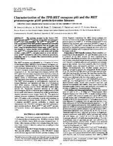
Patients, Methods and Results In June 2012, a 55-year-old, non-obese female patient with a medical history of left adrenalectomy for PCC 18 years ago, was admitted to the Endocrinology Clinic with permanent hypertension. Once every few days, symptoms worsened with blood pressure reaching 210/110 mmHg, concomitant headache, chest pain, tachycardia, sweating and intense panic. Ischemic stroke was documented one year prior to hospital admission. Laboratory evaluation revealed normal serum fasting glucose (108 mg/dL), increased serum cholesterol (224 mg/dL) and normal blood cells count and serum electrolytes, including ionized calcium (iCa) levels. Thyroid status evaluation showed serum thyrotropin (TSH) level of 0.76 mU/L (normal range 0.5–4 mU/L) and serum free thyroxin (FT4) level of 0.81 ng/dL (normal range 0.8–1.4 ng/dL). However, the anti-thyroid peroxidase antibodies (anti-TPO) titer was elevated by more than 1400% (700.3 U/mL vs. normal 50 U/mL), confirming a chronic autoimmune thyroiditis, which was previously suggested by the inhomogeneous, hypo-echoic and multinodular thyroid on neck ultrasound examination. Some enlarged cervical lymph nodes were also found (Figure 1).
Figure 1 – Inhomogeneous, hypo-echoic and multinodular thyroid on ultrasound examination.
Endocrine evaluation of the adrenal medulla function revealed elevated plasma metanephrines levels of 275.8 pg/mL (normal T mutation.
data. Since at genetic testing, MEN2A and FMTC share similar RET gene mutations (e.g., exon 11 – codon 634)
394
Alina Simona Şovrea et al.
and because in our case, none of the more specific mutations [2, 6] were identified (e.g., exon 13 – codons 768, 790, 791; exon 14 – codon 804 and exon 15 – codon 891), the differentiating criterion used to exclude the diagnosis of FMTC was the presence of hereditary PCC. The difference between MEN2A and MEN2B syndromes was given by the absence of specific phenotypic MEN2B features, together with the late MTC clinical onset, its lack of aggressiveness and the absence of the typical RET mutations [6] (exon 16 – codon 918 or exon 15 – codon 883) at genetic testing. Discussion As it has previously been shown, MEN2A syndrome consists of several neuroendocrine origin abnormalities: MTC, PCC and HPT. Diagnosis of any of these tumors implies correlative analyses and undertakes further investigations for the detection of the other possible associated neoplasia [5]. MTC, arising from parafollicular thyroid C-cells, currently accounts for 5–10% of all thyroid cancers [5, 13]. Despite its extreme versatile morphology, MTC diagnosis benefits from the identification of specific markers, both genetic (somatic or germline mutations of RET proto-oncogene) and serological (C-cell secreted CT level) [1, 5, 7]. The majority of MTC cases are sporadic, but the hereditary ones (30%) are present in virtually all cases of MEN2 syndromes [1, 5], MTC being diagnosed in more than 90% of carriers of different germline RET mutations. The strong genotype/phenotype correlation between MEN2 and germline RET mutations [1, 5, 7] defined the mutational screening as a mandatory test for affected families. Genetic screening for RET mutations has certain advantages over the biochemical or clinical screening, allowing the early detection of C-cell hyperplasia and microscopic MTC, before any clinical or laboratory tests become positive; this means that C-cells are more sensitive to RET gene mutations than adrenal medullary or parathyroid cells [6, 14]. Prophylactic surgery should be performed only if RET gene analysis is positive; so this test should be offered to all patients and their relatives, therefore improving the cure rate after thyroidectomy and the long-term prognosis [15]. According to the literature data, RET gene has 21 exons spanning over 50 kb [16] and encodes a transmembrane tyrosine kinase receptor. Its endogenous ligand appears to be the glial cell-derived neurotrophic factor (GDNF) family, which is critical for normal enteric and renal nervous system development [3, 17]. The RET protein consists of three parts: the extracellular region with six domains (four cadherin-like, a calcium-binding site and a cysteine-rich domain), the transmembrane region, and the intracellular region with two tyrosine kinase domains [6]. Mutations of RET gene reported in MEN2A act in a “two-hit” model [18] and concern the cysteine-rich extracellular domain of the receptor, mostly in exons 10 and 11 (codons 609, 611, 618, 620 and 634, respectively). These missense mutations result in RET gain of function and activation of downstream signaling
pathways [3]. Less frequently, exons 5, 8 and 13–16 may be also involved (codons 790, 791, 804 and 918) [1, 18, 19]. In hereditary syndromes, RET mutations were previously classified on a three-level risk scale. Recently, the American Thyroid Association (ATA) [11] categorized mutations on four levels (A, B, C, D) according to their risk for aggressive MTC: level D (codons 883, 918) – the highest risk; level B (codons 609, 611, 618, 620, 630, 631) and C (codon 634) – high risk and level A (codons 321, 515, 533, 600, 603, 606, 635, 649, 666, 768, 776, 790, 791, 804, 819, 833, 844, 861, 891, 912) – moderate risk [1, 6, 8]; apparently, mutations affecting the extracellular domains of RET cause the most severe phenotypes [20] and prophylactic thyroidectomy is recommended as early as possible, in many cases before the age of five years (level C) or even within the first six months of life (level D) [1, 5]. Approximately 98% of MEN2A patients have a RET mutation in either exons 10 or 11 [1]. Most frequently (more than 80%), mutations involve codon 634 [1] affecting one of the six Cys residues from the extracellular domain; the result is a permanent activation of the receptor through homodimerization [3]. In 50% of MEN2A patients, there is a specific point mutation replacing the Cys residue with Arg (C634R) [20]; these patients present more distant metastases at diagnosis, nodal metastases being reported even at the age of 5 [10] and also HPT is more common [6]. In cases when the molecular screening of common exons (10, 11, 13, and 14) is negative, RET gene sequencing [9] or analysis of microsatellites is recommended [5]. For carriers of p.Cys634Arg/Gly/Phe/Ser/Trp/Tyr mutation, there is a high age-related risk for aggressive MTC (level C) [4], which can become malignant in very young children (even at one year of age) [10], distant metastases, bilateral PCC and HPT [6, 21]. In such cases, prophylactic total thyroidectomy (with or without central node dissection) is recommended, whenever possible, before the age of 5 [5] and screening for PCC and HPT should be started as soon as possible [4, 6]. In cases when surgery is not possible at a younger age, the cure rates depend on the lymph node metastases (i.e., less than 50% for 1–10 affected nodes to less than 4% when more than 10 nodes are involved) [18]. Level D RET mutations in codons 918 or 883 (exons 16 and 15, respectively) are associated with MEN2B, so the genetic analysis is very important for the differential diagnosis from MEN2A [6]. In case of FMTC, mutations span the entire RET gene, especially in exons 10 and 11 (codons 618, 620, 634), 13 (codons 768, 790, 791), 14 (codon 804) and 15 (codon 891) [2, 6]. Since MEN2A and FMTC share similar RET gene mutations, the differential diagnosis can be difficult in the absence of PCC or HPT [6]; recently, it was proposed that FMTC should be considered as a subtype of MEN2A [22]. In our study, patients presented level C RET mutation (C634F), notably not the most frequent one (C634R). According to general consensus, total thyroidectomy should be performed prior to the age of five years (in case of early diagnosis); in our case, due to late
Diagnostic correlation between RET proto-oncogene mutation, imaging techniques, biochemical markers…
diagnosis, it was indicated to all RET mutation carriers. Although the identified RET mutation indicated a possible aggressive evolution of MTC, in our case there was a slow development and no distal metastases in all three patients. Since the recurrence risk for MEN2A is 50% [13, 23, 24], there is a high risk for hereditary transmission but in our case only two of the children inherited the RET C634F mutation, the 26-year-old daughter being free of this mutation. Usually, the clinical picture in MTC is not a reliable diagnostic element. The clinical course is rather discrete in the beginning (with irrelevant diffuse neck pain), frequently being masked or altered by emerging signs from an associated PCC or HPT [4], as it was found in our case. Moreover, in our study, both mother and daughter presented with overlapping of other thyroid related symptoms and morphological signs (chronic autoimmune thyroiditis, multinodular goiter), which completely hid the tumor features. Only the son had a 3 mm hypoechoic thyroid micronodule, which was, up to a point, suggestive for the diagnosis. Anyway, in his case the diagnosis confirmation came from the correlations with increased basal CT levels in the context of hereditary transmission. Yet, according to literature data [1, 4–6, 9], hoarseness, dysphagia or respiratory distress may appear in cases of posterior region tumors, compressing or invading local structures. Weight loss, neuroendocrine features such as flushing or diarrhea or in rare cases skin lesions or intestinal problems (i.e., cutaneous lichen amyloidosis – correlated with exon 11 RET gene mutations, codon 634 or Hirschsprung’s disease – correlated with exon 10 RET gene mutations, codons 609, 611, 618, 620) are linked to poor prognosis. Unlike sporadic MTC, which is considered a slowly developing tumor, hereditary syndromes are characterized by clinically aggressive MTC associated with a high mortality rate. The tumor onset can be very early (sometimes before 5-year-old and generally prior the age of 35) with multifoci and bilateralism [5], but in the case of our patients the onset mimicked the time of diagnosis for sporadic MTC. Local cervical lymph nodes or even distal metastases in a reduced number of cases (mediastinal lymph nodes, liver, bones, lungs) can occur in the fifth or sixth decade of life [4, 5]. In most occasions, the diagnosis of MTC is primarily suggested by ultrasound neck examinations that can evidentiate thyroid nodules, which are sometimes bilateral. The ultrasound can also be used for diagnosis of cervical adenopathy or for detecting tumor recurrences after thyroidectomy [5]. Part of the metastatic evaluation of a patient with an initial diagnosis of MTC, a contrastenhanced computed tomography of the chest, mediastinum and abdomen can also be recommended [5]. Following the imaging techniques, possible biochemical abnormalities (such as increased serum CT or CEA levels) are verified parallel to a FNA of the thyroid nodules [5, 13]. In our study, the female patient underwent an ultrasound neck examination that confirmed the presence of chronic autoimmune thyroiditis. The thyroid parenchyma was globally inhomogeneous, hypoechoic and multinodular, initially masking the MTC. The
395
diagnosis was reconsidered only after endocrine evaluation (elevated serum CT values) and FNA. Serum CT, a hallmark of this tumor, is generally used for MTC early detection (highlighting the precursor lesion, C-cell hyperplasia) and diagnosis, but also for prognosis and follow-up after surgical resection [5, 13, 23, 24]. Unlike sporadic MTC, where C-cell hyperplasia appears only in small foci, in hereditary cases this precursor lesion is important, enabling the prevention of the tumor [4]. High serum CT levels (>100 pg/mL) are well correlated with tumor presence. Slightly elevated CT levels (10–40 pg/mL vs. normal 90 pg/mL) and normetanephrine (normal 400 mg; serum Ca >1 mg/dL above upper limit of normal; creatinine clearance >30% below normal for patient’s age; bone density >2.5 standard deviations for below peak (i.e., T-score of -2.5); patient age