diagnostics Review
The Role of Ultrasound in Screening Dense Breasts—A Review of the Literature and Practical Solutions for Implementation Denise Thigpen *, Amanda Kappler and Rachel Brem Department of Radiology, The George Washington University, Washington, DC 20037, USA;
[email protected] (A.K.);
[email protected] (R.B.) * Correspondence:
[email protected]; Tel.: +1-202-741-3003 Received: 7 February 2018; Accepted: 13 March 2018; Published: 16 March 2018
Abstract: Breast cancer is the second leading cause of cancer death in women. Estimates indicate a nearly 40% breast cancer mortality reduction when screening women annually starting at age 40. Although mammography is well known to be a powerful screening tool in the detection of early breast cancer, it is imperfect, particularly for women with dense breasts. In women with dense breast tissue, the sensitivity of mammography is reduced. Additionally, women with dense breasts have an increased risk of developing breast cancer while mammography has a lower sensitivity. Screening ultrasound, both handheld and automated, is effective in detecting mammographically occult cancer in women with dense tissue. Studies have shown that ultrasound significantly increases detection of clinically important, small, largely invasive, node-negative cancers. The purpose of this review article is to summarize the literature to date regarding screening breast ultrasound, emphasizing differences in cancer detection in high risk and intermediate risk women, and to discuss practical ways to implement screening ultrasound in clinical practice, including automated whole breast ultrasound, as a viable solution to the increasing need for additional screening. Keywords: screening breast ultrasound; breast cancer; breast density; automated breast ultrasound; mammography
1. Introduction In the United States, an estimated 252,710 new cases of invasive breast cancer will have been diagnosed in 2017 and 40,610 women will have died of their disease. Breast cancer is the second leading cause of cancer death in women [1]. It is well established that early detection reduces breast cancer deaths [2]. Estimates indicate a nearly 40% breast cancer mortality reduction when screening women annually starting at age 40 [3]. Although mammography is well known to be a powerful screening tool in the detection of early breast cancer, it is imperfect, particularly for women with dense breasts. Breast density refers to the relative amounts of fat and glandular tissue in the breast. This ranges from nearly all fat to nearly all glandular tissue and affects the mammographic appearance of the breast (Figure 1). Breast density description has been standardized by the American College of Radiology (ACR) Breast Imaging Reporting and Data System (BI-RADS) Atlas [4]. The descriptions of these categories have changed over the years. The 5th Edition, published in 2013, emphasizes the text descriptions of density and also allows for the tissue composition categories to be referred to as a–d, while the 4th Edition, published in 2003, emphasized the numeric percentage of dense tissue and referred to the categories as 1–4 (Table 1) [5]. For the purposes of this article, all density classifications will be referred to by their text description or by the 5th edition categories a–d. Conventionally, the two least dense categories (fatty
Diagnostics 2018, 8, 20; doi:10.3390/diagnostics8010020
www.mdpi.com/journal/diagnostics
Diagnostics 2018, 8, 20 Diagnostics 2018, 8, x FOR PEER REVIEW
2 of 14 2 of 13
and scattered) areand referred to asare “non-dense” the two mostand dense categories (fatty scattered) referred toand as “non-dense” thecategories two most(heterogeneously dense categories dense and extremely dense) are referred to as “dense.” (heterogeneously dense and extremely dense) are referred to as “dense.”
(a)
(b)
(c)
(d)
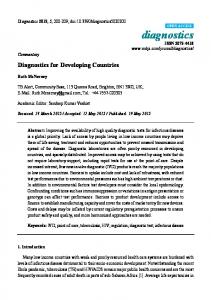
Figure 1. tissue, (c) Figure 1. Breast Breast density density (a) (a) almost almost entirely entirely fatty, fatty, (b) (b) scattered scattered fibroglandular fibroglandular tissue, (c) heterogeneously heterogeneously dense, and (d) extremely dense, as determined by the BI-RADS Atlas [4]. dense, and (d) extremely dense, as determined by the BI-RADS Atlas [4]. Table 1. Tissue composition descriptions descriptions used used in in the the BI-RADS BI-RADS Atlas Atlas [4]. [4]. Table 1. Tissue composition
Tissue Composition 4th Edition 5th Edition Tissue Composition 4th Edition 5th Edition Almost entirely fatty 1 a Almost entirely fattytissues 12 ab Scattered fibroglandular Scattered fibroglandular tissues 2 b Heterogeneously dense 3 c Heterogeneously dense 3 c Extremely dense 4 d Extremely dense 4 d Overall, the sensitivity of mammography for the detection of breast cancer is 85%; however, in Overall, the sensitivity of mammography for the detection of breast cancer is 85%; however, women with dense breast tissue, the sensitivity of mammography is reduced to 47.8–64.4% [6]. Not in women with dense breast tissue, the sensitivity of mammography is reduced to 47.8–64.4% [6]. only is mammography less sensitive in women with dense breasts, women with extremely dense Not only is mammography less sensitive in women with dense breasts, women with extremely dense breasts have a 4.7-fold increased risk of developing breast cancer [7]. Therefore, women with dense breasts have a 4.7-fold increased risk of developing breast cancer [7]. Therefore, women with dense breasts have a higher risk of breast cancer, yet mammography is less effective. Cancers detected in breasts have a higher risk of breast cancer, yet mammography is less effective. Cancers detected in women with dense breasts are larger and more often node positive [8]. Interval cancers, which have women with dense breasts are larger and more often node positive [8]. Interval cancers, which have a worse prognosis than screen-detected cancers, are 18 times more likely to occur in women with adense worsebreasts prognosis than is screen-detected cancers, since are 18more timesthan morehalf likely to occur inwomen women have with dense dense [7]. This even more significant of American breasts [7]. This even the more significant than halfand of American women have dense cancer breast breast tissue [9]. is Given prevalence ofsince densemore breast tissue the challenges of identifying tissue [9]. Given the prevalence of dense breast tissue and the challenges of identifying cancer in dense in dense breasts with mammography, additional imaging modalities to detect mammographically breasts with mammography, additional imaging modalities to detect mammographically occult breast occult breast cancer are needed. cancer Asare of needed. January 2018 in the United States, 30 states have “density notification” laws requiring As of 2018 in United 30 states “density notification” requiring women to January be informed of the their breastStates, density, many have mandating that women be laws informed that women to be informed of their breast density, many mandating that women be informed that additional additional screening can detect cancer not visible with mammography [10]. The issue of dense breast screening detect cancer notbreast visible with risk mammography [10]. The issue of dense breast tissue and tissue andcan its impact on both cancer and mammographic limitation is increasingly being its impact on both breast cancer risk and mammographic limitation is increasingly being featured in featured in the lay press and media. The concept of individualized, risk-based screeningthe is lay press and media. The concept of individualized, risk-based screening is increasingly taking hold. increasingly taking hold. At present, there risk-stratified screening place in in the the United United States. States. Mammography Mammography At present, there is is aa risk-stratified screening model model in in place is the mainstay of screening for women aged 40 and over. High-risk populations (womenwith witha is the mainstay of screening for women aged 40 and over. High-risk populations (women alifetime lifetimerisk riskofofgreater greaterthan than20–25%) 20–25%)are are advised advised to to undergo undergo additional additional annual annual surveillance surveillance with with magnetic resonance imaging (MRI) [11–13] or if they cannot undergo MRI, the ACR now magnetic resonance imaging (MRI) [11–13] or if they cannot undergo MRI, therecommends ACR now they should consider screening breast ultrasound (SBU) [14]. However, thereHowever, is a gap inthere the approach recommends they should consider screening breast ultrasound (SBU) [14]. is a gap to intermediate riskintermediate women (women with a lifetime of a15–20%), may not qualify in screening the approach to screening risk women (womenrisk with lifetime who risk of 15–20%), who may not qualify for high risk screening with MRI. Women with dense breasts constitute the largest
Diagnostics 2018, 8, 20
3 of 14
Diagnostics 2018, 8, x FOR PEER REVIEW
3 of 13
for high risk screening with MRI. Women with dense breasts constitute the largest portion of this intermediate and stand to benefit fromto early detection usingdetection adjunct screening approaches in portion of thisgroup intermediate group and stand benefit from early using adjunct screening addition to screening mammography. approaches in addition to screening mammography. Dense breast tissue as does does breast breast cancer, cancer, which Dense breast tissue appears appears white white on on mammograms, mammograms, as which is is why why dense dense tissue can sometimes obscure a cancer. In contrast, dense tissue is echogenic on ultrasound, while tissue can sometimes obscure a cancer. In contrast, dense tissue is echogenic on ultrasound, breast while cancer is hypoechoic. Ultrasound leverages the differences in tissue to improve cancer breast cancer is hypoechoic. Ultrasound leverages the differences incharacteristics tissue characteristics to improve detection in women with dense (Figure(Figure 2). cancer detection in women withbreasts dense breasts 2).
(a)
(b)
(c)
Figure 2. 2. A A 43-year-old 43-year-old woman woman with with extremely extremely dense dense breast breast tissue. tissue. Dense obscures aa breast breast Figure Dense tissue tissue obscures cancer that is easily visible with ultrasound imaging. (a,b) Craniocaudal (CC) and mediolateral cancer that is easily visible with ultrasound imaging. (a,b) Craniocaudal (CC) and mediolateral oblique obliquedigital (MLO) digital mammography. (c) Handheld high-resolution demonstrates a 1.2 (MLO) mammography. (c) Handheld high-resolution ultrasound ultrasound demonstrates a 1.2 cm irregular cm irregular denoted by image, caliperswhich in thewas image, which was biopsied proven to be invasive mass, denotedmass, by calipers in the biopsied and proven to be and invasive ductal carcinoma. ductal carcinoma.
Screening ultrasound, both handheld (Figure 3) and automated (Figure 4), is effective in Screening ultrasound, both handheld (Figure 3) and automated (Figure 4), is effective in detecting mammographically occult cancer in women with dense tissue. Studies have shown that detecting mammographically occult cancer in women with dense tissue. Studies have shown that ultrasound significantly increases detection of clinically important, small, invasive, node-negative ultrasound significantly increases detection of clinically important, small, invasive, node-negative cancers. The purpose of this review article is to summarize the literature to date regarding screening cancers. The purpose of this review article is to summarize the literature to date regarding screening breast ultrasound (SBU), emphasizing differences in cancer detection in high risk and intermediate breast ultrasound (SBU), emphasizing differences in cancer detection in high risk and intermediate risk screening ultrasound ultrasound in in clinical clinical practice, practice, risk women, women, and and to to discuss discuss practical practical ways ways to to implement implement screening including automated whole breast ultrasound (ABUS), as a viable solution to the increasing need for for including automated whole breast ultrasound (ABUS), as a viable solution to the increasing need additional screening. additional screening.
Diagnostics 2018, 8, 20
4 of 14
Diagnostics 2018, 8, x FOR PEER REVIEW Diagnostics 2018, 8, x FOR PEER REVIEW
(a) (a)
4 of 13 4 of 13
(b) (b)
(c) (c)
Figure3.3.A A 53-year-old woman dense breasts and a palpable abnormality presents for Figure woman withwith dense a palpable presents for evaluation. Figure 3.53-year-old A 53-year-old woman with breasts dense and breasts and a abnormality palpable abnormality presents for evaluation. (a,b) Implant displaced CC and ML digital mammograms fail to reveal a mass (triangle (a,b) Implant displaced CC displaced and ML digital fail to reveal mass (triangle evaluation. (a,b) Implant CC andmammograms ML digital mammograms faila to reveal a massdenotes (triangle denotes abnormality). palpable abnormality). (c) High resolution handheld ultrasound easilya depicts a 0.9 cm palpable (c) High resolution depicts 0.9depicts cm spiculated denotes palpable abnormality). (c) Highhandheld resolutionultrasound handheld easily ultrasound easily a 0.9 cm spiculated mass due to invasive ductal carcinoma. mass due to invasive ductal carcinoma. spiculated mass due to invasive ductal carcinoma.
(a) (a)
(b) (b)
Figure 4. Cont.
Diagnostics 2018, 8, 20
5 of 14
Diagnostics 2018, 8, x FOR PEER REVIEW
5 of 13
(c)
(d)
(e) Figure4.4.(a,b) (a,b)CC CC and and MLO MLO digital digital mammograms mammograms in Figure in aa 56 56 year yearold oldwoman womanwith withheterogenously heterogenouslydense dense breasts with an occult breast cancer. Automated whole breast ultrasound (ABUS). (c) transverse breasts with an occult breast cancer. Automated whole breast ultrasound (ABUS). (c) transverse image ABUS and (d) reconstructed coronal image demonstrate irregularmass. hypoehoic mass. ofimage ABUSofand (d) reconstructed coronal image demonstrate an irregularan hypoehoic (e) Handheld (e) Handheld ultrasound 0.8 cm irregular, heterogenous hypoechoic mass. Pathology ultrasound confirms a 0.8 cmconfirms irregular,a heterogenous hypoechoic mass. Pathology demonstrated mixed demonstrated mixed invasive ductal and invasive lobular carcinoma, grade 2, ER invasive ductal and invasive lobular carcinoma, grade 2, ER positive, PR and HER2 positive, negative.PR and HER2 negative.
2. Review of the Literature—Handheld Screening Breast Ultrasound 2. Review of the Literature—Handheld Screening Breast Ultrasound When analyzing the body of literature regarding screening breast ultrasound, it is paramount When analyzing the body of literature regarding screening breast ultrasound, it is paramount to to evaluate the patient population studied. The incremental cancer detection of screening breast evaluate the patient population studied. The incremental cancer detection of screening breast ultrasound in a high-risk population (>20% lifetime risk of cancer) will undoubtedly be higher than ultrasound in a high-risk population (>20% lifetime risk of cancer) will undoubtedly be higher than that This difference difference is is due due to to higher higher prevalence prevalenceof ofoccult occult thatfound foundin inan anintermediate intermediate risk risk population. population. This breast cancer in high risk populations than in women with lower risk of the disease. Here, we will breast cancer in high risk populations than in women with lower risk of the disease. Here, we will review landmark trials (Table 2) demonstrating the feasibility and utility of screening breast ultrasound, review landmark trials (Table 2) demonstrating the feasibility and utility of screening breast which populations is most useful and how it can integrated screening ultrasound, whichit populations it for, is most useful for,beand how it into can practical be integrated into programs. practical screening programs.
Diagnostics 2018, 8, 20
6 of 14
Table 2. Summary of Findings in Reviewed Literature.
Study
Study Description
Method
No. of Screening US Exam
No. of US-Only Cancers
Kaplan [15]
BI-RADS c-d density; patients with negative clinical examination and mammographic findings; in patients with focal abnormal mammographic findings or palpable abnormalities, all areas of the breast outside of the quadrant with abnormalities were evaluated with ultrasound
Tech HHUS
1862
6
Kolb et al. [6]
BI-RADS b-d density; patients with no clinical symptoms
MD HHUS
13,547
37
Mammography Plus Ultrasound Sensitivity (%)
Specificity (%)
Positive Predictive Value (%)
Additional Cancer Yield from US per 1000 Women Screened
-
-
-
3.2
97.3
-
-
2.73
-
-
3.8
B1-2 density: MA = 80 US = 88
BI-RADS a-d density; palpable abnormalities were excluded from analysis
MD HHUS
4236
16
Berg et al. [17]
BI-RADS c-d density in at least one quadrant and at high risk; radiologist blinded to mammography and physical examination findings
MD HHUS
2809
12
77.5
-
11.2
4.2
Berg et al. [18]
BI-RADS c-d density in at least one quadrant and at high risk; radiologist blinded to mammography and physical examination findings
MD HHUS
2809
32
76
84
16
3.7
Hooley et al. [19]
BI-RADS c-d density; patients with no clinical symptoms; mammographic findings were excluded
Tech HHUS
935
3
-
-
6.5
3.2
Weigert and Steenberge [20]
BI-RADS c-d density; patients with normal mammograms; no clinical symptoms
Tech HHUS
8647
28
96.6
94.9
6.7
3.25
Weigert and Steenbergen [21]
BI-RADS c-d density; patients with normal mammograms; no clinical symptoms
Tech HHUS
10,282
24
-
96
9
2.3
Brem et al. [22]
BI-RADS c-d density; patients with normal mammograms and no clinical symptoms
Tech ABUS
15,318
30
100
72
2.6
1.9
Tagliafico et al. [23]
BI-RADS c-d density; patients with no clinical symptoms; mammography-negative; radiologist who performed ultrasound aware of negative 2D mammography and blinded to tomosynthesis
MD HHUS
3231
11 (not seen on 2D or 3D)
-
-
-
3.4 (not seen on 2D or 3D)
Leconte et al. [16]
23 (seen also on 3D)
B3-4 density: MA = 56 US = 88
7.1 (seen also on 3D)
Diagnostics 2018, 8, 20
7 of 14
Table 2. Cont.
Specificity (%)
Positive Predictive Value (%)
Additional Cancer Yield from US per 1000 Women Screened
67
-
-
-
1.8
185
31
74.1
76.1
1668
4
-
-
33.3
Study Description
Method
No. of Screening US Exam
No. of US-Only Cancers
Ohuchi et al. [24]
BI-RADS a-d density; intervention group included mammography and ultrasound on all patients; control group included mammography only; radiologists blinded to mammography and ultrasound findings
Tech HHUS
36,752
Giger et al. [25]
BI-RADS c-d density; patients with no clinical symptoms; retrospective study design
Tech ABUS
Wilczek et al. [26]
BI-RADS c-d density; patients with no clinical symptoms; first reader interpreted mammogram and ultrasound; second reader interpreted ultrasound only
Tech ABUS
Weigert [27]
BI-RADS c-d density; patients with normal mammograms; no clinical symptoms; 4 year retrospective study design
Tech HHUS
Destounis et al. [28]
BI-RADS c-d density; patients with no clinical symptoms
HHUS
Mammography Plus Ultrasound Sensitivity (%)
Study
-
2.4
Year 1: 2706
11
-
-
7.3
4
Year 2: 3351
9
-
-
5
2.7
Year 3: 4128
11
-
-
7.4
2.7
Year 4: 3331
10
-
-
18.9
3
5434
18
-
-
18
3.3
Note—Dashes indicate parameter was not reported in cited article. US—Ultrasound, MD—Radiologist, HHUS—Handheld ultrasound, Tech—Technologist, MA—Mammography, ABUS—Automated whole breast ultrasound.
Diagnostics 2018, 8, 20
8 of 14
In a single institution study in 2001, Kaplan and colleagues evaluated the performance of screening ultrasound in patients with heterogeneously dense or extremely dense BI-RADS categories with negative findings at clinical examination and negative mammography results [15]. In this study which included 1862 women, 57 biopsies were recommended in 56 patients with six breast cancers detected. This resulted in a diagnostic yield of three additional cancers per 1000 women. Notably, the sonographically detected cancers were mostly small, invasive, early stage cancers with mean size of 9 mm, all stage 0 or 1. In this study, technologists with experience in breast ultrasound performed the examination with the average time to perform the examination approximately 10 min. In 2003, Leconte et al. compared the sensitivities of mammography with subsequent sonography for the detection of non-palpable breast cancers in patients with non-dense tissue (almost entirely fatty and scattered fibroglandular tissue) versus dense tissue (heterogeneously and extremely dense). In patients with non-dense tissue, the sensitivities of mammography and sonography were 80% and 88% respectively and this difference was not statistically significant [16]. In patients with dense tissue, however, the sensitivities were 56% for mammography and 88% for mammography plus ultrasound, a statistically significant finding, thereby determining the group for which SBU was most beneficial—women with dense breasts. Screening breast ultrasound had been shown to find additional cancers in women with dense breasts. But how effective could it be for a screening program? Interval cancer rates can be used as a metric for assessing the effectiveness of a screening program. In 2011, Corsetti, et al. reported that adding SBU brought the interval cancer rate in dense breasted patients down to a similar level as non-dense patients, suggesting an improved screening benefit and paving the way for largescale randomized trials [29]. The initial randomized multi-center trial investigating the utility of screening breast ultrasound was the American College of Radiology Imaging Network (ACRIN) 6666 trial. This trial investigated the increase in cancer detection using handheld SBU in high risk women with dense tissue in at least one quadrant of the breast. The results of the first year were published in 2008 and demonstrated that the addition of ultrasound to screening mammography detected an additional 4.2 cancers per 1000 patients than were detected by mammography alone [17]. What is important to understand about this study was that the patients were not only dense, but they were also high risk, with at least one additional risk factor: elevated risk (lifetime risk ≥25% as assessed by either the Gail or Claus model), 5-year Gail model risk ≥2.5% or ≥1.7% and extremely dense breasts, personal history of breast cancer, prior atypical breast biopsy, history of chest, mediastinal, or axillary adenopathy, and/or BRCA1/2 mutations. There was, however, a decrease in specificity from 96% with mammography alone to 89% with mammography plus ultrasound. Notably, ultrasound examinations were performed by radiologists and took an average of 19 min. In 2012, Berg and colleagues reported years 2 and 3 follow up mammography and ultrasound screening findings of the ACRIN 6666 trial. In years 2 and 3, an additional 3.7 cancers were detected with screening breast ultrasound per 1000 women screened [18]. The sensitivity of mammography combined with ultrasound was higher than that for mammography alone (76% vs. 52%). Importantly, the specificity of combined screening increased from 74% in the first year to 84% in years 2–3, while maintaining a similar cancer detection rate. Studies confirmed that the addition of ultrasound to mammography in women with increased breast density as well as increased risk of cancer resulted in a substantial increase in the detection of mammographically occult breast cancer. However, the question as to the impact of screening breast ultrasound in women with dense breast tissue without requiring additional risk factors remained. This was answered as states began implementing breast density notification laws with the increasing use of adjunct screening ultrasound in asymptomatic women with normal mammograms and dense breast tissue. Connecticut became the first state to pass a “density notification” law in 2009 requiring physicians to advise women of their breast density. Following the implementation of breast density reporting
Diagnostics 2018, 8, 20
9 of 14
laws, Weigert reported the incremental cancer detection rate in all women who accepted SBU with dense breasts. In 2012, initial results of ultrasound screening in 12 practices in Connecticut which included 72,030 screening mammograms and 8647 screening ultrasound examinations over a one-year period were reported, which Weigert termed “The Connecticut Experiment”. In women with >50% breast density, the addition of screening breast ultrasound yielded an additional 3.25 breast cancers per 1000 women screened [20]. Similar to Weigert, Hooley and colleagues reported on their first year experience in Connecticut after the implantation of the density reporting law. In this study, 935 women were included, a majority either intermediate or low risk (81.6%), and all of which had heterogeneously dense or extremely dense breast tissue. Technologist performed handheld SBU yielded a cancer detection rate of 3.2 cancers per 1000 women screened [19]. Notably, both Weigert and Hooley had similar results despite different practice settings, the former a private practice and the latter at an academic institution. In 2015, Weigert reported on the second year of the Connecticut Experiments. In this study, the addition of screening ultrasound in women with mammographically normal but dense breasts continued to improve breast cancer detection by finding an additional 2.3 cancers per 1000 women screened. If high risk lesions are included, a total of 3.8 cancers/high risk lesions per 1000 women screened were detected [21]. After the fourth year of the Connecticut Experiments, Weigert reported that the positive predictive value (PPV) had doubled (from 7.3% in year 1 to 20.1% in year 4) while maintaining a stable rate of cancer/high risk lesions [27]. These results suggest there is a learning curve in determining which lesions to biopsy, which can be refined with clinical experience while maintaining a lesion detection rate similar to other published studies. In 2016, Tagliafico and colleagues reported the interim results of a prospective screening trial Adjunct Screening with Tomosynthesis or Ultrasound in Women with Mammography-Negative Dense Breasts (ASTOUND). The goal of this study was to compare incremental breast cancer detection by tomosynthesis and handheld physician performed ultrasound in mammographically negative dense breasts. In this study which included 3231 screening participants, 24 additional breast cancers were detected, the incremental cancer detection rate for tomosynthesis detected breast cancers versus ultrasound detected breast cancers was found to be 4.0 per 1000 screens versus 7.1 per 1000 screens respectively [23]. In this study, the false positive recall rate did not differ between tomosynthesis and ultrasound. Tomosynthesis has enjoyed widespread adoption with a similar recall rate, and yet, these results demonstrate SBU outperforms tomosynthesis in incremental cancer detection rates. In 2017, Destounis confirmed the continued success of their screening ultrasound program by retrospectively reviewing 5434 screening ultrasounds performed on 4898 women with heterogeneously or extremely dense tissue. 95.7% of these screening exams resulted in BI-RADS 1 or 2 designations, with a postitive predictive value of 18%, an overall biopsy rate of 2.0% and an additional cancer detection rate of 3.3 per 1000. This study utilized handheld ultrasound and detected mostly small, node-negative cancers. Notable, Destounis offered screening breast ultrasound to all women with dense breasts but noted that many patients who chose to undergo additional screening had additional self-reported high risk factors [28]. This may reflect that women who perceive themselves as high-risk, will opt for additional screening, emphasizing the need for a personalized approach. While there is ample literature demonstrating the improved cancer detection rate with the addition of SBU, there is no empirical evidence that SBU reduces breast cancer mortality [30]. In general, analysis of mortality requires long-term study. Typically, shorter-term studies use surrogate endpoints such as stage to determine benefits to the population. The Japan Strategic Anti-Cancer Randomized Trial (J-START) is the world’s first large-scale randomized controlled trial investigating the efficacy of SBU in addition to mammography in 72,998 healthy Japanese women ages 40–49, which aims to determine effects on mortality [24]. Initial results reported by Ohuchi in 2016 demonstrate that the addition of SBU increased sensitivity (91.1% compared to 77.0% for mammography alone), decreased specificity (87.7% compared to 91.4% for mammography alone) and increased cancer detection rate (5.0/1000 compared to 3.2/1000 for mammography alone) for an additional yield of 1.8 cancers/1000 women
Diagnostics 2018, 8, 20
10 of 14
screened, with cancers more frequently stage 0 and 1 [24]. It is important to note that this trial included women of all breast densities and only women aged 40–49. Enrolling a relatively young cohort will allow long-term follow-up, but will also likely have fewer cancers detected initially, due to their young age. Although it remains to be seen how results from this population may be applied to other cohorts, further reports from this trial looking at specific density groups will be important, and mortality results from this trial may eventually influence recommendations regarding combined screening approaches with SBU. 3. Review of the Literature—Automated Screening Breast Ultrasound and Integrating SBU into Clinical Practice Although studies have shown screening breast ultrasound in women with dense breast tissue to be effective in detecting mammographically occult predominantly small node- negative breast cancer, several barriers exist limiting its implementation as a screening modality. Handheld screening ultrasound, requires a great deal of resources to screen large numbers of women as the scanning is performed by the technologist. Furthermore, the identification of the sonographically detected abnormality is made by the technologist. Furthermore, the time, required for a bilateral handheld whole breast ultrasound, can range from 10 to nearly 20 min, making it challenging to implement in a clinical practice [15]. Additionally, handheld ultrasound is well-known to be operator dependent, and if a technologist is performing the exam, the radiologist must rely only on the representative images obtained by the technologist. Automated whole breast ultrasound allows for uncoupling of image acquisition from interpretation. The entirety of the breast can be imaged and subsequently the entire data set can be reviewed by the radiologist. This allows for more reliable and reproducible imaging of the entirety of the breast, more extensive images for annual comparison, and allows the radiologist to interpret the entire data set as opposed to representative images obtained by a technologist. Image acquisition for ABUS takes 60 s per view with a total exam time of about 15 min. Study interpretation time, performed by the radiologist is only 2.9 min [22]. Of note interpretation time does not include the time to compare to prior examinations or to generate a report. In a large cohort study of 1886 women, Vourtsis and colleagues found ABUS to be comparable to hand-held ultrasound; the overall agreement between the two modalities was found to be 99.8%. Moreover, ABUS seemed to outperform hand held ultrasound in the detection of architectural distortion, particularly with the use of the coronal plane [31]. Implementation of ABUS, too, has barriers. The cost of the machine (which is similar to the cost of a new handheld ultrasound unit) and dedicated workstation, along with relatively low reimbursement has thwarted its widespread adoption. Medicare reimbursement for bilateral whole breast ultrasound averages around $165 [32]. Currently, a dedicated workstation is required for viewing ABUS datasets, although future picture archiving and communications systems (PACS) developments could allow for seamless integration. Finally, with the addition of any new modality, there are there is required training time and expense for both physicians and technologist. Several studies to date have investigated the use of automated breast ultrasound in women with dense breasts. In 2014, Brem and colleagues in a multi-center observational study of 15,318 women with heterogeneously dense or extremely dense breast tissue demonstrated an increased detection of 1.9 cancers per 1000 screening examinations with the use of supplemental ABUS regardless of further risk characterization [22]. Moreover, of the additional cancers detected by ABUS, 93.3% were invasive and node-negative cancers, suggesting this technology detects clinically significant cancers. In 2016, Giger and colleagues in a reader study set out to assess and compare radiologists’ performance in the detection of breast cancer using mammography alone versus mammography with ABUS [25]. In this study, all patients were asymptomatic with heterogeneously dense or extremely dense breast tissue. The analysis included 185 cases with 133 non-cancers and 52 biopsy proven cancers. Readers first interpreted mammography alone and subsequently mammography with
Diagnostics 2018, 8, 20
11 of 14
ABUS. Performance was compared in terms of area under the curve (AUC) for the receiver operating characteristics (ROC) curve, sensitivity and specificity. For mammographically negative cancers, mammography with ABUS yielded a statistically significant 25% relative improvement in the AUC. Notably, this study also demonstrated overall clinically insignificant decrease in specificity of 78.1% for mammography alone and 76.1% for mammography with ABUS. In 2012, the Food and Drug Administration approved automated whole breast ultrasound for use as supplemental screening in women with heterogeneously and extremely dense breasts [33]. While improving cancer detection rate of clinically important cancers, the early trials utilizing automated ultrasound as an adjunct to mammography demonstrated an increase in recall rate and a decrease in specificity (−13.4% in the SomoInsight Study) [22]. These were expected results with the addition of a new modality. With continued clinical experience, as well as subsequent rounds of screening, these parameters are expected to improve. Indeed, when using automated ultrasound combined with mammography, Wilczek reported in 2016 only 9 additional recalls per 1000 women screened and a decrease in specificity of only −0.7%, while still maintaining an additional 2.4 cancers found per 1000 women screened with combined automated ultrasound and mammography [26]. This is similar to the improvement in specificity using hand held SBU over the 4 years of the Connecticut Experiments [19]. The addition of computer-aided detection (CAD) software for ABUS is a newer technology which has the potential to improve the screening performance of radiologists. Jan C.M. van Zelst and colleagues investigated the effect of CAD software for ABUS on reading time, sensitivity, specificity and positive predictive value for eight radiologists [34]. With the addition of CAD, average reading time was significantly shorter (133.4 s/case CAD-ABUS vs. 158.3 s/case ABUS). On average, sensitivity of CAD based ABUS was similar to that of unaided conventional ABUS reading (84% for each). Although not statistically significant, the average specificity increased from 67% to 71% with the addition of CAD. Although this study showed CAD software for ABUS has the potential to improve efficiency of reading ABUS, more research is necessary to investigate the effect of CAD on breast cancer detection and recall rate in a screening program. [34]. When implementing a SBU program into a practice, one has to determine when to offer the exam. First, a woman’s density must be evaluated to determine if she should be offered the exam. For the patient, the ideal time to have the exam would likely be on the same day she receives her screening mammogram. Should a practice devote resources and manpower to determining her density on the spot in order to offer her additional SBU on the same day? Cohen and Margolies assessed the efficiency of using prior mammogram density information to allow discussion of supplemental SBU before the mammogram. They found that 81.4–90.9% of patients would be correctly counseled on their density using a mammogram result from the last 3 years, thereby reducing the barriers of additional appointments, wait-times and anxiety [35]. Finally, a concern many practices may face is how to manage the volume of additional studies for women with dense breasts. Studies have demonstrated that only 30% of patients offered SBU avail themselves of supplemental screening [27]. This allows practices to slowly learn how to incorporate breast ultrasound and by leveraging the workflow advantages offered by ABUS, many practices will likely be able to incorporate the volume of the densely breasted population. The radiology community has an opportunity to advance supplemental screening which will allow for the detection of earlier cancers in women with dense breasts, while not being overwhelmed by the learning curve. 4. Conclusions In summary, women with dense breasts suffer from an increased risk of breast cancer combined with decreased sensitivity of mammography alone. Adding ultrasound screening can increase breast cancer detection rates by 1.9–4.2%, depending on the population. Automated ultrasound devices can mitigate the challenges posed by handheld screening programs, namely with faster scan times, reduced operator dependence, and improved workflow and datasets. Automated screening ultrasound,
Diagnostics 2018, 8, 20
12 of 14
however, also has barriers to implementation, including the need for additional training, cost of the device, and possible integration into pre-existing PACS. Continued experience with this modality, however, demonstrates acceptable recall rate and sensitivity while maintaining improved cancer detection rates of clinically important cancers. Acknowledgments: The authors would like to thank Dr. Christel Velasco for her assistance with manuscript preparation. Author Contributions: Denise Thigpen and Amanda Kappler analyzed the literature; Denise Thigpen, Amanda Kappler and Rachel Brem wrote the paper. Conflicts of Interest: Denise Thigpen and Amanda Kappler declare no conflict of interest. Rachel Brem is a consultant for Delphinius and on the Board of directors for ICAD and Dilon.
References 1. 2. 3. 4. 5.
6.
7.
8.
9.
10. 11.
12.
13.
14.
American Cancer Society. About Breast Cancer. Available online: https://www.cancer.org/content/dam/ CRC/PDF/Public/8577.00.pdf (accessed on 30 December 2017). Etzioni, R.; Urban, N.; Ramsey, S.; McIntosh, M.; Schwartz, S.; Reid, B.; Radich, J.; Anderson, G.; Hartwell, L. The case for early detection. Nat. Rev. Cancer 2003, 3, 243–252. [CrossRef] [PubMed] Arleo, E.K.; Hendrick, R.E.; Helvie, M.A.; Sickles, E.A. Comparison of recommendations for screening mammography using CISNET models. Cancer 2017, 123, 3673–3680. [CrossRef] [PubMed] D’Orsi, C.J.; Sickles, E.A.; Mendelson, E.B. ACR BI-RADS® Atlas, Breast Imaging Reporting and Data System; American College of Radiology: Reston, VA, USA, 2013. Irshad, A.; Leddy, R.; Ackerman, S.; Cluver, A.; Pavic, D.; Abid, A.; Lewis, M.C. Effects of changes in BI-RADS density assessment guidelines (fourth versus fifth edition) on breast density assessment: Intra and interreader agreements and density distribution. AJR 2016, 207, 1366–1371. [CrossRef] [PubMed] Kolb, T.M.; Lichy, J.; Newhouse, J.H. Comparison of the performance of screening mammography, physical examination, and breast US and evaluation of factors that influence them: An analysis of 27,825 patient evaluations. Radiology 2002, 225, 165–175. [CrossRef] [PubMed] Boyd, N.F.; Guo, H.; Martin, L.J.; Sun, L.; Stone, J.; Fishell, E.; Jong, R.A.; Hislop, G.; Chiarelli, A.; Minkin, S.; et al. Mammographic density and the risk and detection of breast cancer. N. Engl. J. Med. 2007, 356, 227–236. [CrossRef] [PubMed] Yaghjyan, L.; Colditz, G.A.; Rosner, B.; Tamimi, R.M. Mammographic breast density and subsequent risk of breast cancer in postmenopausal women according to the time since the mammogram. Cancer Epidemiol. Biomarkers Prev. 2013, 22, 1110–1117. [CrossRef] [PubMed] Pisano, E.D.; Hendrick, R.E.; Yaffe, M.J.; Baum, J.K.; Acharyya, S.; Cormack, J.B.; Hanna, L.A.; Conant, E.F.; Fajardo, L.L.; Bassett, L.W.; et al. Diagnostic accuracy of digital versus film mammography: Exploratory analysis of selected population subgroups in DMIST. Radiology 2008, 246, 376–383. [CrossRef] [PubMed] Dense Breast-Info, Legislation and Regulations—What is Required? Available online: http://densebreastinfo.org/legislation.aspx (accessed on 2 February 2018). Leach, M.O.; Boggis, C.R.; Dixon, A.K.; Easton, D.F.; Eeles, R.A.; Evans, D.G.; Gilbert, F.J.; Griebsch, I.; Hoff, R.J.; Kessar, P.; et al. Screening with magnetic resonance imaging and mammography of a UK population at high familial risk of breast cancer: A prospective multicentre cohort study (MARIBS). Lancet 2005, 365, 1769–1778. [PubMed] Kriege, M.; Brekelmans, C.T.; Boetes, C.; Besnard, P.E.; Zonderland, H.M.; Obdeijn, I.M.; Manoliu, R.A.; Kok, T.; Peterse, H.; Tilanus-Linthorst, M.M.; et al. Efficacy of MRI and mammography for breast-cancer screening in women with a familial or genetic predisposition. N. Engl. J. Med. 2004, 351, 427–437. [CrossRef] [PubMed] Lehman, C.D.; Isaacs, C.; Schnall, M.D.; Pisano, E.D.; Ascher, S.M.; Weatherall, P.T.; Bluemke, D.A.; Bowen, D.J.; Marcom, P.K.; Armstrong, D.K.; et al. Cancer yield of mammography, MR, and US in high-risk women: Prospective multi-institution breast cancer screening study. Radiology 2007, 244, 381–388. [CrossRef] [PubMed] Monticciolo, D.; Newell, M.S.; Moy, L.; Niell, B.; Monsees, B.; Sickles, E.A. Breast Cancer Screening in Women at Higher-Than-Average Risk: Recommendations from the ACR. J. Am. Coll. Radiol. 2018, in press.
Diagnostics 2018, 8, 20
15. 16.
17.
18.
19.
20. 21. 22.
23.
24.
25.
26.
27. 28. 29.
30.
31.
13 of 14
Kaplan, S.S. Clinical utility of bilateral whole-breast US in the evaluation of women with dense breast tissue. Radiology 2001, 221, 641–649. [CrossRef] [PubMed] Leconte, I.; Feger, C.; Galant, C.; Berlière, M.; Berg, B.V.; D’Hoore, W.; Maldague, B. Mammography and subsequent whole-breast sonography of nonpalpable breast cancers: The importance of radiologic breast density. AJR Am. J. Roentgenol. 2003, 180, 1675–1679. [CrossRef] [PubMed] Berg, W.A.; Blume, J.D.; Cormack, J.B.; Mendelson, E.B.; Lehrer, D.; Böhm-Vélez, M.; Pisano, E.D.; Jong, R.A.; Evans, W.P.; Morton, M.J.; et al. Combined screening with ultrasound and mammography vs mammography alone in women at elevated risk of breast cancer. JAMA 2008, 299, 2151–2163. [CrossRef] [PubMed] Berg, W.A.; Zhang, Z.; Lehrer, D.; Jong, R.A.; Pisano, E.D.; Barr, R.G.; Böhm-Vélez, M.; Mahoney, M.C.; Evans, W.P.; Larsen, L.H.; et al. Detection of breast cancer with addition of annual screening ultrasound or a single screening MRI to mammography in women with elevated breast cancer risk. JAMA 2012, 307, 1394–1404. [PubMed] Hooley, R.J.; Greenberg, K.L.; Stackhouse, R.M.; Geisel, J.L.; Butler, R.S.; Philpotts, L.E. Screening US in patients with mammographically dense breasts: Initial experience with Connecticut Public Act 09-41. Radiology 2012, 265, 59–69. [CrossRef] [PubMed] Weigert, J.; Steenbergen, S. The Connecticut experiment: The role of ultrasound in the screening of women with dense breasts. Breast J. 2012, 18, 517–522. [CrossRef] [PubMed] Weigert, J.; Steenbergen, S. The Connecticut experiments second year: Ultrasound in the screening of women with dense breasts. Breast J. 2015, 21, 175–180. [CrossRef] [PubMed] Brem, R.F.; Tabar, L.; Duffy, S.W.; Inciardi, M.F.; Guingrich, J.A.; Hashimoto, B.E.; Lander, M.R.; Lapidus, R.L.; Peterson, M.K.; Rapelyea, J.A.; et al. Assessing improvement in detection of breast cancer with three-dimensional automated breast US in women with dense breast tissue: The SomoInsight Study. Radiology 2015, 274, 663–673. [CrossRef] [PubMed] Tagliafico, A.S.; Calabrese, M.; Mariscotti, G.; Durando, M.; Tosto, S.; Monetti, F.; Airaldi, S.; Bignotti, B.; Nori, J.; Bagni, A.; et al. Adjunct screening with tomosynthesis or ultrasound in women with mammography-negative dense breasts: Interim report of a prospective comparative trial. J. Clin. Oncol. 2016, 34, 1882–1888. [CrossRef] [PubMed] Ohuchi, N.; Suzuki, A.; Sobue, T.; Kawai, M.; Yamamoto, S.; Zheng, Y.F.; Shiono, Y.N.; Saito, H.; Kuriyama, S.; Tohno, E.; et al. Sensitivity and specificity of mammography and adjunctive ultrasonography to screen for breast cancer in the Japan Strategic Anti-cancer Randomized Trial (J-START): A randomised controlled trial. Lancet 2016, 387, 341–348. [CrossRef] Giger, M.L.; Inciardi, M.F.; Edwards, A.; Papaioannou, J.; Drukker, K.; Jiang, Y.; Brem, R.; Brown, J.B. Automated Breast Ultrasound in Breast Cancer Screening of Women With Dense Breasts: Reader Study of Mammography-Negative and Mammography-Positive Cancers. AJR Am. J. Roentgenol. 2016, 206, 1341–1350. [CrossRef] [PubMed] Wilczek, B.; Wilczek, H.E.; Rasouliyan, L.; Leifland, K. Adding 3D automated breast ultrasound to mammography screening in women with heterogeneously and extremely dense breasts: Report from a hospital-based, high-volume, single-center breast cancer screening program. Eur. J. Radiol. 2016, 85, 1554–1563. [CrossRef] [PubMed] Weigert, J.M. The Connecticut Experiment; The Third Installment: 4 Years of Screening Women with Dense Breasts with Bilateral Ultrasound. Breast J. 2017, 23, 34–39. [CrossRef] [PubMed] Destounis, S.; Arieno, A.; Morgan, R. New York state breast density mandate: Follow-up data with screening sonography. Ultrasound Med. 2017, 36, 2511–2517. [CrossRef] [PubMed] Corsetti, V.; Houssami, N.; Ghirardi, M.; Ferrari, A.; Speziani, M.; Bellarosa, S.; Remida, G.; Gasparotti, C.; Galligioni, E.; Ciatto, S. Evidence of the effect of adjunct ultrasound screening in women with mammography-negative dense breasts: Interval breast cancers at 1 year follow-up. EUR J. Cancer 2011, 47, 1021–1026. [CrossRef] [PubMed] Uematsu, T. The need for supplemental breast cancer screening modalities: A perspective of population-based breast cancer screening programs in Japan. Breast Cancer 2017, 24, 26–31. [CrossRef] [PubMed] Vourtsis, A.; Kachulis, A. The performance of 3D ABUS versus HHUS in the visualization and BI-RADS characterization of breast lesions in a large cohort of 1,886 women. Eur. Radiol. 2017, 28, 592–601. [CrossRef] [PubMed]
Diagnostics 2018, 8, 20
32. 33. 34.
35.
14 of 14
Berg, W. Current status of supplemental screening in dense breasts. J. Clin. Oncol. 2016, 34, 1840–1843. [CrossRef] [PubMed] Brem, R.F.; Lenihan, M.J.; Lieberman, J.; Torrente, J. Screening breast ultrasound: Past, present, and future. AJR Am. J. Roentgenol. 2015, 204, 234–240. [CrossRef] [PubMed] Van Zelst, J.C.M.; Tan, T.; Clauser, P.; Domingo, A.; Dorrius, M.D.; Drieling, D.; Golatta, M.; Gras, F.; de Jong, M.; Pijnappel, R.; et al. Dedicated computer-aided detection software for automated 3D breast ultrasound; an efficient tool for the radiologist in supplemental screening of women with dense breasts. Eur. Radiol. 2018. [CrossRef] [PubMed] Cohen, S.L.; Maroglies, L.R.; Schwager, S.J.; Zuckerman, S.; Patel, N.; Szabo, J.; Sonnenblick, E. Early discussion of breast density and supplemental breast cancer screening: Is it possible? Breast J. 2014, 20, 229–234. [CrossRef] [PubMed] © 2018 by the authors. Licensee MDPI, Basel, Switzerland. This article is an open access article distributed under the terms and conditions of the Creative Commons Attribution (CC BY) license (http://creativecommons.org/licenses/by/4.0/).