1470 Bull. Korean Chem. Soc. 2012, Vol. 33, No. 5 http://dx.doi.org/10.5012/bkcs.2012.33.5.1470
Jianbo Chen et al.
Metabolic Pharmacokinetics in Rats: Differences between Pure Amygdalin and Amygdalin in a Decoction of Peach Seeds Jianbo Chen, Xitao Yan, Tae Jin Kim, Sang Hyuck Kim, Kyung Tae Kim, Young Keun Lee,§ Cheong Weon Cho, Jongsuep Baek, Yong Ki Park,† Young Ho Kim, Wonjae Lee,‡,* and Jong Seong Kang* College of Pharmacy, Chungnam National University, Daejeon 305-764, Korea. *E-mail:
[email protected] (J. S. Kang) † College of Oriental Medicine, Dongguk University, Gyeongju 780-714, Korea ‡ College of Pharmacy, Chosun University, Gwangju 501-759, Korea. *E-mail:
[email protected] (W. J. Lee) § Daejeon Health Sciences College, Daejeon 300-711, Korea Received September 26, 2011, Accepted January 31, 2012 The pharmacokinetics of prunasin after oral administration of amygdalin or a decoction of peach seeds was determined and compared in rats. A C18 column was used for separation at a column temperature of 25 oC. The mobile phase consisted of 20% aqueous acetonitrile, and the flow rate was 0.5 mL/min. After oral administration of a decoction of peach seeds, prunasin was absorbed rapidly, reaching a maximum plasma concentration (Cmax) of 62.1 mg/L within 45 min. After oral administration of amygdalin, the absorption of prunasin was delayed. The Cmax of prunasin was 42.9 mg/L and was reached at 60 min. Values for the pharmacokinetic parameters of prunasin, including Tmax, Cmax, AUC, T1/2, CL/F, and V1/F, were significantly different for the oral administration of amygdalin compared with that of a decoction of peach seeds. Key Words : Amygdalin, Prunasin, Peach seed, High performance liquid chromatography, Pharmacokinetic studies
Introduction Seeds of the peach tree (Prunus persica)1,2 are widely used in traditional Chinese medicine to promote blood circulation and eliminate phlegm. Previous pharmacological studies have demonstrated their efficacy as anti-inflammatory, antiallergic, and anti-cancer agents.3 The fats and oils contained in peach seeds help moisten and lubricate the intestines and thus can treat constipation; they can also be used to relieve coughing.4,5 Amygdalin (D-mandelonitrile-β-gentiobioside), one of the major active constituents of peach seeds, is a cyanogenic glycoside (Fig. 1), which has been widely used in oriental medicine to treat asthma, aplastic anemia, and tumors.6-9 The metabolism of amygdalin produces hydrogen cyanide, a potent toxin. Beta-glucosidase, one of the enzymes that catalyzes the release of cyanide from amygdalin, is present in the human small intestine and is also found in a variety of common foods.10,11 This leads to an unpredictable and potentially lethal toxicity when amygdalin is taken orally. Methods used to determine the amount of amygdalin in plasma and urine have been published.12-14 However, few studies have been reported on the pharmacokinetics of pure amygdalin compared with amygdalin contained in decoctions
of peach seeds. Herbal medicines are mixtures containing many ingredients, and herb-drug interactions or herb-herb interactions are common. The multitude of pharmacologically active compounds obviously increases the likelihood that interactions will occur. Case reports and clinical studies have highlighted the existence of a number of clinically important interactions. For example, the metabolic half-life (T1/2) of pure paeoniflorin is significantly greater than paeoniflorin contained in decoctions of cortex moutan (Paeonia suffruticosa) and red sage root (Radix salviae miltiorrhizae).15 Similarly, the absorption of tetramethylpyrazine from Chuanxiong (Ligusticum wallichii) rhizoma was postponed and its bioavailability reduced when the Chuanxiong rhizome was combined with R. salviae miltiorrhizae.16 In rabbits, the bioavailability of glycyrrhizin is significantly greater from licorice than from pure glycyrrhizin.17 The aim of the present study was to compare the pharmacokinetics of amygdalin absorption in rats after oral administration of pure amygdalin versus a decoction of peach seeds. We explored whether some herbal ingredients in a decoction of peach seeds might affect the pharmacokinetic behavior of amygdalin in rats. This study of the pharmacokinetic parameters of amygdalin may provide useful information for clinical reference. Materials and Methods
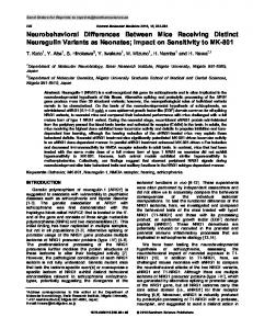
Figure 1. Chemical structures of (1) amygdalin and (2) prunasin.
Chemicals and Materials. High-pressure liquid chromatography (HPLC)-grade methanol and acetonitrile were purchased from Burdick & Jackson (NJ, USA). Water was
Metabolic Pharmacokinetics in Rats
purified with an ultra filtration system (Sinhan, Korea). Amygdalin and prunasin standards were purchased from Sigma (MO, USA). Peach seeds were purchased from KwangMyungDang (Daejeon, Korea) and identified by Prof. Yong Ki Park, College of Oriental Medicine, Dongguk University. The voucher specimen was deposited in the herbarium at the College of Chungnam National University. Rats. Sprague-Dawley rats (220-250 g) were obtained from the Department of Laboratory Animal Medicine (Seoul, Korea) and housed with unlimited access to food and water, except for a 12-h fasting period immediately before the experiment. The animals were maintained on a 12 h light/12 h dark cycle (light on at 8:00 am) at ambient temperature (22-25 oC) and 60% relative humidity. Instrumentation and Conditions. HPLC with diode array detection (HPLC-DAD) was carried out using a Shimadzu LC-10AD series HPLC system (Japan). A C18 column (4.6 × 150 mm, 5 μm, Phenomenex, CA, USA) was used for separation at a column temperature of 25 oC. The mobile phase consisted of 20% acetonitrile, and the flow rate was 0.5 mL/min. The monitoring wavelength was 215 nm. HPLC-mass spectrometry (MS) analyses were carried out on a Shimadzu LCMS-2010-EV linked to an electrospray ionization (ESI) source operating in both negative and positive mode. The liquid chromatography (LC) conditions for MS were same as those for HPLC above. LC-MS solution software was used to control the instruments for data acquisition and processing. The LC-MS was operated with a nebulizing gas flow rate of 1.4 L/min, a curved desolvation line (CDL) temperature of 250 oC, a heat block temperature of 200 oC, a detector voltage of 1.50 kV, and a CDL voltage of 15.0 V. Preparation of Peach Seed Extract. Dried peach seeds (50 g) were crushed and then extracted twice under thermal reflux with 500 mL water for 2 h. The solutions obtained were filtered and then combined and concentrated to approximately 50 mL under vacuum at 30 oC. The concentration of amygdalin in the decoction of peach seed was determined by HPLC. Pharmacokinetic Study. Experiments were performed on 18 rats that were randomly divided into three groups. In the first group, an aqueous solution of amygdalin was intravenously injected by vena caudalis at a dose of 30 mg per 100 g body weight. In the second and third groups, an aqueous solution of amygdalin or a decoction of peach seeds, respectively, were orally administered by gavage using a gauge syringe at a dose of 30 mg amygdalin per 100 g body weight or 1 g of peach seed (equal to 1 mL of decoction) per 100 g body weight. Blood samples (~0.5 mL) were collected via the orbital vein before administration and at 5, 10, 20, 30, 45, 60, 90, 120, 180, 240, and 360 min after administration. The blood samples were centrifuged at 17,000 rpm for 10 min at 4 oC. The supernatants (i.e., plasma) were decanted and mixed with 0.4 mL methanol. After 30 min, the precipitated plasma proteins were separated by centrifugation at 17,000 rpm for 10 min at 4 oC. The upper solvent layer was dried under a stream of N2. The precipitated residue was
Bull. Korean Chem. Soc. 2012, Vol. 33, No. 5
1471
dissolved in 0.1 mL methanol and stored at −20 oC for further analysis. Data from these samples were used to construct pharmacokinetic profiles by plotting drug concentration versus time. The pharmacokinetic parameters were calculated from the plots using WinNonlin Standard Edition software (Ver. 2.1). A comparison of the pharmokinetic parameters obtained with pure amygdalin and amygdalin in a decoction of peach seeds was carried out using a t-test. All experimental data and pharmacokinetic parameters were expressed as means ± standard deviation (SD). Calibration Curves and Quality Control Samples. Stock solutions of amygdalin and prunasin were prepared in methanol, and a mixture containing 1.0 mg/mL each was also prepared. Calibration samples were prepared by spiking plasma samples with the mixed stock solution. The final concentrations of amygdalin and prunasin in the calibration samples were 6.3, 12.5, 25.0, 50.0, and 100.0 mg/L. Quality control (QC) samples were also prepared in the same way using the individual stock solutions (12.5, 25.0, 50.0 mg/L for amygdalin and prunasin, separately). All solutions were stored at 4 oC before use. Linearity was determined for the two standards using HPLC. Each standard was injected in triplicate. Calibration curves in the 6.3-100 mg/L range were constructed by plotting the ratios of peak area against concentration. The concentrations of amygdalin and prunasin in the test samples were calculated using regression parameters obtained from the standard curve. Precision and Accuracy. Three concentrations (high, medium, and low) of the standard solution mixture were prepared using the blank rat plasma, and the concentrations of amygdalin and prunasin were measured as an accuracy variation test; the test was performed three times on each day that the compounds were administered to the rats. The precision of the measurements was determined as the coefficient of variation (RSD). The accuracy was calculated from the mean value of the observed concentrations (Cobs) and the theoretical concentrations (Cthe) according to the equation: % = [Cobs/Cthe] × 100. The limits of detection (LOD) and limits of quantification (LOQ) for the standard solutions were calculated with a signal to noise ratio of 3 (S/N = 3) and 10 (S/N = 10), respectively. Results and Discussion Optimization of Chromatographic Conditions. Different mobile phases were tested to determine the optimal separation of amygdalin and prunasin from endogenous-related substances. An acetonitrile-water solution (20:80, v/v) at a flow rate of 0.5 mL/min on an analytical C18 column proved to be the best chromatographic method. The maximum absorption wavelength was 215 nm. Under these conditions, amygdalin and prunasin gave optimum peak shapes, yielded an excellent response, and could be eluted within 15 min. Identification of Amygdalin and Prunasin. Amygdalin and prunasin were clearly separated from the matrix with retention times of 7.8 and 12.9 min, respectively (Figs. 2(a) and (b)), in the blank rat plasma. Amygdalin was detected in
1472
Bull. Korean Chem. Soc. 2012, Vol. 33, No. 5
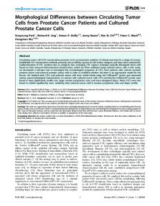
Figure 2. Representative HPLC chromatograms of (a) rat plasma; (b) rat plasma spiked with amygdalin and prunasin; (c) a rat plasma sample obtained 10 min after intravenous injection of amygdalin; and (d) a rat plasma sample obtained 45 min after oral administration of amygdalin. Peak identifications: 1 amygdalin, 2 prunasin.
the plasma samples obtained from rats that were administered intravenous amygdalin injections. However, prunasin and not amygdalin was detected in the plasma samples obtained from rats that received oral administration of amygdalin or oral administration of the decoction of peach seeds (Figs. 2(a) and (b)). Amygdalin and prunasin in the plasma samples were confirmed by HPLC-MS. In positive mode, the peaks were observed at m/z = 480 [M+Na]+ for amygdalin (MW = 457) and at m/z = 296 [M+H]+ (MW = 295) for prunasin; in negative ion mode, the peaks were observed at m/z = 456 [M-H]− for amygdalin (MW = 457) and at m/z = 294 [M-H]− for prunasin (MW = 295). Methods Validation. Blank plasma samples spiked with various concentrations of standard solution were analyzed as described in Materials and methods. Calibration curves were constructed from the ratios of the peak areas to the standard concentrations. The calibration curves were linear within the concentration range assayed. The mean regression values of the calibration curves were y = 0.3552x + 1.8897 (r2 =
Jianbo Chen et al.
0.9992) for amygdalin and y = 0.7381x + 1.3878 (r2 = 0.9991) for prunasin. The LOQ for amygdalin and prunasin in plasma were 3.2 mg/L and 4.8 mg/L, respectively, and the LOD were 1.5 mg/L and 2.4 mg/L, respectively. Accuracy and Precision. The precision and accuracy of the assay were estimated by analyzing QC samples containing known concentrations of amygdalin and prunasin at low, middle, and high values. The concentrations of analytes in the QC samples were then determined by HPLC and calculated from the calibration curves determined on the same day (Table 1). From a comparison of the calculated versus known values, the precision and accuracy of the determination method was calculated as described in Materials and methods. The intra-day precision (RSD) ranged from 4.2 to 5.5%, and the inter-day precision (RSD) ranged from 5.4 to 7.1%. The intra-day accuracy ranged from 97.6 to 100.1%, and inter-day accuracy ranged from 95.2 to 105.0%. Application to Pharmacokinetic Study. The established method was applied to the analysis of plasma samples after intravenous injection of amygdalin or oral administration of amygdalin or a decoction of peach seeds. Amygdalin is one of the major active constituents in peach seeds, and prunasin is one of its major metabolites; in plasma samples obtained after oral administration of amygdalin or peach-seed decoction, prunasin and not amygdalin was detected. We compared the difference in pharmacokinetics between the oral administration of pure amygdalin and amygdalin contained in the decoction of peach seeds. The concentration of amygdalin in plasma versus time after intravenous injection of amygdalin is shown in Figure 3(a). The concentration of prunasin in plasma versus time after oral administration of amygdalin or peach-seed decoction is shown in Figure 3(b). After comparing the metabolic pharmacokinetics between pure amygdalin and amygdalin in decoctions of peach seeds using the t-test, it was concluded that significant differences (P < 0.01) exist between the important pharmacokinetic parameters (V1/F, AUC, T1/2, CL/F) of the two groups. The pharmacokinetic parameters of amygdalin after intravenous injection of amygdalin are presented in Table 2. The distribution processes of amygdalin after intravenous injection could be fitted to a one-compartment model. These results indicate that after intravenous injection, amygdalin was absorbed into the blood rapidly. After oral administration of amygdalin or a decoction of peach seeds, virtually
Table 1. Precision and accuracy of amygdalin and prunasin in rat plasma Intra-day (n = 5)
Inter-day (n = 5)
Component
Spiked (mg/L)
Measured (mg/L)
Accuracy (%)
Precision (RSD%)
Measured (mg/L)
Accuracy (%)
Precision (RSD%)
Amygdalin
12.5 25.0 50.0 12.5 25.0 50.0
12.4 24.4 50.4 12.1 24.5 49.5
99.2 97.6 100.1 96.8 98.0 99.0
5.0 5.2 4.6 4.2 5.5 4.7
12.9 24.6 51.1 11.9 25.5 52.5
103.2 98.4 102.2 95.2 102.0 105.0
7.1 6.3 6.2 5.4 7.1 6.0
Prunasin
Metabolic Pharmacokinetics in Rats
Bull. Korean Chem. Soc. 2012, Vol. 33, No. 5
1473
concentration (Cmax) of 62.1 mg/L within 45.3 min. After oral administration of aqueous amygdalin solution, the absorption of prunasin was delayed. Prunasin was absorbed and reached a maximum concentration of 42.9 mg/L in 60.1 min. The AUC (14196 mg min/L), T1/2α (26.8 min), and T1/2β (245.1 min) values of prunasin from oral administration of a decoction of peach seeds were markedly greater than the corresponding AUC (9871 mg min/L), T1/2α (16.2 min), and T1/2β (122.6 min) values from the oral administration of pure amygdalin; only the CL/F value showed a marked decline. There may be two reasons for these results. First, amygdalin must be hydrolyzed by β-glucuronidase to create prunasin in the intestine subsequent to intestinal absorption.11 β-glucosidase, which may exist in the decoction of peach seeds, would accelerate the release of cyanide from amygdalin. Second, there may have been interactions between different chemical ingredients. The decoction of peach seeds contains liposomes, cyanogenetic glycosides, amino acids, essential oils, sterides, and flavones, each of which could produce certain pharmacological effects and may have impacts on the metabolism of amygdalin. Figure 3. (a) Concentration of amygdalin in rat plasma as a function of time after intravenous injection of amygdalin at a dose of 30 mg per 100 g body weight. (b) Concentration of prunasin in rat plasma as a function of time after oral administration of (1) amygdalin at a dose of 30 mg per 100 g body weight (open circles) or (2) a decoction of peach seeds at a dose of 1 g per 100 g body weight (closed circles). Table 2. Pharmacokinetic parameters of prunasin (p.o.) and amygdalin (i.v.) in rat plasma (n = 6) after an oral administration of amygdalin and decoction of peach seed and intravenous injection of amygdalin Parameters
Peach seed decoction, p.o.a
Amygdalin, p.o.
Amygdalin, i.v.
Cmax (mg/L) Tmax (min) AUC (mg min/L) T1/2 (min) CL/F (min−1)
62.1 ± 8.2** 45.3 ± 5.3* 14196 ± 5438** 26.8 ± 10.8** 0.85 ± 0.42
42.9 ± 6.8 60.1 ± 8.3 9871 ± 3422 16.2 ± 8.8 0.98 ± 0.48
82.7 ± 9.6 − 7238 ± 3215 11.4 ± 4.3 1.2 ± 0.8
**
*
P < 0.01; P < 0.05 when compared with an oral administration of amygdalin.
all of the amygdalin was metabolized to prunasin before being absorbed into the blood. The pharmacokinetic parameters of prunasin derived from the plasma profiles after oral administration of amygdalin solution or a decoction of peach seeds is presented in Table 2. The distribution processes of prunasin could be fitted to a two-compartment model. By comparison, the pharmacokinetic parameters of prunasin varied greatly between pure amygdalin and amygdalin contained in a decoction of peach seeds. After oral administration of the decoction of peach seeds, prunasin was absorbed at a fast rate and reached a maximum plasma
Conclusions In this study, we developed a simple and rapid HPLC/ ultraviolet (UV)-MS method for identification and determination of prunasin in plasma after the oral administration of aqueous amygdalin solution or a decoction of peach seeds. Based on the results, the parameters of Cmax and AUC for prunasin were remarkably greater after oral administration of the peach-seed decoction compared with pure amygdalin. Consequently, we suggest that there should be a warning about the interaction of amygdalin with the herbal ingredients in peach seeds. The knowledge gained from this study can be used to evaluate the efficacy and safety of clinical applications of peach seeds. Further studies are needed to clarify the effect(s) of other herbal ingredients on prunasin. People taking a decoction of peach seeds should be alerted to the possibility of herb-drug interactions. Acknowledgments. This work was supported by the Priority Research Centers Program through the National Research Foundation of Korea (NRF) funded by the Ministry of Education, Science and Technology (2009-0093815). References 1. The Pharmacopoeia of the People's Republic of China; Part 1. The Pharmacopoeia Commission of PRC: 2010; p 260. 2. Liu, Q. Z. Chin. Pat. Med. Res. 1987, 8, 47. 3. Xu, H. Y.; Yun, C. X.; Wang, Y. X. J. Qiqihar Med. 2004, 5, 487. 4. Rui, H. K. Chin. Pat. Med. 1992, 14, 33. 5. Sun, W. K. Hunan Med. J. 1993, 9, 47. 6. Fang, M. F.; Fu, Z. L.; Wang, Q. L.; Wang, S. X.; Xiao, C. N.; Zhang, X. H. Chin. J. Chin. Mater. Med. 2010, 35, 2684. 7. Wang, Y. L.; Li, H. B.; Hu, Y. Q. Chin. Pharm. 2002, 5, 550. 8. Lian, Y. J.; Chen, D. D.; Xu, T. W.; Zheng, Y. B.; Huang, T.; Ke, M. L. Chin. J. Bas. Clin. Gen. Sur. 2005, 12, 138. 9. Zhuo,Y. Z.; Zhao, L. G.; Liu, J. H. Tianjin J. Trad. Chin. Med.
1474
Bull. Korean Chem. Soc. 2012, Vol. 33, No. 5
2009, 26, 500. 10. Strugala, G. J.; Stahl, R.; Elsenhans, B. Hum. Exp. Toxicol. 1995, 14, 895. 11. Deng, Y.; Guo, Z. G.; Zeng, Z. L.; Wang, Z. Chin. J. Chin. Mater. Med. 2002, 27, 565. 12. Rauws, A. G.; Gramberg, L. G.; Olling, M. Phar. Weekbl. Sci. Edi. 1982, 4, 172. 13. Ge, B. Y.; Chen, H. X.; Han, F. M.; Chen, Y. J. Chromatogr. B
Jianbo Chen et al. 2007, 857, 281. 14. Koo, J. Y.; Hwang, E. Y.; Cho, S. J. Chromatigr. B 2005, 814, 69. 15. Wu, H.; Zhu, Z. Y.; Zhang, G. Q.; Zhao, L.; Zhang, H.; Zhu, D. L. J. Ethnopharmacol. 2009, 125, 444. 16. Huang, X.; Wen, A. D.; Zang, Y. M. Chin. J. Int. Trad. Med. 1994, 14, 288. 17. Hou, Y. C.; Hsiu, S. L.; Ching, H.; Lin, Y. T.; Tsai, S. Y.; Wen, K. C. Life Sci. 2005, 76, 1167.