Mar 29, 1976 - 1 mM EDTA and twice with 10 ml CMF and finally resuspended in ... Takeichi (1976), 03 ml of cell suspension in CMF-MEM (9 x io6 cells).
J. Cell Sci. 23, 685-69S (1976) Printed in Great Britain
685
DIFFERENT CATION REQUIREMENTS FOR AGGREGATION OF BHK CELLS AND THEIR TRANSFORMED DERIVATIVES HIDEKO URUSHIHARA,* MASATOSHI TAKEICHI*, AKIRA HAKURAf AND T. S. OKADA* Laboratory for Cell Differentiation and Morphogenesis, Institute of Biophysics, Faculty of Science, University of Kyoto, Kyoto 606, Japan* and Research Institute for Microbial Diseases, University of Osaka, Suita 564, Japan\
SUMMARY BHK21 cells singly dissociated by trypsin aggregate in medium, with calcium ions, but not in medium with magnesium ions. Their malignant derivatives (pyBHK) induced by polyoma virus, after dissociation with trypsin, aggregate equally well in medium with either calcium or magnesium ions. When EDTA is used for dissociation of cells from culture on a substrate, neither BHK21 nor pyBHK cells require addition to the medium of divalent cations for rapid aggregation. Trypsin-dissociated BHK21 cells become aggregative in medium without divalent cations, when they are incubated for 60 min in dispersion with medium containing calcium ions before aggregation. In the case of pyBHK cells, incubation in dispersion in the medium, with calcium or magnesium ions is effective in this respect. Calcium and magnesium ions are equally effective for adhesion of both BHK21 and pyBHK cells to non-cellular substrate or to a cell monolayer. We discuss the different cation requirements for aggregation of BHK21 and their transformed derivatives in relation to the recovery process of cell surfaces after exposure to trypsin.
INTRODUCTION
Transformation of cells to the malignant state causes various changes on cell surfaces (Abercrombie & Ambrose, 1962; Inbar, Ben-Basset & Sachs, 1972; Comoglio, Tarone, Prat & Bertini, 1975), which necessarily alter cellular interactions such as specific recognition among different cell types and communication between apposed cells (Dorsey & Roth, 1973). Aggregation by cell-to-cell contact also seems to be influenced by malignant transformation (Edwards & Campbell, 1971). In spite of the recent increased interest in the study of cell contact, however, information, about this problem in transformed cells is still rather scanty. Detailed comparison between normal cells and their malignant derivatives is necessary under well-controlled experimental conditions. The aim of the present study is to compare properties of BHK cells and their derivatives transformed with polyoma virus by 2 methods of measuring cell adhesion: adhesion to the substrate and to other cells. Recently, qualitatively different effects of calcium and magnesium ions on cell adhesion have been suggested (Takeichi & Okada, 1972; Okada, Takeichi, Yasuda & Ueda, 1974; Ueda & Takeichi, 1976). Therefore, the adhesive properties of cells have been carefully examined with respect to the
686
H. Urushihara and others
different cation requirements of normal and transformed cells. Since the culture density of growing cells in vitro has been known to affect their aggregative ability (Edwards & Campbell, 1971), adhesive properties have been compared between cells at different stages of growth. MATERIALS AND METHODS Cell culture BHK21 cells, clone 13 (BHK) and polyoma virus-transformed BHK (pyBHK) were used in the present experiments. The latter cells were prepared by infecting the former with Dulbecco's LP-strain of polyoma virus in the Institute of Microbial Diseases, Osaka University by A. H. Their malignancy was often confirmed by both in vitro and in vivo assays. The cells were grown in Eagle's MEM supplemented with 6 % foetal calf serum. For subculturing, cells in the confluent state were detached by trypsinization (see later) and washed with culture medium (see later), then 1 x 10s cells were inoculated into each fresh 10-cm glass Petri dish. The medium was changed every day after the initial 2 days of subculturing. Cell preparation Trypsinized cells. Confluent cultures were washed twice with Ca1"1" — and Mgs+— free saline (CMF) and incubated with 0-25 % trypsin (Difco, 11250) inCMF at 37 CC for 15 min. Detached cells, usually 3x10' cells, were filtered through 4 layers of gauze, washed once with 10 ml of 1 mM EDTA and twice with 10 ml CMF and finally resuspended in Caa+— and Mg1+-free Eagle's MEM buffered with 10 mM HEPES, pH 74 (CMF-MEM) to give a cell density of 3 x io6 cells/ml. EDTA-treated cells. Confluent cultures were washed twice with CMF and incubated with 1 mM EDTA (in CMF buffered with HEPES, pH 74) at 37 °C for 15 min. The cells were dissociated into single cells by pipetting, filtered through gauze, washed twice with CMF and suspended in CMF-MEM. Assays for cell adhesion Cell-to-substrate adliesion. To each gelatinized, 5-cm Falcon plastic dish prepared as described by Ueda & Takeichi (1976), 03 ml of cell suspension in CMF-MEM (9 x io6 cells) was inoculated into 27 ml of CMF-MEM. After incubation for 15 min at 37 °C, a given amount of Mg1+ and/or Ca1+ as chloride was added and incubation continued without agitation at 37 °C. After an appropriate time, the cells were fixed in 5 % glutaraldehyde solution, and the unattached cells were collected and counted with a Coulter Counter (Ueda & Takeichi, in Press). Cell-to-cell adliesion. Plastic dishes (Jintan, Tokyo) 25 cm in diameter, which had previously been coated with bovine serum albumin (BSA) to prevent adhesion of cells to the bottom as described by Takeichi & Okada (1972), were used for the assay. Cells, 9 x io6 in 3 ml of CMF-MEM, were inoculated into each coated dish. After adding a given amount of divalent cations, dishes were incubated on a gyratory shaker (90 rev/min) at 37 °C. At appropriate times, the cells werefixedas above and the total particle number (TPN) in the medium counted with a Coulter Counter. Cell-to-cell monolayer adhesion. Confluent cell cultures in 3-cm Falcon plastic dishes (2 x 10' cells per dish) were washed 3 times with CMF-MEM containing either calcium or magnesium ions. To the washed cell sheet 5 x io5 cells in 2 ml of MEM with either calcium or magnesium ions were added gently. To estimate the number of cells spontaneously detached during incubation, MEM without cells was added to the monolayer in other dishes as a control. The dishes were incubated without agitation at 37 °C and the number of floating cells counted.
Aggregation of BHK cells
687
Recovery from trypsinization Trypsinized cells in 5-cm BSA-coated dishes were incubated on a gyratory shaker (90 rev/ min) at 37 °C in medium with or without divalent cation. In large dishes, cell-to-cell adhesion was mostly prevented. After 1 h to 3 h of incubation in dispersion the cells were collected, filtered, washed once with EDTA and twice with CMF and finally resuspended in CMF-MEM. RESULTS
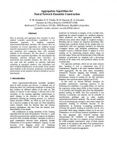
Trypsinized cells Cell-to-cell aggregation. In the medium containing 1 mM calcium, both BHK and pyBHK cells only started to aggregate after a lag of 1 h, and TPN after 3 h was about 50% of initial TPN, thereafter remaining constant during further incubation with gyration. In medium with 1 mM magnesium, aggregation of pyBHK cells occurred in the same way as with calcium ions, while BHK cells failed to aggregate (Fig. 1 A, B). Both BHK and pyBHK cells aggregated more extensively in medium containing both 1 mM calcium and 1 mM magnesium ions. No aggregation of either cells occurred in CMF-MEM.
10
10
~r 05 z
I 05
Z
60
180
Incubation time, min
300
60
120
180
300
Time, mm
Fig. 1 A, B. Effects of calcium and magnesium ions on aggregation of BHK (A) and pyBHK (B) right: x — x , in CMF, O—O, in J mM magnesium chloride; # — # , in 1 mM calcium chloride; and O—O, in 1 mM magnesium chJoride+i mM calcium chloride. These symbols are also used in Figs, 3, 4, 7, 8. Each point represents the mean of 3 plates. Five independent experiments were performed and the results were reproducible. The size of the experimental points represents an approximate limit of error throughout all the figures.
Cell-to-cell substrate attachment. In medium with either 1 mM calcium or 1 mM magnesium ions, both BHK and pyBHK cells attached to gelatinized plastic substrate, though magnesium was more effective in enhancing attachment than calcium (Fig. 2); with magnesium, approximately 75 % of the inoculated cells attached after incubation for 90 min, while with calcium the corresponding level was less than 55 %. Practically
H. Urushihara and others
688
no adhesion of either BHK or pyBHK cells occurred in CMF-MEM, whereas in the medium containing both calcium and magnesium ions more than 80 % of the inoculated BHK or pyBHK cells attached. 10
10
£ 0.5
? 05 Z
30
60
90
60
Time.min
120
240
Time, min
Fig. 2. Effects of calcium and magnesium ions on cell attachment to gelatin-coated plastic substrate. Each point represents the mean of 2 plates and the results were reproduced in 2 other experiments. A, BHK cells; B, pyBHK. 10
10 -
z I? 05
I 30
I 60 Time, mm
I 90
I 30
60
90
Time, min
Fig. 3. Attachment of BHK (left) and pyBHK (right) cells to the homotypic cell monolayer. Cells spontaneously detached from the cell monolayer were subtracted as background, and each point represents the mean value of 2 plates. The experiment was repeated two further times with similar results.
Cell-to-cell monolayer attachment. Both BHK and pyBHK cells rapidly attached to the homotypic cell monolayer in medium containing either 1 mM magnesium or 1 mM calcium ions. Both types of cells adhered approximately with the same rapidity and to the same extent in either medium (Fig. 3). Measurement of attachment in CMF-
Aggregation of BHK cells
689
MEM was not possible, because the monolayer came off easily from the plate under such conditions. Culture density and adhesion. Cells taken from cultures at different stages of growth were subjected to assay for their adhesiveness. Aggregation of pyBHK cells in presence of either magnesium or calcium ions occurred similarly irrespective of the stage at which cells were harvested. However, as shown in Fig. 4, aggregation of BHK cells in presence of calcium ions occurred more extensively with increase in cell density of the cultures. Cell-to-substrate attachment was independent of culture density in both BHK and pyBHK cells.
10
"-= 05
8 60
120
I 180
240
300
Time, min
Fig. 4. Aggregation of BHK cells in 1 mM calcium ions at various culture densities. Growth curve of BHK cells is shown in the inset, lower left. Cells were harvested for assay at approximately 2, 3, 4, and 6 days. The result was confirmed in 2 other experiments.
EDTA-dissociated cells BHK cells dissociated by EDTA aggregated much more rapidly without a lag in 3 types of media including CMF-MEM compared with cells dissociated with trypsin (Fig. 5). Aggregation was, however, enhanced by 1 mM calcium and still more by 1 mM magnesium. Dissociated BHK cells aggregated more extensively than pyBHK cells when divalent cations were not present. Thus the promoting effect of calcium or magnesium ions was more prominent in aggregation of pyBHK than of BHK cells.
690
H. Urtishihara and others 10
10
I 0.5
% OS 2
I 30
60
90
20
Time, min
40 Time, min
80
Fig. 5. Aggregation of BHK (A) and pyBHK (B) cells dissociated with 1 mM EDTA. Each point shows the mean of 2 plates. The result was reproduced in 2 other experiments. 10
-
30
60
120
Preincubation time, mm
Fig. 6. Relation between the period of previous incubation in dispersion and the aggregative ability of BHK cells. Cells were incubated in dispersion in medium with 1 mM calcium chloride for an appropriate length of time, routinely washed and aggregated in CMF for 120 min. Each point represents the mean of 2 plates. The result was confirmed by another experiment.
Aggregation of BHK cells
691
Recovery from trypsinization After incubation for 120 min in dispersion in the medium containing 1 mM calcium ions, trypsinized BHK cells aggregated without a lag in medium with calcium, and in contrast to freshly dissociated cells, also in medium containing magnesium ions and even in CMF-MEM. As shown in Fig. 6, incubation in dispersion for 60 min is
x
10
*—-
Z^~~——x
^
0-5
-
05
-
B
60 Time, mm
1
1
1
30
120
30
60 Time, mm
120
Fig. 7. Effect of previous incubation of cells in dispersion for 120 min on the aggregation of BHK cells. After incubation in dispersion in medium with 1 mM calcium chloride (A) and with 1 mM magnesium choride (B). Each point represents the mean of 2 plates. The results were reproducible in 2 other experiments.
1 0
10
-=
% Z
05
05
J_ 20
40 Time, mm
80
20
40 Time, mm
80
Fig. 8. Effect of previous incubation in dispersion for 120 min on the aggregation of pyBHK cells. After incubation in dispersion in medium with 1 mM calcium chloride (A) and with 1 mM magnesium chloride (B). Each point represents the mean of 2 plates. These experiments were repeated twice and the results confirmed. 45-2
692
H. Uiushihara and others
sufficient for BHK cells to become fully adhesive. The results were different when BHK cells dissociated with trypsin were incubated in dispersion in medium containing 1 mM magnesium instead of calcium. Unlike the latter case, this treatment did not alter the aggregation of the cells. They scarcely aggregated in medium with magnesium or in CMF-MEM, though a low rate of aggregation with a lag of 60 min occurred in medium containing calcium (Fig. 7). Trypsinized pyBHK cells also became aggregative in CMF-MEM as well as in medium with either calcium or magnesium ions, after previous incubation in dispersion for 120 min. The results, however, were different from those of a similar experiment using trypsinized BHK cells in that incubation in dispersion with magnesium ions as well as with calcium allowed aggregation of trypsinized pyBHK cells in CMFMEM (Fig. 8).
DISCUSSION
There are a number of characteristics of the cell surface which distinguish experimentally transformed cells from their normal predecessors (Hakomori & Murakami, 1968; Inbar et al. 1972; Noonan & Burger, 1973; Comoglio et al. 1975). A reflexion of the altered properties of the cell surface arises in the adhesion of the transformed malignant cells, showing an increase (Halpern, Pejsachowicz, Febvre & Barski, 1966; Cassiman & Bernfield, 1975) or a decrease (Edwards, Campbell & Williams, 1971) in aggregation and a decrease in intercellular recognition (Roth, 1968; Dorsey & Roth, 1973) compared with normal cells. Using very similar material to ours, Edwards et al. (1971) and Shields & Pollock (1974) reported that py-transformed BHK cells are much less adhesive than their normal counterparts. In the present study, the overall decrease in aggregation of the transformed cells reported by Edwards et al. (1971) could not be confirmed using the physiological medium containing both calcium and magnesium ions. The difference may lie in choice of medium: Hanks's saline in their assays and Eagle's MEM in ours, and the method of aggregation, namely, the use of a reciprocating shaker in the former and of a gyratory shaker in the present study. In unpublished work, we have found that overall aggregation is much less in Hanks's saline than in MEM in both BHK and pyBHK cells. It may be also possible that the different results could be explained by the induction of dissimilar cell properties by the polyoma infection. In the present study, a marked difference has been shown between transformed and normal cells with respect to their cation requirements for aggregation. After trypsin dissociation, transformed cells can aggregate equally well in medium with either calcium or magnesium, whereas normal cells can do so only in medium containing calcium. While total particle count is considered a valid assessment of aggregation (Edwards, 1973), a theoretical limit has been placed on the use of this type of shaker for the evaluation of cellular adhesiveness (Curtis, 1969). This will certainly make a thorough discussion of the present results in terms of different adhesiveness difficult. However, the present findings of the different cation requirements were
Aggregation of BHK cells
693
very reproducible. Unpublished studies show that differences in the number of passages before assaying and in the duration of freezing before cell growth in vitro do not influence the results. The results are consistent with those of Edwards, Campbell, Robson & Vicker (1975). Thus, the difference in the cation requirements of cell aggregation can be considered to be a rather stable characteristic for distinguishing normal BHK cells from their transformed derivative. Our previous studies with cultured fibroblasts of chick embryos showed that the action of calcium and magnesium ions on cell adhesion is qualitatively different (Okada et al. 1974), and a tentative assumption of the presence of 2 different mechanisms, one calcium-dependent and the other magnesium-dependent, has been proposed. Thus, it could be conceivable that only the calcium-dependent mechanism is well developed in BHK cells, while both mechanisms are present in pyBHK cells. It has often been suggested that there are specific sites on the cell surface for holding together adjacent cells in contact (Moscona & Moscona, 1966; Oppenheimer, Edidin, Orr & Roseman, 1969). Several workers considered that tryptic digestion for dissociating cells damages key molecules at the cell surface or modifies their arrangement on the surface, and thus alters adhesive properties of cells (Steinberg, Armstrong & Granger, 1973; Nicolson, 1972; Edwards & Campbell, 1971). The present results seem to substantiate such a view, since previous incubation in dispersion is necessary to render trypsinized cells adhesive in CMF-MEM. A requirement for protein synthesis during the period of incubation in dispersion has been indicated in other experiments from our laboratory (Yasuda, in preparation) and by other workers (Steinberg et al. 1973; Grinnell, Milam & Srere, 1973). In BHK cells such a recovery process much depends on the presence of calcium ions in the medium used, whereas with pyBHK cells the same process occurs equally well in medium containing either calcium or magnesium ions. Since cells dissociated with EDTA or incubated in dispersion after trypsin dissociation can aggregate in CMF-MEM, it is rather difficult to consider that divalent cations participate in aggregation as 'bridges' between cells. Bearing this in mind, the following speculation may be set forth to interpret the present results. Tryptic digestion may remove from the cell surface some components which are responsible for aggregation. For renewal of such components, divalent cations could be needed either metabolically or by being integrated directly into them. With regard to the relationship between growth density and aggregative ability of BHK cells, our data were similar to those of Edwards et al. (1971) in that the extent of aggregation increased progressively with density of the cultures from which the suspensions were prepared. Since there was no significant decrease in viability of cells prepared from the lower-density cultures, the difference may be in sensitivity to trypsin. With respect to cell adhesion to non-cellular substrate or monolayer sheets of cells, in which magnesium ions are more effective than calcium ions, there are few differences between BHK and pyBHK cells. It is possible that totally different mechanisms function in this type of cell adhesion compared with those involved in cell-to-cell aggregation. On the other hand, an explanation of these 2 types of cell adhesion in
694
H. Urushihara and others
terms of common mechanisms is also possible, if we assume that BHK cells still have sufficient magnesium-binding sites to enable adhesion to substrates, though not enough to ensure adhesion to other cells in suspension. The authors gratefully acknowledge the valuable comments and critical reading of the manuscript of Dr R. B. Kemp. We thank Mr S. Matsuura for his collaboration in some parts of the present experiments and Miss Reiko Takayasu for helping in the preparation of this manuscript. The work was in part supported by a grant for basic cancer reasearch from the Japanese Ministry of Education, Culture and Science.
REFERENCES ABERCROMBIE, M. & AMBROSE, E. J. (1962). The surface properties of cancer cells: a review. Cancer Res. 22, 525-548. CASSIMAN, J.-J. & BERNFIELD, M. R. (1975). Transformation-induced alteration in fibroblast adhesion: masking by trypsin treatment. Expl Cell Res. 91, 31-35. COMOGLIO, P. M., TARONE, G., PRAT, M. & BERTINI, M. (1975). Studies on the outer surface of normal and RSV-transformed BHK fibroblast plasma membrane. Expl Cell Res. 93, 402410.
A. S. G. (1969). The measurement of cell adhesiveness by an absolute method. J. Embryol. exp. Morph. 22, 305-325. DORSEY, J. K. & ROTH, S. (1973). Adhesive specificity in normal and transformed mouse fibroblasts. Devi Biol. 33, 249-256. EDWARDS, J. G. & CAMPBELL, J. A. (1971). The aggregation of trypsinized BHK21 cells. J. Cell Sci. 8, 53-71. EDWARDS, J. G., CAMPBELL, J. A., ROBSON, R. T. & VICKER, M. G. (1975). Trypsinized BHK21 cells aggregate in the presence of metabolic inhibitors and in the absence of divalent cations. J. Cell Sci. 19, 653-667. EDWARDS, J. G., CAMPBELL, J. A. & WILLIAMS, J. F. (1971). Transformation by polyoma virus affects adhesion of fibroblasts. Nature, New Biol. 231, 147-148. GRINNELL, F., MILAM, M. & SRERE, P. A. (1973). Studies on cell adhesion. III. Adhesion of baby hamster kidney cells. J. Cell Biol. 56, 659-665. EDWARDS, J. G. (1973). Intercellular adhesion. In New Techniques in Biophysics and Cell Biology (ed. T. Pain & B. J. Smith), p. 1. New York: Wiley. HAKOMORI, S. & MURAKAMI, W. T. (1968). Glycolipids of hamster fibroblasts and derived malignant-transformed cell lines. Proc. natn. Acad. Sci. U.S.A. 59, 254-261. HALPERN, B., PEJSACHOWICZ, B., FEBVRE, H. L. & BARSKI, G. (1966). Differences in patterns of aggregation of malignant and non-malignant mammalian cells. Nature, Lond. 209, 157-159. INBAR, M., BEN-BASSAT, H. & SACHS, L. (1972). Membrane changes associated with malignancy. Nature, New Biol. 236, 3. MOSCONA, M. H. & MOSCONA, A. A. (1966). Inhibition of cell aggregation in vitro by puromycin. Expl Cell Res. 41, 703-706. NICOLSON, G. L. (1972). Topography of membrane concanavalin A sites modified by proteolysis. Nature, New Biol. 239, 193. NOONAN, K. D. & BURGER, M. M. (1973). Binding of fH]Concanavalin A to normal and transformed cells. J. biol. Chem. 248, 4286-4292. OKADA, T. S., TAKEICHI, M., YASUDA, K. & UEDA, M. J. (1974). The role of divalent cations in cell adhesion. Adv. Biophys. 6, 157-181. OPPENHEIMER, S. B., EDIDIN, M., ORR, C. W. & ROSEMAN, S. (1969). An L-glutamine requirement for intercellular adhesion. Proc. natn. Acad. Sci. U.S.A. 63, 1395-1462. ROTH, S. A. (1968). Studies on intercellular adhesive selectivity. Devi Biol. 18, 602-631. SHIELDS, R. & POLLOCK, K. (1974). The adhesion of BHK and pyBHK cells to the substratum. Cell 3, 3i-38. STEINBERG, M. S., ARMSTRONG, P. B. & GRANGER, R. E. (1973). On the recovery of adhesiveness by trypsin-dissociated cells. J. Membrane Biol. 13, 97-128. CURTIS,
Aggregation of BHK cells
695
M. & OKADA, T. S. (1972). Roles of magnesium and calcium ions in cell-to-substrate adhesion. Expl Cell Res. 74, 51-60. UEDA, M. J. & TAKEICHI, M. (1976). Two mechanisms in cell adhesion revealed by effects of divalent cations. Cell Struct. Func. 1 (in Press). TAKEICHI,
(Received 29 March 1976)