BIOLOGY OF REPRODUCTION 70, 1010–1017 (2004) Published online before print 3 December 2003. DOI 10.1095/biolreprod.103.022061
Different Testicular Gene Expression Patterns in the First Spermatogenic Cycle of Postnatal and Vitamin A-Deficient Rat Testis1 Kai-Fai Lee,2–4 William S.B. Yeung,3,4 Judy F.C. Chow,3,4 Cathy K. Shum,5 and John M. Luk5 Department of Obstetrics and Gynecology,3 Center of Human Reproduction,4 and Department of Surgery,5 The University of Hong Kong, Pokfulam, Hong Kong, China ABSTRACT Spermatogenesis is a complicated process of germ cell differentiation, involving programmatic expression of diverse cell type- and developmental stage-specific genes. To date, the vitamin-A-deficiency (VAD) rats and postnatal rats are two models commonly used to study spermatogenesis. In the present study, we studied the expression of 1185 known genes in the vitaminA-deficient and retinol-reinitiated spermatogenesis of rat testis using Clontech Atlas rat cDNA expression arrays. The mRNA expression patterns of post-vitamin-A (PVA) testis on Days 15 and 35 were compared with those of the spermatogenic arrested rat testis on Day 0. About 9% (110/1185) of the genes studied were highly expressed. When compared with VAD rat testis on Day 0, 20 and 31 genes were differentially expressed by a factor of twofold or greater on Days 15 and 35, respectively. Four genes (cytochrome P450 17, sulfated glycoprotein 2, protein kinase inhibitor, and cathepsin L) that play important roles in spermatogenesis were selected and their gene expression patterns were confirmed by semiquantitative reverse transcription-polymerase chain reaction. Comparison of the expression patterns of these genes between PVA-VAD and postnatal rat testis in developmentally matched stages revealed substantial differences during the early stages of spermatogenesis. This discrepancy could be caused by either the presence of arrested but mature somatic cells in the PVA-VAD testis that may contribute to a unique gene expression pattern in this model or the direct effect of retinol on spermatogenesis. Therefore, caution is needed in interpreting the gene expression data using the PVA-VAD and postnatal rat models in studying spermatogenesis in rat testes.
gene regulation, male reproductive tract, spermatogenesis, testis
INTRODUCTION
Spermatogenesis is a complicated process of germ-cell differentiation, involving programmatic expression of diverse cell types and developmental stage-specific genes. Although the morphological changes of germ cells in spermatogenesis have been well described, the molecular mechanisms of gene regulation involved in these important reproductive events are poorly known. Spermatogenesis occurs in a cyclic manner in the seminiferous tubules. In rats, it can be divided into 14 stages (I–XIV), with each stage consisting of a specific complement of male germ cells. All the 14 stages can be found in This study was supported in part by a CRCG grant to W.S.B.Y. and a RGC grant (HKU7272/01M) to J.M.L. 2 Correspondence. FAX: 852 2816 1947; e-mail:
[email protected] 1
Received: 6 August 2003. First decision: 26 August 2003. Accepted: 26 November 2003. Q 2004 by the Society for the Study of Reproduction, Inc. ISSN: 0006-3363. http://www.biolreprod.org
the mature testis at any time. This makes the purification of sufficient germ cells at specific spermatogenic stages from a normal testis for the study of gene regulation extremely tedious and difficult. This difficulty is aggravated by the complicated interaction between germ cells and somatic cells [1]. Synchronization of the spermatogenic stages is an approach to simplifying the difficulty encountered in the study of spermatogenesis. Vitamin-A-deficiency (VAD) rat model is a commonly used model in this regard. Vitamin A is important for the proper function of a number of biological processes, such as vision, reproduction, cellular growth and differentiation, embryonic development, and immune response [2–4]. VAD in male rats leads to an arrest in spermatogenesis [5, 6], resulting in seminiferous tubules that contain only Sertoli cells, spermatogonia, and some spermatocytes [7, 8]. Spermatogenesis in these rats can be reinitiated by administration of retinol, resulting in the formation of a synchronized epithelium [9–11]. In VAD rat testes, the spermatogonia, mainly type A1, that survive in the vitamin-A-deficient treatment repopulate the regenerated testis after retinol replacement [10, 12, 13]. These spermatogonia respond quickly to vitamin A treatment and undergo cell division as shown by increased thymidine uptake [13] and H1 histone kinase activity [14]. The rate of development of the spermatogenic cells after vitamin A replacement is similar to that in normal rats [10]. In such synchronized testes, the seminiferous tubules contain primarily a few successive spermatogenic stages [12, 15] instead of the entire 14 stages seen at any given time in the seminiferous epithelium of normal rat testis. These rats with synchronized spermatogenesis have normal fertility [16]. The study of spermatogenesis using the VAD model can be further simplified by investigating the change in cellular physiology in the first synchronized spermatogenic cycle after vitamin A replacement, when the somatic cells interact with germ cells at a limited number of developmental stages. An alternative to the VAD model is the first spermatogenic cycle in the postnatal animal. Similar to the VAD model, the spermatogenetic event in the postnatal model is fairly synchronized, though the amount of sample that can be obtained from the postnatal animals is more limited than that of VAD-treated animals. Changes in cellular physiology are usually associated with alterations of gene expression. Two of the most commonly used methods for comparative gene expression analysis are 1) mRNA differential display [17] and 2) cDNA expression array analysis [18]. Using mRNA differential display to compare the gene expression pattern in the testis from mature (.60-day-old) and prepubertal (15- to 16-dayold) mice, Catalano and coworkers [19] identified a dozen genes that were expressed during spermatogenesis in mature but not in prepubertal mouse testis. The cDNA ex-
1010
1011
VAD AND POSTNATAL RAT TESTIS GENE EXPRESSION
pression array is a newer method to compare the expression of a large number of genes simultaneously and has been successfully used to study the gene expression profiles in different stages of mouse spermatogenic cells [20]. This is done by hybridizing radioactively-labeled cDNA onto nylon membranes and visualizing by x-ray autoradiography. Recently, the development of chip technology has increased the number of genes that can be studied, but the equipment for making the microarrays and for detecting the signals is expensive. We have recently used the VAD model and differential display to study the gene regulation in spermatogenesis and have identified a number of novel genes [21]. To further extend our search for differentially expressed genes during spermatogenesis, we report here our results in VAD rat testis using cDNA expression array technology. The second objective of this study was to compare the gene expression pattern in the first spermatogenic cycle in the VAD model and in the postnatal model. MATERIALS AND METHODS
Animals and Histology Male Sprague-Dawley rats at 20 days old were purchased from Charles River Laboratories (Andover, MA) and housed in Sealsafe individually ventilated cages (Tecniplast, Buguggiate, Italy). Vitamin-A-deficient diet was obtained from ICN Biomedicals Inc. (Aurora, OH). All rats were fed with VAD diet for 60–70 days to synchronize spermatogenesis as described previously [21]. After histological verification of spermatogenic arrest from one killed animal per group (n 5 4), the VAD rats were injected i.p. with retinol (Sigma, St. Louis, MO) dissolved in sesame oil at a dosage of 7.5 mg per head on Day 0 of post-vitamin A (PVA) treatment. The animals were fed with a normal diet from the same day. In the following 6 days, the animals received 1 mg retinol/head by i.p. injection. After vitamin A treatment, the testes (n 5 2–4) were collected from each group of animals at different time points. One part of the testes was snap frozen in liquid nitrogen for RNA analysis and another part was fixed in 10% buffered formaldehyde for histological examination. The testicular tissues were paraffin-embedded, cut (4 mm thick), and stained with hematoxylin and eosin. The Committee on the Use of Live Animals in Teaching and Research, of the University of Hong Kong approved this study protocol.
RNA Isolation Total RNA from rat testis samples was extracted using TRIzol reagent (Life Technologies, Grand Island, NY) as described [22]. Messenger RNA was purified from total RNA with the oligotex mRNA mini kit (Qiagen, Hilden, Germany). The RNA quantity and integrity were determined by absorbance at 260 nm and 0.8% agarose gel electrophoresis, respectively.
Complementary DNA Array Hybridization Broad-scale expression profiling of the PVA-VAD rats was performed using Atlas Rat 1.2 cDNA Expression Arrays (cat. #7854-1; Clontech Lab., Palo Alto, CA). Each array is a positively charged nylon membrane (8 3 12 cm) that is spotted with cDNA fragments representing 1176 known genes and 9 housekeeping genes or control sequences (list of genes available at http://atlasinfo.clontech.com/atlasinfo/array-info-action.do? catalog no57854-1). The manufacturer’s protocol of probe synthesis and hybridization was followed. Briefly, in a 10-ml reaction volume, 2 mg of
total RNA from the rat testis was reverse transcribed using 3.5 ml of [a32P]-dATP (3000 Ci/mmol; Amersham Pharmacia Biotech, Arlington Heights, IL), 0.5 mM each dCTP, dGTP, and dTTP, 10 mM dithiothreitol, CDS primer mix, and 13 reaction buffer provided in the Atlas cDNA Expression Array kit. The 32P-radiolabeled cDNA probes (2–10 3 106 cpm) were column purified and used for membrane hybridization. Hybridization was carried out at 688C overnight in a hybridization oven. The membranes were washed thrice with 250 ml of 2 3 SSC, 1% SDS solution at room temperature for 30 min and twice with 250 ml of 1 3 SSC, 0.1% SDS solution at 458C for 30 min. Then the membranes were wrapped and exposed to x-ray film (BioMax MS; Kodak, Rochester, NY) for autoradiography. Each VAD rat testis RNA will hybridize three times onto different membranes and the averaged signal intensities were calculated for array comparison.
Analysis of Array Data The images for the hybridized Atlas Arrays were captured by computer scanning at 200-dpi resolution and quantified using AtlasImage 1.5 analysis computer software (Clontech Lab., Palo Alto, CA) in accordance with the manufacturer’s instruction. The adjusted intensity equals the intensity of each gene minus the background value. The genes with an adjusted intensity less than twofold the background value were not detected. Expression data from triplicate membranes were normalized and compared with other groups. In the present study, the ratio threshold was set at 2.0. Only those genes that showed an increase or decrease of twofold or greater were considered to be differentially expressed.
Semiquantitative Reverse Transcription-Polymerase Chain Reaction Comparison of the gene expression of the two models based on days after retinol administration in PVA-VAD model versus the same postnatal days might only give differences reflecting a temporal shift in the gene expression pattern because the spermatogenic event in PVA-VAD testes proceeds faster than that in postnatal testes (see below). To better determine the difference in testicular gene expression of the two models, the spermatogenic event was divided into four groups. Group 1 consisted of samples with spermatogonia undergoing mitosis (PVA-VAD, Days 3–7; Postnatal Day 7), group 2 with samples in their early meiosis when zygotene spermatocytes were passing through the Sertoli tight junction (PVA-VAD, Days 9–15; Postnatal Day 15), group 3 with samples in the main meiosis event (PVA-VAD, Days 20–25; Postnatal Day 25), and group 4 with tissue undergoing spermiogenesis (PVA-VAD, Days 35–43; Postnatal Day 45). There were 2–4 animals in each group. The relative changes of mRNA transcripts were determined using a semiquantitative reverse transcription-polymerase chain reaction (RT-PCR) assay as previously described [23]. In brief, mRNAs from the PVA-VAD and postnatal (and adult) rat testis were isolated as described above and each RNA sample was subjected to semiquantitative RT-PCR using the one-tube ACCESS RT-PCR System (Promega, Madison, WI). One microliter of mRNA was reverse transcribed and PCR amplified in 50 ml reaction volume containing 13 avian myeloblastosis virus (AMV)/thermos flavus (Tfl) buffer, 5 U AMV reverse transcriptase, 5 U Tfl DNA polymerase, 1 mM MgSO4, 200 mM dNTP, and 1 mM gene-specific primers (Table 1). The reverse transcription was carried out at 488C for 45 min, followed by 2 min at 948C to inactivate the AMV reverse transcriptase. The PCR conditions were 948C for 30 sec, 608C for 1 min, and 688C for 2 min for 18–26 cycles. The housekeeping GAPDH mRNA was used for normalization for PCR reaction. PCR products were separated on 2.5% NuSieve 3:1 agarose gel (FMC BioProducts, Rockland, ME) with a 1-kb plus DNA ladder marker (Invitrogen Life Tech., Carlsbad, CA).
TABLE 1. Primer pairs for semiquantitative PCR. Clone CYP17 SGP2 pKib Cath L GAPDH
Sense primer (59 to 39) GACTCCAGCATTGGAGAGTT TACAGTTCCCGGATGTGGAT TCTGTAATCAGCAGCTTCGC CGGTCTTTGGCTATTTTGATG ACCACAGTCCATGCCATCAC
Antisense primer (59 to 39) AGTCAAACCTCTGCAGTAGC CACGAGAGGGGACTTCTGAG AGAGGTTACCATGGGTTACC TCTGGACTCAGAGGAGTCTT TCCACCACCCTGTTGCTGTA
Size (base pairs)
PCR cycles
262 402 310 369 452
24 26 26 18 22
1012
LEE ET AL.
TABLE 2. Genes up-/downregulated in expression from Day 0 to Day 15 PVA-VAD rat testis. Array position
Ratio GenBank no.
Upregulated A13b A14e
2.3 3.3
D37880 J02592
3.6 2.1 2.9 2.1 2.1 2.4 2.7 2.7
M21208 M18335 X87106 M64723 J03624 AF010464 D16554 X59051
Sky proto-oncogene; Tyro3; Rse; Dtk Glutathione S-transferase Yb subunit; GST subunit 4 mu (GSTM2) Cytochrome P450 17 (CYP17) Cytochrome P450 2C7 (CYP2C7); P450F; PTF1 Ribosomal protein L10 Clusterin; TRPM2; sulfated glycoprotein 2 (SGP2) Galanin precursor (GALN; GAL) Interleukin-7 (IL-7) Polyubiquitin 40S ribosomal protein S29 (RPS29)
D45201
Neurofibromin; neurofibromatosis protein type I
C06j C07e C11i C12l D01n E05c G11 G47
Downregulated A13i 0.38 B07l B08k D13m D14l F01j F03h F04g F05a
0.24 0.44 0.49 0.43 0.43 0.40 0.10 0.38
U15098 S68944 M16410 D79215 M20637 M96159 M64092 D29766
F09g
0.20
Y00697
Name of gene/protein
Gene classification Protein kinase receptors Metabolism of cofactors Complex lipid metabolism Complex lipid metabolism Ribosomal proteins Other stress response proteins Hormone receptors Interleukins and interferons Other stress-response proteins Ribosomal proteins
Intracellular transducers, effectors, and modulators GluT and GluT-R glutamate transporter Symporters and antiporters Sodium/chloride neurotransmitter transporter Symporters and antiporters Neurokinin B precursor (Neuromedin K) Neuropeptides Fibroblast growth factor 10 precursor (FGF10) Growth factors, cytokines, and chemokines Phospholipase C delta 1 (PLC delta-1); PLC-III Phospholipases and phosphoinositol kinases Adenylyl cyclase type V Adenylate/guanylate cyclases and diesterases PKI-beta; cAMP-dependent protein kinase inhibitor (testis form) Kinase activators and inhibitors Crk-associated substrate (Cas); focal adhesion kinase substrate; Kinase activators and inhibitors p130 Cathepsin L Cysteine proteases
Statistical Analysis All the data were expressed as mean 6 SD. Pair comparisons were performed using a Student t-test. Groups were considered significantly different if P , 0.05. All statistics were calculated with the help of SigmaStat statistical software (Jandel Scientific, San Rafael, CA).
RESULTS
VAD Synchronized and Postnatal Rat Testes
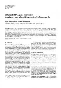
Twenty-day-old Sprague-Dawley rats were treated with VAD diet for 2 mo. Symptoms of vitamin A deficiency (hairlessness, weight loss, or cessation of weight gain and discharges from eyes) were observed in the treated groups at the end of 2 mo. Testes from the PVA-VAD rats were fixed, sectioned, and hematoxylin/eosin stained (Fig. 1). On Day 0 (n 5 3), all the rat testes collected contained mainly type A spermatogonia and few preleptotene spermatocytes, indicating the arrest of spermatogenesis after VAD treatment. Spermatogenesis resumed after retinol treatment followed by consumption of normal diet. We observed the presence of type B spermatogonia and the presence of zygotene primary spermatocytes on Days 6 and 15, respectively. On Day 25, pachytene spermatocytes were the major germ cell population in the seminiferous tubule and spermatids started to appear. Elongated spermatids were formed on Day 35 at stages VI–VII. On Day 43, spermatogenesis was at stages I–II, with differentiation of the spermatozoa close to completion. This developmental schedule was essentially similar to that of the postnatal rat testis (Fig. 1) in this study as well as in a previous report [24]. However, some differences between the PVA-VAD and postnatal testis were noted. Gonocytes were present in the center of the seminiferous tubules of Day 1 postnatal testis but not in PVA-VAD testis (Fig. 1). Spermatogenesis appeared to be proceeding slightly faster in PVA-VAD testis than that in postnatal testis. While round spermatids were fairly abundant in PVA-VAD testis on Day 25, they were rare in the Day 25 postnatal testis. Moreover, the number of elongated
spermatozoa in PVA-VAD testis on Days 35 and 43 was much more than that in postnatal testis at similar day. Array Analysis
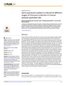
RNA from the PVA-VAD rat testis was used to hybridize the rat cDNA expression arrays and to identify differences in the expression pattern of 1185 known genes. Among the 1185 genes, 1075 of them showed weak hybridization signal (less than twofold different from the background threshold value) after 3 days of exposure. The remaining 110 (9%) genes were either constantly expressed (90 and 79 genes on Days 15 and 35, respectively) or differentially expressed with or greater twofold changes (20 and 31 genes on Days 15 and 35, respectively) in their expression levels when compared with Day 0 (control, Fig. 2). There were 10 and 9 upregulated and 10 and 21 downregulated genes on Days 15 and 35, respectively (Tables 2 and 3). Among them, glutathione S-transferase Yb subunit (GSTM2), cytochrome P450 17 (CYP17), and sulfated glycoprotein 2 (SGP2) were found to be upregulated, while fibroblast growth factor 10 precursor (FGF10), phospholipase C delta 1 (PLC delta-1), cAMP-dependent protein kinase inhibitor (pKib), and cathepsin L (Cath L) were found to be downregulated on both days. It was noted that two housekeeping genes (polyubiquitin and 40S ribosomal protein S29) were found to be upregulated on Days 15 and 35. Semiquantitative RT-PCR on PVA-VAD and Postnatal Rat Testis
To confirm differential expression of these genes in PVA-VAD rat testis as determined by cDNA array, and to find out whether similar expression patterns of genes occur in postnatal rat testis, we adopted semi-quantitative RTPCR to analyze two upregulated (Cyp17 and SGP2) and two downregulated genes (pKib and Cath L) that are related to spermatogenesis. RT-PCR analysis confirmed the expression patterns of the selected genes in PVA-VAD with
1013
VAD AND POSTNATAL RAT TESTIS GENE EXPRESSION TABLE 3. Genes up-/downregulated in expression from Day 0 to Day 35 PVA-VAD rat testis. Array position
Ratio
Upregulated A01c 2.2
GenBank no. M10072
A14e
3.7
J02592
C02j C03i C06j C08k C12l G11 G47
2.3 2.3 3.0 3.2 3.5 2.6 2.2
M64797 X14209 M21208 M22642 M64723 D16554 X59051
Downregulated A04h 0.46
L33413
A12c A12m B08b
0.41 0.31 0.28
X06942 U09793 D12771
B10b C14k
0.38 0.33
J02701 U69278
D01m D05a D14h D14l E06k
0.25 0.50 0.41 0.40 0.41
X77797 U94708 M36589 D79215 M91590
E09b E11c E13b F01j F02g F03a F03e F04g F07m F09g F10a
0.39 0.43 0.23 0.38 0.50 0.32 0.43 0.40 0.33 0.33 0.32
M18332 U28356 L35921 M20637 D83538 M80633 J04563 M64092 L05175 Y00697 D10755
Name of gene/protein
Gene classification
Leukocyte common antigen precursor (LCA); CD45 antigen; PTPRC Glutathione S-transferase Yb subunit; GST subunit 4 mu (GSTM2) Testis fructose-6-phosphate 2-kinase Cytochrome c oxidase, subunit IV, mitochondrial Cytochrome P450 17 (CYP17); P450C17; CYPXVII Cytosolic thymidine kinase (TK1) Clusterin (CLU); TRPM2; sulfated glycoprotein 2 (SGP2) Polyubiquitin 40S ribosomal protein S29 (RPS29) Advanced glycosylation end product-specific receptor precursor; RAGE A-raf proto-oncogene c-K-ras 2b proto-oncogene; transforming G-protein p21 Fibroblast ADP/ATP carrier protein; ADP/ATP translocase 2; ANT2 Sodium/potassium-transporting ATPase beta 1 subunit (ATP1B1) Rek4 Eph-related receptor tyrosine kinase; ephrin type-A receptor 3 Interleukin 8 receptor Prostaglandin E2 receptor EP2 subtype; prostanoid EP2 receptor Beta-nerve growth factor precursor (beta-NGF) Fibroblast growth factor 10 precursor (FGF10) Beta-arrestin 2 (ARRB2) Protein kinase C zeta type (PKC-zeta) HEP; LC-PTP protein-tyrosine phosphatase; HEPTP GTP-binding protein G(i)/G(s)/G(o) gamma-9 subunit; Ggamma8 Phospholipase C delta 1 (PLC delta-1); PLC-III PI4-K; phosphatidylinositol 4-kinase (230 kDa) Adenylyl cyclase 4 DPDE4; cAMP-dependent 39,59-cyclic phosphodiesterase 4B PKI-beta; cAMP-dependent protein kinase inhibitor (testis form) Granzyme M precursor (GZMM); MET-ASE; RNK-MET-1 Cathepsin L Proteasome iota subunit; 27-kDa prosomal protein (PROS27); PSMA6
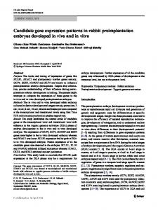
the cDNA array result (Fig. 3). Both CYP17 and SGP2 transcripts were upregulated on Days 9–15 in PVA-VAD samples when compared with Day 0 (Fig. 3A). In general, there was a gradual increase and then decrease of CYP17 transcript in PVA-VAD model. Interestingly, low expression levels of CYP17 transcript were observed in Days 7 and 15 postnatal rat testes. The expression of CYP17 transcript peaked on Days 25 and 35–45 and then dropped to a low level in adult testis. On the contrary, we observed an increase of SGP2 mRNA in PVA-VAD testis from Day 3 to Day 25. A more significant increase of SGP2 mRNA was observed in postnatal testis when compared at birth (Day 1). The expression levels of SGP2 transcripts in PVAVAD at Day 43 and in adult testes were high. In PVA-VAD rat, the pKib mRNA was highly expressed on Days 0, 20–25,and 35–43 but weakly expressed on Days 3–15. RT-PCR confirmed the cDNA array result that the expression level of pKib mRNA on Day 15 was low. In postnatal rat testis, pKib transcript was almost undetectable up to Day 15 (Fig. 3B). An increase in expression of pKib was found between Days 25 and 45. Nevertheless, a strong expression of pKib was observed in adult rat testis. Cath L was found to be downregulated from Day 20 to Day 43 in PVA-VAD rat testis. However, in postnatal and adult rat testis, the expression of Cath L remained fairly
Cell surface antigens Metabolism of cofactors Intracellular transducers, effectors, and modulators Energy metabolism Complex lipid metabolism Nucleotide metabolism Other stress-response proteins Other stress-response proteins Ribosomal proteins Cell surface antigens Oncogene and tumor suppressors Oncogene and tumor suppressors Symporters and antiporters ATPase transporters Growth factors, cytokines, and chemokines Interleukins and interferon receptors Hormone receptors Growth factors, cytokines, and chemokines Growth factors, cytokines, and chemokines Intracellular adaptors and receptor-associated proteins Intracellular kinase network members Intracellular kinase network members G proteins Phospholipases and phosphoinositol kinases Phospholipases and phosphoinositol kinases Adenylate/guanylate cyclases and diesterases Adenylate/guanylate cyclases and diesterases Kinase activators and inhibitors Serine Proteases Cysteine proteases Proteosomal proteins
constant at different time points (Fig. 3B). The housekeeping gene GAPDH was used as loading controls for the present study (data not shown). DISCUSSION
Synchronization of spermatogenesis by treating the animal with a vitamin-A-deficient diet provides an alternative model for the investigation of stage-dependent changes of gene expression during spermatogenesis. In vitamin-A-deficient animals, injection of vitamin A resulted in synchronization of spermatogenesis in over 80% of the seminiferous tubules with 3–5 stages of the spermatogenic cycle [15]. It has been reported that only Sertoli cells, spermatogonia, and a few spermatocytes were found in the seminiferous epithelium of VAD rats [7, 8]. The disappearance of other cell types in the testes is mainly caused by apoptosis or selective loss of the germ cells [3]. The development of PVA-VAD testis of Sprague-Dawley rat in the present study is comparable with an early VAD study on Wistar rat [16] with successive formation of more advanced types of spermatogenic cells in a synchronized manner in the VAD testis, and the majority of the seminiferous tubules were at 3–4 spermatogenic stages (Fig. 1). The first spermatogenic cycle of the PVA-VAD rat con-
1014
LEE ET AL.
FIG. 1. Histological examination of PVAVAD rat testis on Days 0, 6, 15, 25, 35, and 43 and postnatal testis on Days 1, 7, 15, 25, 35, and 45 [See text for description of histology]. A, A Spermatogonia; B, B spermatogonia; ES, elongated spermatid; G, gonocyte; In, intermediate spermatogonia; P, pachytene; Pl, preleptotene spermatocytes; RS, round spermatid; S, spermatid; Ser, Sertoli cell; and Z, zygotene. Original magnification 3200.
tains germ cells at fewer spermatogenic stages. This provides a simplified model to delineate the regulation of spermatogenesis. Another model for studying the spermatogenesis is the first spermatogenic cycle of the postnatal animals. Similar to the VAD model, the developmental stages of the germ cells are limited and spermatogenesis is fairly synchronized. However, the amount of testicular samples obtained from the VAD model far exceeds that of the postnatal rat, as the former are collected at an age of over 80 days. Thus, more experiments and analyses can be performed using the VAD model. The spermatogenic event in the PVA-VAD testis seemed to proceed slightly faster than that in the postnatal testis such that the histology of Day 43 PVA-VAD testis was comparable with that of Day 45 postnatal rat. Besides, the slightly earlier appearance of round spermatids on Day 25 and the presence of more abundant elongated spermatozoa on Day 35 in PVA-VAD testis supported the above obserFIG. 2. Selected cDNA array images of the expression pattern of PVA-VAD rat testis on Days 0 and 35. Differential hybridization of two identical Atlas rat cDNA expression arrays was shown. A region of three arrays is shown to illustrate downregulation of cathepsin L (arrowhead) on PVA-VAD rat testis. Each array contains 1176 known genes divided into six regions, with 9 putative housekeeping genes located at the bottom row. A complete list of gene names and their locations is available at Clontech’s Web page (see Materials and Methods). Each PVA-VAD sample was hybridized to three membranes to obtain the mean values for analysis.
vation. The exact reason for the faster spermatogenesis in PVA-VAD rat is unclear. This could be an effect of vitamin A treatment on spermatogenesis. Another possibility is the different physiological status of the somatic cells in the two models. This was reflected in part by their differential gene expression levels between PVA-VAD and postnatal rat testis at Day 0 and Day 1, respectively (Fig. 3). Sertoli cells and Leydig cells of rat undergo proliferation and differentiation in the postnatal period [25, 26]. These events are partially completed when the rats are started on a vitaminA-deficient diet. Thus, the developmental stages of these somatic cells in the PVA-VAD model are more advanced than those in the postnatal model and may explain the more efficient spermatogenesis. In this study, the gene expression of PVA-VAD testis on Days 15 and 35 after vitamin A administration were studied using the rat cDNA expression array. We found that 9% (110/1176) of the genes were highly and differentially ex-
VAD AND POSTNATAL RAT TESTIS GENE EXPRESSION
1015
FIG. 3. Semiquantatitive RT-PCR analysis of the selected clones from the cDNA expression array. The mRNA samples from PVA-VAD rat testis on Days 0, 3–7, 9–15, 20–25, and 35–43, and postnatal rat testis on Days 1, 7, 15, 25, 35–45, and adult (A), were amplified by gene-specific primers (Table 1). The PCR products were resolved in 2.5% Nuseive 3:1 agarose gel. A) CYP17: cytochrome P450 17 (M21208) and SGP2: sulfated glycoprotein 2 (M64723). B) pKib: testis form of cAMPdependent protein kinase inhibitor (M64092) and Cath L: cathepsin L (Y00697). The signal intensities were normalized with a housekeeping gene glyceraldehyde 3-phosphate dehydrogenase (GAPDH) for comparisons. Each bar represented mean 6 SD for three individual experiments. Asterisks denote significant difference (P , 0.05) from the controls (PVAVAD, and Postnatal, at Day 0 and Day 1, respectively).
pressed in the PVA-VAD rat testis (Tables 2 and 3). To confirm the differential expression of these genes in PVAVAD rat testis as determined by cDNA expression array and to compare the expression pattern of genes with that in postnatal rat testis, we selected two upregulated (CypP17 and SGP2) and two downregulated (pKib and Cath L) genes that are related to spermatogenesis for further study. Semiquantitative RT-PCR was used to detect the changes in steady-state mRNA levels produced by genes in the two models tested. The biosynthesis of testosterone requires the activities of cytochrome P450 17 (CYP17). The Leydig cell of the testis is the only cell in the male that has the capacity to synthesize testosterone from cholesterol. Testosterone is critical during fetal development for male sexual differentiation and postnatally for initiation and maintenance of spermato-
genesis and the expression of the male secondary sex characteristics [27]. Coincidentally, a high expression of CYP17 transcript was found in PVA-VAD testis, presumably produced from the mature Leydig cells of the testis. It was noted that a substantial increase in CYP17 transcripts on Days 25–40 in postnatal rat testis confirms the role of this gene on sexual differentiation (Fig. 3A). Differential screening of testis cDNA libraries from VAD rats before and after (6 h later) administration of alltrans retinoic acid (ATRA) have been successfully used to isolate genes coding for cytochrome c oxidase (COX) and sulfated glycoprotein 2 (SGP2), which are upregulated (3.9fold) and downregulated (70%) in PVA-VAD testis, respectively [28]. It is speculated that the increase in COX expression is due to an increase in energy demand of testicular cells in PVA-VAD testis, while the downregulation
1016
LEE ET AL.
of SGP2 is the direct effect of ATRA treatment on the Sgp2 promoter [28]. However, this study only addressed the short-term changes (up to 30 h) of these transcripts in rat testis. In the present study, we detected both upregulation of the COX by 2.3-fold during spermiogenesis (Table 3) and SGP2 by 2.1- and 3.5-fold in early meiosis and in spermiogenesis, respectively (Tables 2 and 3), in the PVA-VAD rat testis. Northern blot analysis demonstrated that SGP2 transcript increased to a detectable level between Days 7 and 14 of postnatal rat testis and the relative level of expression did not change significantly after Day 14 [29]. In agreement with the previous study, we demonstrated that SGP2 mRNA increases dramatically from Day 1 to Day 15 in postnatal rat testes (P , 0.05) and maintains a fairly constant level after Day 15 (Fig. 3A), as determined by a more sensitive RT-PCR technique. The protein kinase inhibitor (pKi) family includes three genes (a, b, and g), coding small, heat-stable inhibitors of the cAMP-dependent kinase PKA. The pKib isoform, also known as testis pKi, is highly expressed in the testis, while the pKia isoform is highly expressed in skeletal muscle, heart, cerebral cortex, and cerebellum [30]. In postnatal rat testis, pKib mRNA is almost undetectable before Day 25 (P , 0.05). There was a gradual increase of pKib transcript from Day 25 onward. Similar patterns had previously been reported [31]. Compared with those on Day 0, the pKib mRNA level in PVA-VAD testis decreased (P , 0.05), when the spermatogonia were undergoing mitotic division and in early meiosis. The relationship between decrease in pKib expression and mitosis of spermatogonia is unclear. The level of pKib increased thereafter, reaching peak expression when spermatids were formed. In line with this observation, the association of pKib mRNA expression in situ with spermatid formation has been reported [31]. In postnatal rats, the testicular mRNA levels of Cath L did not change very much and they agree with a previous report [32]. The expression pattern of Cath L in the PVAVAD rat was similar but showed a decreasing trend with development; its expression in PVA-VAD testis in meiosis and spermiogenesis was comparatively low (P , 0.05 to control). In adult rat testis, Cath L expression in Sertoli cells was regulated by germ cells in a stage-specific manner [33]. Using isolated seminiferous tubules, it was demonstrated that the transcription of the Cath L gene by Sertoli cells was sevenfold higher at stages VI–VII than at stages IX–XII [34]. A more recent study using laser-capture microdissection and real-time polymerase chain reaction confirmed the high expression of Cath L at stages VII–VIII and a decrease at stages IX–XIII [35]. Although there are variable reports on the expression of Cath L at stages I–V [35, 36], it is generally accepted that the expression of the gene is highest at around stages VI–VIII. In postnatal rat, Cath L expression remained fairly constant. It has been suggested that the change in Cath L expression in the rat testis is related to spermatid development [32, 33]. Apart from the four genes discussed above, the expression of a number of other genes in the cDNA expression array changes during spermatogenesis. Glutathione S-transferase, which was increased by 3.3-fold at Day 15 and 3.7fold at Day 35, is involved in testicular eicosanoid biosynthesis for prostaglandin D production [37]. In the developing testis, prostaglandin D may act as a paracrine factor to induce Sertoli cell differentiation [38]. On the other hand, both prostaglandin E2 receptor (EP2 subtype, 0.50fold) and adenylyl cyclase (0.32-fold) were downregulated at PVA-VAD testis at Day 35. Coincidentally, PGE2 recep-
tors, EP2 and EP4, are coupled to adenylyl cyclase pathway to generate cAMP in the bovine endometrium during the estrous cycle [39]. Whether a similar regulatory mechanism was involved in rat spermatogenesis needs further investigation. One obvious difference between the PVA-VAD and postnatal rat model is the gene expression of Day 0 and Day 1 testis, respectively. The expression of SGP2 and pKib were high in PVA-VAD testis but were very low in the postnatal testes. This difference is likely due to the more advanced development of the somatic cells in the former. The chronological age of the cells in PVA-VAD rat testis is that of a mature animal. The Sertoli cells and Leydig cells proliferate and differentiate with changes in function and morphology during the postnatal period [25, 26]. In conclusion, substantial differences in the gene expression exist between the PVA-VAD and postnatal rat model on the first cycle of spermatogenesis. This discrepancy could be caused by either the presence of arrested but mature somatic cells in the PVA-VAD testis that may contribute to a unique gene expression pattern in this model or by the direct effect of retinol on spermatogenesis. Therefore, caution is needed on interpreting the gene expression data using the PVA-VAD and postnatal rat models on studying spermatogenesis in rat testes. REFERENCES 1. Jegou B, Pineau C, Dupaix A. Paracrine control of testis function. In: Wang C (ed.), Male Reproductive Function. London: Kluwer Academic Publishers; 1999:41–64. 2. Napoli JL. Retinoic acid biosynthesis and metabolism. FASEB J 1996; 10:993–1001. 3. Akmal KM, Dufour JM, Vo M, Higginson S, Kim KH. Ligand-dependent regulation of retinoic acid receptor alpha in rat testis: in vivo response to depletion and repletion of vitamin A. Endocrinology 1998; 139:1239–1248. 4. White JC, Highland M, Kaiser M, Clagett-Dame M. Vitamin A deficiency results in the dose-dependent acquisition of anterior character and shortening of the caudal hindbrain of the rat embryo. Dev Biol 2000; 220:263–284. 5. Wolbach BS, Howe PR. Tissue changes following deprivation of fatsoluble A vitamin. J Exp Med 1925; 42:753–777. 6. Mason KE. Differences in testes injury and repair after vitamin Adeficiency, vitamin E deficiency and inanition. Am J Anat 1933; 52: 153–239. 7. Mitranond V, Sobhon P, Tosukhowong P, Chindaduangrat W. Cytological changes in the testes of vitamin-A-deficient rats. I. Quantitation of germinal cells in the seminiferous tubules. Acta Anat (Basel) 1979; 103:159–168. 8. Unni E, Rao MR, Ganguly J. Histological and ultrastructural studies on the effect of vitamin A depletion and subsequent repletion with vitamin A on germ cells and Sertoli cells in rat testis. Indian J Exp Biol 1983; 21:180–192. 9. Morales C, Griswold MD. Retinol-induced stage synchronization in seminiferous tubules of the rat. Endocrinology 1987; 121:432–434. 10. van Pelt AMM, de Rooij DG. The origin of the synchronization of the seminiferous epithelium in vitamin-A-deficient rats after vitaminA replacement. Biol Reprod 1990; 42:677–682. 11. van Pelt AM, de Rooij DG. Retinoic acid is able to reinitiate spermatogenesis in vitamin-A-deficient rats and high replicate doses support the full development of spermatogenic cells. Endocrinology 1991; 128:697–704. 12. Griswold MD, Bishop PD, Kim KH, Ping R, Siiteri JE, Morales C. Function of vitamin A in normal and synchronized seminiferous tubules. Ann N Y Acad Sci 1989; 564:154–172. 13. Ismail N, Morales C, Clermont Y. Role of spermatogonia in the stagesynchronization of the seminiferous epithelium in vitamin-A-deficient rats. Am J Anat 1990; 188:57–63. 14. Wang Z, Kim KH. Vitamin-A-deficient testis germ cells are arrested at the end of S phase of the cell cycle: a molecular study of the origin of synchronous spermatogenesis in regenerated seminiferous tubules. Biol Reprod 1993; 48:1157–1165.
VAD AND POSTNATAL RAT TESTIS GENE EXPRESSION 15. Bartlett JM, Weinbauer GF, Nieschlag E. Quantitative analysis of germ cell numbers and relation to intratesticular testosterone following vitamin A-induced synchronization of spermatogenesis in the rat. J Endocrinol 1989; 123:403–412. 16. Huang HFS, Hembree WC. Spermatogenic response to vitamin A in vitamin A deficient rats. Biol Reprod 1979; 21:891–904. 17. Liang P, Pardee AB. Differential display of eukaryotic messenger RNA by means of the polymerase chain reaction. Science 1992; 257: 967–971. 18. Zhao N, Hashida H, Takahashi N, Misumi Y, Sakaki Y. High-density cDNA filter analysis: a novel approach for large-scale, quantitative analysis of gene expression. Gene 1995; 156:207–213. 19. Catalano RD, Vlad M, Kennedy RC. Differential display to identify and isolate novel genes expressed during spermatogenesis. Mol Hum Reprod 1997; 3:215–221. 20. Yu Z, Guo R, Ge Y, Ma J, Guan J, Li S, Sun X, Xue S, Han D. Gene expression profiles in different stages of mouse spermatogenic cells during spermatogenesis. Biol Reprod 2003; 69:37–47. 21. Luk JM, Mok BW, Shum CK, Yeung WS, Tam PC, Tse JY, Chow JF, Woo J, Kam K, Lee KF. Identification of novel genes expressed during spermatogenesis in stage-synchronized rat testes by differential display. Biochem Biophys Res Commun 2003; 307:782–790. 22. Chow JFC, Lee KF, Chan STH, Yeung WSB. Quantification of transforming growth factor b1 (TGF b1) mRNA expression in mouse preimplantation embryos and determination of TGFb receptors (Type I and Type II) expression in mouse embryos and reproductive tract. Mol Hum Reprod 2001; 7:1047–1056. 23. Lee KF, Chow JFC, Xu JS, Chan STH, Ip SM, Yeung WSB. A comparative study of gene expression in murine embryos developed in vivo, cultured in vitro, and cocultured with human oviductal cells using messenger ribonucleic acid differential display. Biol Reprod 2001; 64:910–917. 24. Malkow M, Fisher Y, Don J. Developmental schedule of the postnatal rat testis determined by flow cytometry. Biol Reprod 1998; 59:84–92. 25. Sharpe RM, McKinnell C, Kivlin C, Fisher JS. Proliferation and functional maturation of Sertoli cells, and their relevance to disorders of testis function in adulthood. Reproduction 2003; 125:769–784. 26. Mendis-Handagama SM, Ariyaratne HB. Differentiation of the adult Leydig cell population in the postnatal testis. Biol Reprod 2001; 65: 660–671. 27. Payne AH, Youngblood GL. Regulation of expression of steroidogenic enzymes in Leydig cells. Biol Reprod 1995; 52:217–225. 28. Gaemers IC, van Pelt AMM, Themmen APN, de Rooij DG. Isolation and characterization of all-trans-retinoic acid-responsive genes in the rat testis. Mol Reprod Dev 1998; 50:1–6.
1017
29. Zakeri Z, Curto M, Hoover D, Wightman K, Engelhardt J, Smith FF, Kierszenbaum AL, Gleeson T, Tenniswood M. Developmental expression of the S35-S45/SGP-2/TRPM-2 gene in rat testis and epididymis. Mol Reprod Dev 1992; 33:373–384. 30. van Patten SM, Howard P, Walsh DA, Maurer RA. The alpha- and beta-isoforms of the inhibitor protein of the 39,59-cyclic adenosine monophosphate-dependent protein kinase: characteristics and tissueand developmental-specific expression. Mol Endocrinol 1992; 6: 2114–2122. 31. van Patten SM, Donaldson LF, McGuinness MP, Kumar P, Alizadeh A, Griswold MD, Walsh DA. Specific testicular cellular localization and hormonal regulation of the PKIa and PKIb isoforms of the inhibitor protein of the cAMP-dependent protein kinase. J Biol Chem 1997; 272:20021–20029. 32. Mathur PP, Grima J, Mo MY, Zhu LJ, Aravindan GR, Calcagno K, O’Bryan M, Chung S, Mruk D, Lee WM, Silvestrini B, Cheng CY. Differential expression of multiple cathepsin mRNAs in the rat testis during maturation and following lonidamine induced tissue restructuring. Biochem Mol Biol Int 1997; 42:217–233. 33. Wright W, Zabludoff SD, Penttila TL, Parvinen M. Germ cell-Sertoli cell interactions: regulation by germ cells of the stage-specific expression of CP-2/cathepsin l mRNA by Sertoli cells. Dev Genet 1995; 16:104–113. 34. Zabludoff SD, Charron M, DeCerbo JN, Simukova N, Wright WW. Male germ cells regulate transcription of the cathepsin L gene by rat Sertoli cells. Endocrinology 2001; 142:2318–2327. 35. Sluka P, O’Donnell L, Stanton PG. Stage-specific expression of genes associated with rat spermatogenesis: characterization by laser-capture microdissection and real-time polymerase chain reaction. Biol Reprod 2002; 67:820–828. 36. Kim GH, Wright WW. A comparison of the effects of testicular maturation and aging on the stage-specific expression of CP-2/cathepsin L messenger ribonucleic acid by Sertoli cells of the brown Norway rat. Biol Reprod 1997; 57:1467–1477. 37. Anuradha D, Reddy KV, Kumar TC, Neeraja S, Reddy PR, Reddanna P. Purification and characterization of rat testicular glutathione S-transferases: role in the synthesis of eicosanoids. Asian J Androl 2000; 2: 277–282. 38. Adams IR, McLaren A. Sexually dimorphic development of mouse primordial germ cells: switching from oogenesis to spermatogenesis. Development 2002; 129:1155–1164. 39. Arosh JA, Banu SK, Chapdelaine P, Emond V, Kim JJ, MacLaren LA, Fortier MA. Molecular cloning and characterization of bovine prostaglandin E2 receptors EP2 and EP4: expression and regulation in endometrium and myometrium during the estrous cycle and early pregnancy. Endocrinology 2003; 144:3076–3091.