www.nature.com/scientificreports
OPEN
received: 30 September 2015 accepted: 18 March 2016 Published: 07 April 2016
Differential coding of uncertain reward in rat insular and orbitofrontal cortex Suhyun Jo1,2 & Min Whan Jung1,2,3 Anterior insular and orbitofrontal cortex (AIC and OFC, respectively) are known to play important roles in decision making under risk. However, risk-related AIC neural activity has not been investigated and it is controversial whether the rodent OFC conveys genuine risk signals. To address these issues, we examined AIC and OFC neuronal activity in rats responding to five distinct auditory cues predicting water reward with different probabilities. Both structures conveyed significant neural signals for reward, value and risk, with value and risk signals conjunctively coded. However, value signals were stronger and appeared earlier in the OFC, and many risk-coding OFC neurons responded only to the cue predicting certain (100%) reward. Also, AIC neurons tended to increase their activity for a prolonged time following a negative outcome and according to previously expected value. These results show that both the AIC and OFC convey neural signals related to reward uncertainty, but in different ways. The OFC might play an important role in encoding certain reward-biased, risk-modulated subjective value, whereas the AIC might convey prolonged negative outcome and disappointment signals. Humans and animals often choose a safe over a risky option even when the safe option has a lower expected value, indicating that subjective value of an expected outcome is affected by risk1–3. Neural systems associated with decision making under risk are not completely understood, but studies so far suggest anterior insular cortex (AIC) and orbitofrontal cortex (OFC) as two key structures in this process. Numerous brain imaging studies have found activation of the AIC4–10 and OFC4,11,12 in association with risk. Lesion studies in humans11,13,14 and animals15–18 have also found altered choice behavior during decision making under risk following lesions to the AIC or OFC. In studies comparing AIC and OFC lesion effects in the same behavioral task, distinct lesion effects were observed, suggesting distinct roles of the AIC and OFC in risky decision making. In humans, damages in the OFC were associated with increased betting regardless of the odds of winning, whereas patients with damages in the AIC failed to adjust their bets by the odds of winning13. In rats, OFC and AIC inactivation increased and decreased risk preference, respectively16. Lesion effects are not consistent across studies, however. For example, contrary to the above finding16, OFC lesions promoted preference for certain reward in rats15,17. Compared to brain imaging and lesion studies, progress has been relatively slow in revealing neural correlates of risky decision making in the AIC and OFC. Single neuron recording studies have found risk-coding neurons in the monkey OFC19,20. However, in the rat OFC, neural correlates of risk have not been clearly demonstrated. OFC neurons in rats have been found to encode ‘decision confidence’ before choice outcome was revealed21 and to show differential responses to large uncertain vs. small certain rewards in risk-preferring, but not in risk-neutral animals22. However, few OFC neurons signaled risk information in rats responding to olfactory cues predicting reward with different probabilities23, and OFC neural activity seemingly responsive to risk has been proposed to represent ‘acquired salience24’ rather than risk25. In the AIC, there has been no neurophysiological study investigating risk-related neural activity so far. Thus, although there exists a substantial body of evidence for the involvement of the AIC and OFC in decision making under risk, their specific roles and the nature of information processed in each brain structure are unclear. The following two issues are therefore important to understand how the AIC and OFC contribute to decision making under risk: 1) determining whether and how OFC neurons in rats convey neural signals related to risk, and 2) examining neuronal activity related to risk in the AIC. Further, it would be desirable to compare AIC and 1
Center for Synaptic Brain Dysfunctions, Institute for Basic Science, Daejeon 305-701, Korea. 2Neuroscience Graduate Program, Ajou University School of Medicine, Suwon 443-721, Korea. 3Department of Biological Sciences, Korea Advanced Institute of Science and Technology, Daejeon 305-701, Korea. Correspondence and requests for materials should be addressed to M.W.J. (email:
[email protected]) Scientific Reports | 6:24085 | DOI: 10.1038/srep24085
1
www.nature.com/scientificreports/
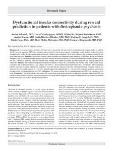
Figure 1. Animal behavior and recording sites. (a) Behavioral apparatus. N, nose poke hole; W, water-delivery nozzle. One of five different auditory cues, signaling five different reward probabilities (0, 25, 50, 75 and 100%), was delivered for 1 s during the animal’s nose poke (minimum 1 s to complete a trial). A blue LED (the small circle above the water-delivery nozzle) was turned on at nose poke exit, and the animal’s arrival at the reward port triggered probabilistic water delivery. (b) Behavioral performance. The proportion of complete trials (left), nose poke duration (middle), and reaction time (the time interval between nose poke exit and water-port entry; right) as a function of reward probability. The lines were determined with linear regression analyses. Error bars, SEM. (c) Single units were recorded from the AIC and lateral OFC. The photomicrographs are coronal sections of the rat brain showing marking lesions (arrows) in the AIC (left) and OFC (right). (d) The diagram is a coronal section view of the rat brain at 3.2 mm anterior to bregma. Each circle represents one recording site that was determined based on histology and electrode advancement history. Different colors represent recordings from different rats. One to five units were recorded simultaneously from each site. Modified with permission from Paxinos and Watson, 1998.
OFC neural activity in the same subject in the same behavioral paradigm. In the present study, to address these issues, we compared single unit activity in the AIC and OFC in rats responding to five distinct cues predicting reward with different probabilities. We found both similarities and differences in neural signals related to uncertain reward between the AIC and OFC, which might underlie their distinct contributions to decision making under risk.
Results
Behavior. Three rats were trained in a probabilistic Pavlovian appetitive conditioning task in an open cham-
ber (Fig. 1a). The animal’s nose poke following trial onset (signaled by a blue LED inside the nose poke hole) triggered one of five auditory tones that lasted for 1 s. The five different auditory tones (1.2, 2, 5, 9 and 14 KHz) were associated with five different reward probabilities (0, 25, 50, 75 and 100%) with cue-reward probability combinations varied across rats. The animals were required to maintain nose poke ≥ 1 s (i.e., until the offset of the auditory cue). A blue LED at the water port was turned on at the time of nose poke exit, and the animal’s arrival at the water port triggered water delivery (30 μl) with a given probability. In incomplete trials (nose poke duration