Diffusion- and Perfusion-Weighted MRI Patterns in Borderzone Infarcts Claudia J. Chaves, MD; Brian Silver, MD; Gottfried Schlaug, MD; John Dashe, MD; Louis R. Caplan, MD; Steve Warach, MD, PhD
Downloaded from http://stroke.ahajournals.org/ by guest on April 26, 2018
Background and Purpose—The pathophysiology of borderzone infarcts is not well understood. We investigated whether combined diffusion-weighted imaging (DWI) and perfusion-weighted imaging (PWI) could identify pathophysiologically meaningful categories of borderzone infarcts. Methods—Seventeen patients with borderzone infarcts were identified from the Beth Israel Deaconess Medical Center Stroke Database. All patients had DWI and PWI, the majority of them within the first 24 hours of symptom onset. Results—Three patterns of perfusion abnormalities were associated with the diffusion lesions: 1, normal perfusion (5 patients); 2, localized perfusion deficits matching the area of restricted diffusion (5 patients); and 3, extensive perfusion deficits involving 1 or more vascular territories (7 patients). All but 1 patient with pattern 1 had transient peri-infarct hypotension as the presumed stroke mechanism. Two patients with pattern 2 had cardiac or aortic embolic sources; none had large-artery disease or arterial hypotension. Reperfusion was detected in all patients with this pattern who submitted to a follow-up study. All patients with pattern 3 had severe stenosis or occlusion of a large artery: the internal carotid, anterior cerebral, or middle cerebral. Conclusions—We postulate that the perfusion abnormality varies according to the mechanism of the borderzone infarction. Transient perfusion deficits occurring with hypotension in the absence of significant large-artery disease may not be revealed by PWI. Embolism may cause some cases of small borderzone perfusion deficits. Critical large-artery disease may cause large territorial perfusion deficits and predispose to borderzone infarction. (Stroke. 2000;31:1090-1096.) Key Words: cerebral infarction 䡲 magnetic resonance imaging, diffusion-weighted 䡲 magnetic resonance imaging, perfusion-weighted
B
orderzone infarcts are ischemic lesions that occur at junctions between 2 or 3 arterial territories. Approximately 10% of all brain infarcts are located in borderzone regions.1 Their pathogenesis is controversial and may involve various mechanisms, such as systemic hypotension, critical carotid stenosis, or occlusion and microemboli.2 Delineation of the stroke mechanisms is important in order to guide specific treatment. Two new MR techniques, diffusion-weighted imaging (DWI) and perfusion-weighted imaging (PWI), are able to show areas of ischemic neuronal injury and hypoperfusion, respectively, within minutes of cerebral ischemia in animal models.3–7 DWI detects changes in the self-diffusion of water molecules associated with ischemic injury from the first minutes of critical ischemia. PWI can be used to calculate maps of the mean transit time and the relative cerebral blood flow and volume, thus identifying hypoperfused brain tissue.8 The superiority of these 2 techniques over conventional MRI in the detection of early abnormalities in patients with acute
stroke9 –11 and in prediction of stroke outcome12- 16 have been well established. We investigated the contributions of DWI and PWI to the pathophysiological mechanisms underlying borderzone infarcts.
Subjects and Methods Patients We retrospectively identified 17 patients with borderzone infarcts selected from a total of 331 patients in the Beth Israel Deaconess Medical Center (BIDMC) Stroke Database, collected from 1995 to 1998. The patients selected met the following inclusion criteria: detailed clinical information, including vascular risk factors, complete neurological examination, hospital course, and outcome; documentation of the borderzone infarction by MRI, including DWI and PWI, according to the BIDMC stroke protocol; and investigation of the stroke mechanism by cardiac studies, carotid and vertebral artery ultrasound, MR angiography (MRA) of the extracranial and intracranial arteries, or cerebral angiogram. There were 17 patients, 11 men and 6 women, ranging in age from 56 to 91 years, average 73.8 years. Fourteen patients were Caucasian,
Received November 22, 1999; final revision received February 16, 2000; accepted February 16, 2000. From the Stroke Division, Department of Neurology, Beth Israel Deaconess Medical Center (C.J.C., G.S., L.R.C.), and the Stroke Division, Department of Neurology, New England Medical Center Hospital (J.D.), Boston, Mass; the Department of Clinical Neurological Sciences, London Health Sciences Center, London, Ontario, Canada (B.S.); and the National Institute of Neurological Disorders and Stroke, Stroke Branch (S.W.), Bethesda, Md. Correspondence to Claudia J. Chaves, MD, Stroke Division, Dana 710, Beth Israel Deaconess Medical Center, 330 Brookline Ave, Boston, MA 02115. E-mail
[email protected] © 2000 American Heart Association, Inc. Stroke is available at http://www.strokeaha.org
1090
Chaves et al
DWI and PWI Patterns in Borderzone Infarcts
1091
Downloaded from http://stroke.ahajournals.org/ by guest on April 26, 2018
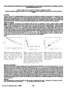
Figure 1. MRI obtained 4 days after symptom onset. DWI showed a left anterior and posterior borderzone infarcts; the rMTT map was normal; MRA showed diffuse intracranial atherosclerotic disease but no area of significant stenosis.
2 African American, and 1 Hispanic. MRAs of the intracranial arteries were obtained in 16 patients; the intracranial vessels of the other patient were studied with a cerebral angiogram. All patients’ extracranial carotid arteries were studied at least by 1 of the following tests: MRA of the neck in 9, duplex of the extracranial
arteries in 8, and cerebral angiograms in 3. Cardiac investigations (ECG and transthoracic echocardiogram) were performed in all patients. Borderzone regions can vary considerably in different patients because of collateral flow.17 In this study, the topography of the
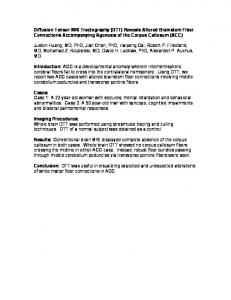
Figure 2. First MRI (TP-1) obtained 8 hours after symptom onset: DWI showed right anterior and posterior borderzone infarcts (arrow) and a small right ACA cortical infarct (notched arrow); analysis of PWI showed prolonged rMTT matching the areas of restricted diffusion; MRA was normal. Follow-up MRI (TP-2) performed 88 days later showed a left anterior borderzone infarct on T2-weighted imaging and normalization of rMTT map.
1092
Stroke
May 2000
Downloaded from http://stroke.ahajournals.org/ by guest on April 26, 2018
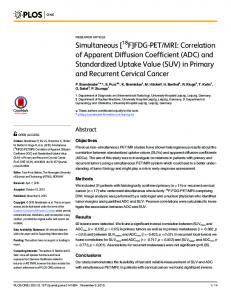
Figure 3. First MRI (TP-1), obtained 5 hours after symptom onset: DWI showed a right anterior borderzone infarct; analysis of the PWI showed prolonged rMTT involving the entire ACA territory; MRA showed right ACA occlusion. Follow-up MRI (TP-2) performed 83 days later showed extension of the area of stroke involving almost the entire ACA territory and persistence of prolonged rMTT.
borderzone infarcts on DWI was assessed according to the mapping guidelines described by Damasio.18 Borderzone infarcts were classified as anterior borderzone, when the infarct occurred between the anterior cerebral artery (ACA) and middle cerebral artery (MCA) territories; posterior borderzone, between the MCA and posterior cerebral artery (PCA) and sometimes the ACA territories; and internal borderzone, between the deep and superficial perforators of the MCA. Determination of the presumed stroke mechanism followed the Harvard Stroke Registry criteria.19 Large-artery disease was defined in the extracranial vessels (carotid artery) as stenosis ⱖ70% and in the intracranial vessels as stenosis ⱖ50%.
MRI Studies The MRI stroke protocol used in our institution include: DWI, Proton density, FLAIR, T2*-weighted, TI-weighted, T2-weighted imaging, PWI and MRA of the intracranial vessels; all of them performed on a 1.5-T MR whole-body system (Siemens AG) with echo planar imaging capability. MRAs of the extracranial vessels were performed using two-dimensional and three-dimensional time of flight (TOF) techniques.
Diffusion-Weighted Imaging For DWI, 2 b values (O and 1000 s/mm2) were used. Other DWI parameters included the following: echo time of 118 ms, matrix size of 128⫻l28, field of view of 260⫻260, and 7-mm slice thickness, with a set of 18 axial slices covering the whole brain. The MR diffusion sequence at b⫽1000 s/mm2 was run 3 times, with diffusion gradients applied to each of the x, y, and z directions. To minimize the effects of diffusion anisotropy, an average of the 3 diffusion directions was calculated to give the trace of the diffusion tensor.
Perfusion-Weighted Imaging The PWI was performed through dynamic first-pass bolus tracking of gadolinium diethylenetriamine pentaacetic acid (0.1 mg/kg) with an echo-planar gradient-echo with an echo time of 60 ms and repetition time of 2 seconds. The dynamic perfusion series were
processed on a pixel-by-pixel basis to produce maps reflecting the relative mean transit time (rMTT), which gave the most distinct boundary between regions of normal perfusion and those with abnormal perfusion.8 DWI and PWI acquired in the acute phase were analyzed without knowledge of patients’ clinical symptoms or the presumed stroke mechanism. Because acute ischemic lesions have slower diffusion, as measured by the apparent diffusion coefficient (ADC) of water, they were identified as areas of hyperintensity on DWI. Hypoperfusion on PWI was identified as an area of increased signal intensity in the rMTT map, and its size was visually assessed and classified as normal; localized perfusion deficit; and extensive perfusion deficit involving 1 or more vascular territories. Examples of these lesions are depicted in Figures 1, 2, and 3, respectively.
Results The following 3 patterns of perfusion abnormality were identified: pattern 1, patients with normal perfusion; pattern 2, patients with localized perfusion deficits matching the area of restricted diffusion; and pattern 3, patients with extensive perfusion deficits involving 1 or more vascular territories. Perfusion defects were larger than DWI defects in this group of patients.
Pattern 1: Normal Perfusion There were 5 patients with this pattern (Figure 1, Table 1). Two of these patients had bilateral borderzone infarcts. The other 3 patients had anterior, posterior, and anterior and posterior borderzone infarcts, respectively. Transient severe systemic arterial hypotension, with systolic blood pressure varying from cardiac arrest to 70 mm Hg, was documented in 4 patients at the time of the initial symptoms and was secondary to cardiac ischemic disease in 2 patients, aortic dissection in 1, and occurred during a
Chaves et al TABLE 1.
Patient
DWI and PWI Patterns in Borderzone Infarcts
1093
Pattern 1: Normal Perfusion
Clinical Presentation
Interval Between Symptom Onset and DWI
DWI
Vascular Studies (MRA/Duplex,Angio)
Cardiac Studies
Downloaded from http://stroke.ahajournals.org/ by guest on April 26, 2018
1
Arterial hypotension during R carotid endarterectomy followed by L hemiparesis
8 hs
R posterior borderzone infarct
70% stenosis RICA
Normal
2
Four episodes of L upper extremity weakness after lunch in the setting of arterial hypotension
2:30 hs
R anterior borderzone infarct
Normal
EF 20%, severe hypokinesis of L ventricle
3
Aphasia and R hemiparesis after plasma exchange for TTP
16:30 hs
R internal and bilateral posterior borderzone infarcts
Normal
Normal
4
Hypotensive arrest due to aortic dissection, followed by R hemiparesis
7 days
Bilateral posterior and internal borderzone infarcts, R internal borderzone infarct
Normal
Normal
5
R hemiparesis and fluent aphasia during an episode of tachycardia, diaphoresis, and arterial hypotension
4 days
L anterior and posterior borderzone infarcts
Generalized intracranial and extracranial atherosclerotic disease
EF 30–35%, akinesis of posterolateral wall
TTP indicates thrombotic thrombocytopenic purpura; Duplex, carotid duplex; Angio, cerebral angiography; and EF, ejection fraction.
carotid endarterectomy in another patient. Two of these patients also had large-artery disease, with generalized intracranial and extracranial atherosclerosis in one and unilateral 70% internal carotid artery (ICA) stenosis in the other. The 1 patient in this group who had no evidence of arterial hypotension, cardiac disease, or large-artery disease was admitted with thrombotic thrombocytopenic purpura and developed bilateral borderzone infarcts shortly after plasma exchange. MR studies, including DWI and PWI, were performed within 24 hours of symptom onset in 3 patients (in 2 of them within the first 8 hours). The other 2 patients had their first MRI 4 and 7 days, respectively, after the initial symptoms. Follow-up DWI and PWI obtained 2 days later in 1 patient did not show any new abnormality.
Pattern 2: Localized Perfusion Deficits Matching the Area of Restricted Diffusion There were 5 patients with this pattern (Figure 2, Table 2). Two patients had ipsilateral anterior and posterior borderTABLE 2.
Patient
zone infarcts. These 2 patients also had small strokes in other vascular territories, one in the ACA and the other the MCA territories. The other 3 patients had anterior, posterior, and internal borderzone infarcts. None of these 5 patients had intracranial or extracranial arterial disease. One patient had an intra-atrial thrombus and another had mitral regurgitation with dilated left atrium. Three other patients had normal cardiac studies; however, 1 of them had aortic atherosclerotic plaques detected during cardiac catheterization. Arterial hypotension was not detected in any of these patients on admission. The infarct mechanism was considered to be embolic in 2 patients, from cardiac sources in one and intra-arterial (related to cardiac catheterization) in the other. In the other 3 patients, the mechanism of the infarct was undetermined. All patients had their first scan within 24 hours of symptom onset, 3 of them within the first 8 hours. Follow-up MRI was obtained in 4 patients between days 3 and 88 (mean 46 days, median 45 days); all of them had complete reperfusion on PWI.
Pattern 2: Localized Perfusion Deficit Matching the Area of Diffusion Abnormality
Clinical Presentation
Interval Between Symptom Onset and DWI
DWI
Vascular Studies (MRA/Duplex/Angio)
Cardiac Studies
1
Sudden onset L motor, sensory, and visual neglect while at work
7:34 h
R anterior and posterior borderzone infarcts, small R ACA cortical infarct
Normal
Normal
2
Sudden onset of mild L hemiparesis and L visual field defect while at work
10 h
R posterior borderzone infarct
Normal
Mitral regurgitation, L atrial enlargement, normal ejection fraction
3
L hemiparesis after cardiac catheterism
4h
R internal borderzone infarct
Normal
Normal heart, aortic atherosclerotic plaques
4
L hemiparesis and slurred speech after waking up
15 h
R anterior borderzone infarct
Normal
Normal
5
Sudden onset of diaphoresis, aphasia, R hemiparesis
2h
R anterior and posterior borderzone infarcts, L frontal cortical infarct
Normal
Intra-atrial thrombus
Duplex indicates carotid duplex; Angio, cerebral angiography.
1094
Stroke
TABLE 3.
Pattern 3: Extensive Perfusion Deficits Involving 1 or More Vascular Territories
Patient
May 2000
Clinical Presentation
Interval Between Symptom Onset and DWI
DWI
Vascular Studies (MRA/duplex,angio)
Cardiac Studies
Downloaded from http://stroke.ahajournals.org/ by guest on April 26, 2018
1
R weakness and aphasia; worsening of symptoms after episode of arterial hypotension
3d
L anterior borderzone infarct
L carotid siphon occlusion
Normal
2
L leg weakness, preceded by 2 similar TIAs
2d
R anterior borderzone infarct
R carotid stenosis (siphon)
Normal
3
After standing up R hemiparesis and field cut in the setting of arterial hypotension
6:47 h
L anterior and posterior borderzone infarcts
L ICA stenosis (supraclinoid portion)
Transient AF
4
Acute onset of L gaze deviation, R hemiparesis, and hemineglect, preceded by 2 similar TIAs
4:17 h
L anterior borderzone infarct
Tight stenosis L ICA bifurcation
EF 45%
5
Acute onset of L hemiparesis while sitting; worsening of symptoms after episode of arterial hypotension
4:45 h
R anterior borderzone infarct
R ACA occlusion
Normal
6
Found by nurse with mild R hemiparesis and aphasia
16:23 h
L internal borderzone infarct
L MCA stenosis
EF 40%
7
Found with R hemiparesis and aphasia; worsening of symptoms after episode of arterial hypotension
8:00 h
L internal borderzone infarct
L ICA occlusion
First-degree AV block
TIA indicates transient ischemic attacks; AF, atrial fibrillation; Duplex, carotid duplex; Angio, cerebral angiography; and EF, ejection fraction.
Pattern 3: Patients With Extensive Perfusion Deficits Involving 1 or More Vascular Territories There were 7 patients with this pattern (Figure 3, Table 3). Four patients had anterior borderzone infarcts, 2 had internal borderzone infarcts, and 1 had unilateral anterior and posterior borderzone infarcts. The mechanism of the stroke was considered to be large-artery disease with hemodynamic ischemia in all 7 patients. Ipsilateral ICA occlusion or tight stenosis was detected in 2 and 3 patients, respectively, and stenosis or occlusion of the ACA or MCA in 1 patient each. Cardiac evaluation was normal in 3 patients and showed moderate decrease in the ejection fraction in 2 patients. One patient had first-degree atrioventricular block, but no significant arrhythmia was observed during Holter monitoring; another patient had transient atrial fibrillation associated with arterial hypotension (90/60 mm Hg) relative to baseline at stroke onset. Transient arterial hypotension with subsequent worsening of the clinical symptoms occurred in 3 patients during the hospitalization. None of the patients had an abnormally high hematocrit. Five patients had their first MRI study within the first 24 hours of symptom onset, 4 of them within the first 8 hours. In the other 2 patients, the first imaging study was done 2 and 3 days after onset of the symptoms, respectively. Follow-up MRI was obtained in 6 patients, between days 3 and 85 (mean 36 days, median 21 days), and showed persistent hypoperfusion in 5 of them.
Discussion Although autopsy results20 –22 and CT studies23–25 have characterized borderzone infarcts, their pathogenesis is still not well understood. Systemic hypotension, microembolism, and critical carotid artery stenosis or occlusion with hypoperfusion are considered to be the probable mechanisms.
In our series, 3 different patterns of perfusion abnormalities were identified among the 17 patients studied: normal perfusion, localized perfusion deficit matching the area of diffusion abnormality, and extensive perfusion deficit involving 1 or more vascular territories exceeding the focal DWI defects. Normal perfusion studies were found in 5 patients. In 4 patients, severe transitory systemic hypotension was well documented at the time of the symptom onset and posited as the probable mechanism of these strokes. The transient nature of the arterial hypotension most likely led to temporary decrease in the cerebral blood flow, which was no longer present by the time the scan was performed in these patients. In the other patient, a hypercoagulable state secondary to thrombotic thrombocytopenic purpura26 probably associated with decreased intravascular volume related to plasma exchange was the inferred mechanism. The role of severe arterial hypotension causing borderzone strokes has been described20,27 and confirmed by experimental studies with primates.28 Adams et al20 identified borderzone infarcts in brains of patients who died shortly after cardiac surgery and ascribed it to 1 or more episodes of abrupt arterial hypotension. Similar findings were reported by Howard et al27 in patients with borderzone infarcts detected by CT scan; in most of his patients the strokes were clearly associated with episodes of severe arterial hypotension, some of them during cardiac surgery. In the majority of these patients, bilateral symmetrical borderzone infarcts were the rule. In our series, transitory arterial hypotension was also associated with unilateral strokes in 3 patients; however, 2 of these patients had large-artery disease: ipsilateral 70% carotid artery stenosis in one and generalized diffuse atherosclerosis in the other. Only 1 patient had unilateral stroke and completely normal intracranial and extracranial arteries.
Chaves et al
Downloaded from http://stroke.ahajournals.org/ by guest on April 26, 2018
The second pattern we observed was localized perfusion deficit that matched the areas of restricted diffusion. None of these patients had intracranial or extracranial arterial occlusive disease or a history of systemic hypotension. In 2 patients, embolism from cardiac source or associated with cardiac catheterism was the presumed mechanism of the stroke. In the other 3 patients, the mechanism was undetermined despite the investigation. The possibility of intra-arterial embolism from an aortic source cannot be excluded in these patients, because this artery was not systematically studied in all of them. Also, the presence of reperfusion in all patients with this pattern, who were submitted to follow-up PWI studies, favors the embolic mechanism. Tumor emboli and showers of cholesterol crystals have been described as causes of borderzone infarcts.21,22 However, the association between platelet emboli and borderzone strokes has been more controversial. Thrombotic occlusions of the pial vessels over conventional watershed infarcts29 –31 were initially thought to be secondary to “stagnation thrombi” due to hypotensive episodes.29,30 More recently, the documentation of occlusion of the leptomeningeal arteries by platelet emboli22 and the detection of intra-arterial emboli by transcranial Doppler monitoring in patients with watershed infarcts32 have provided some evidence for the role of embolism in the pathophysiology of borderzone infarcts. Extensive perfusion deficits involving 1 or more vascular territories was the third pattern observed. All of these patients had severe large-artery disease with hemodynamic ischemia as defined by the Harvard Stroke Registry.19 Several authors23,24,33–35 have postulated the role of hypoperfusion in the pathophysiology of strokes in patients with critical carotid artery stenosis or occlusion. Bogousslavsky and Regli23 studied 51 patients with symptomatic unilateral borderzone infarcts detected by CT and found that the great majority of them had an ipsilateral ICA occlusion or tight stenosis associated with a hemodynamically significant cardiopathy, increased hematocrit, or acute hypotension. Weiller et al33 studied 37 stroke patients with single-photon emission CT scan and transcranial Doppler with CO2 stimulation and observed that the perfusion reserve and vasomotor reactivity were significantly reduced in patients with stroke secondary to carotid artery occlusion. Two different patterns were seen: changes were restricted to the area of the infarct in patients with a territorial stroke and involved a considerably larger area than the infarct in patients with borderzone infarcts, a finding similar to ours. Decreased cerebral blood flow in the anterior and posterior borderzone regions in patients with ICA and MCA occlusions has also been documented by positron emission tomography scan.34,35 Caplan and Hennerici36 have recently postulated the coexistence of hypoperfusion and intra-arterial embolism in patients with borderzone infarcts and carotid artery disease. These authors posited that the reduced perfusion limits the ability of the blood stream to clear (washout) emboli and that the brain borderzones are a favored destination for microemboli that are not cleared. Future investigation with both PWI and transcranial Doppler monitoring for emboli detection will be necessary to clarify this issue.
DWI and PWI Patterns in Borderzone Infarcts
1095
In summary, we propose that the perfusion abnormality detected by PWI varies according to the mechanism of the borderzone infarction. Transient perfusion deficits occurring with hypotension in the absence of a critical large-artery disease is usually accompanied by a normal PWI. Embolism may cause small perfusion deficits in the borderzone territory matching the area of diffusion abnormality. Critical large-artery occlusive disease is usually associated with large territorial perfusion deficits and predisposes to borderzone infarction. Prospective observations are needed to confirm the etiologic significance of these patterns, which may lead to a more rational approach to the management of patients with borderzone infarcts.
References 1. Jorgensen L, Torvik A. Ischaemic cerebrovascular diseases in an autopsy series, part 2: prevalence, location, pathogenesis and clinical course of cerebral infarcts. J Neurol Sci. 1969;9:285–320. 2. Torvik A. The pathogenesis of watershed infarcts in the brain. Stroke. 1984;15:221–223. 3. Moseley ME, Kucharczyk J, Mintorovitch J, Cohen Y, Kurhanewicz J, Derugin N, Asgari H, Norman D. Diffusion-weighted MR imaging of acute stroke: correlation with T2-weighted and magnetic susceptibility enhanced MR imaging in cats. Am J Neuroradiol. 1990;11:423– 429. 4. Minematsu K, Li L, Fisher M, Sotak CH, Davis MA, Fiandaca MS. Diffusion-weighted magnetic resonance imaging: rapid and quantitative detection of focal brain ischemia. Neurology. 1992;42:235–240. 5. Busza AL, Allen KL, King MD, van Bruggen N, Williams SR, Gadian DG. Diffusion-weighted imaging studies of cerebral ischemia in gerbils: potential relevance to energy failure. Stroke. 1992;23:1602–1612. 6. Back T, Hoehn-Berlage M, Kohno K, Hossmann KA. Diffusion nuclear magnetic resonance imaging in experimental stroke: correlation with cerebral metabolites. Stroke. 1994;25:494 –500. 7. Reith W, Hasegawa Y, Latour LL, Dardzinski BJ, Sotak CH, Fisher M. Multislice diffusion mapping for 3-D evolution of cerebral ischemia in a rat stroke model. Neurology. 1995;45:172–177. 8. Schlaug G, Benfield A, Baird AE, Siewert B, Lovblad KO, Parker RA, Edelman RR, Warach S. The ischemic penumbra: operationally defined by diffusion and perfusion MRI. Neurology. 1999; 53:1528 –1537. 9. Warach S, Chien D, Li W, Ronthal M, Edelman RR. Fast magnetic resonance diffusion-weighted imaging of acute human stroke. Neurology. 1992;42:1717–1723. 10. Warach S, Li W, Ronthal M, Edelman RR. Acute cerebral ischemia: evaluation with dynamic contrast-enhanced MR imaging and MRA. Radiology. 1992;182:41– 47. 11. Warach S, Gaa J, Siewert B, Wielopolski P, Edelman R. Acute human stroke studied by whole brain echo planar diffusion-weighted magnetic resonance imaging. Ann Neurol. 1995;37:231–241. 12. Warach S, Dashe JF, Edelman RR. Clinical outcome in ischemic stroke predicted by early diffusion-weighted and perfusion magnetic resonance imaging: a preliminary analysis. J Cereb Blood Flow Metab. 1996;16:53–59. 13. Siewert B, Schlaug G, Edelman R, Warach S. Comparison of EPISTAR and T2*-weighted gadolinium-enhanced perfusion imaging in patients with acute cerebral ischemia. Neurology. 1997;48:673– 679. 14. Schlaug G, Siewert B, Benfield A, Edelman RR, Warach S. Time course of the apparent diffusion coefficient (ADC) abnormality in human stroke. Neurology. 1997;49:113–119. 15. Lovblad K-O, Baird AE, Schlaug G, Benfield A, Siewert B, Voetsch B, Connor A, Burzynski C, Edelman RR, Warach S. Ischemic lesion in acute stroke by diffusion-weighted magnetic resonance imaging correlate with clinical outcome. Ann Neurol. 1997;42:164 –170. 16. Chaves CJ, Silver B, Staroselskaya I, Baird A, Caplan LR, Warach S. Relationship of perfusion-weighted magnetic resonance imaging (MRI) and clinical outcome in patients with ischemic stroke. Stroke. 1999; 30:278. Abstract. 17. Mohr JP. Neurological complications of cardiac valvular disease and cardiac surgery including systemic hypotension. In: Viken P, Bruyn G, eds. Handbook of Clinical Neurology, Vol 38: Neurological Manifestations of Systemic Disease, Part I. Amsterdam, Netherlands: North Holland; 1979:143–171. 18. Damasio H. A computed tomographic guide to the identification of cerebral vascular territories. Arch Neurol. 1983;40:138 –142.
1096
Stroke
May 2000
Downloaded from http://stroke.ahajournals.org/ by guest on April 26, 2018
19. Mohr JP, Caplan LR, Melski JW, Goldstein RJ, Duncan GW, Kistler JP, Pessin MS, Bleich HL. The Harvard Cooperative Stroke Registry. Neurology. 1978;28:754 –762. 20. Adams JH, Brierley JB, Connor RCR, Treip C. The effects of systemic hypotension upon the human brain: clinical and neuropathological observation in 11 cases. Brain. 1966;89:235–268. 21. McKibbin DW, Bulkley BH, Green WR, Gott WL, Hutchins GM. Fatal cerebral atheromatous embolization after cardiac bypass. J Thorac Cardiovasc Surg. 1976;71:741–745. 22. Torvik A, Skullerud K. Watershed infarcts in the brain caused by microemboli. Clin Neuropathol. 1982;1:99 –105. 23. Bogousslavsky J, and Regli F. Unilateral watershed cerebral infarcts. Neurology. 1986;36:373–377. 24. Bogousslavsky J, and Regli F. Borderzone infarctions distal to internal carotid artery occlusion: prognostic implications. Ann Neurol. 1996;20: 346–350. 25. Bladin CF, Chambers BR. Frequency and pathogenesis of hemodynamic stroke. Stroke. 1994;25:2179 –2182. 26. Wade J. Moschcowitz syndrome (thrombotic thrombocytopenic purpura). In: Stroke Syndromes. Bogousslavsky J, Caplan L, eds. New York, NY: Cambridge University Press; 1995:466 – 469. 27. Howard R, Trend P, Russell RWR. Clinical features of ischemia in cerebral arterial border zones after periods of reduced cerebral blood flow. Arch Neurol. 1987;44:934 –940.
28. Brierley JB, Excell BJ. The effect of profound systemic hypotension upon the brain M-rhesus: physiological and pathological observations. Brain. 1966;89:269 –299. 29. Fisher CM. Cerebral thromboangiitis obliterans. Medicine. 1957;36: 169 –209. 30. Romanul FCA, Abramowicz A. Changes in brain and pial vessels in arterial border-zones. Arch Neurol. 1964;11:40 – 65. 29. 31. Torvik A, Jorgensen L. Thrombotic and embolic occlusions of the carotid arteries in an autopsy series, part 2: cerebral lesions and clinical course. J Neurol Sci. 1966;3:410 – 432. 32. Schwartz A, Gass A, Hennerici MG. Is there a need to reclassify acute stroke patients? Cerebrovasc Dis. 1998;8:9 –16 33. Weiller C, Ringelstein EB, Reiche W, Buell U. Clinical and hemodynamic aspects of low-flow infarcts. Stroke. 1991;22:1117–1123 34. Samson Y, Baron JC, Bousser MG, Rey A, Derlon JM, David P, Comoy J. Effects of extra-intracranial arterial bypass on cerebral blood flow and oxygen metabolism in humans. Stroke. 1985;16:609 – 616. 35. Yamauchi H, Fukuyama H, Kimura J, Konishi J, Kameyama M. Hemodynamics in internal carotid artery occlusion examined by positron emission tomography. Stroke. 1990;21:1400 –1406. 36. Caplan LR, Hennerici M. Hypothesis: impaired clearance of emboli (washout) is an important link between hypoperfusion, embolism, and ischemic stroke? Arch Neurology. 1998;55:1475–1482.
Diffusion- and Perfusion-Weighted MRI Patterns in Borderzone Infarcts Claudia J. Chaves, Brian Silver, Gottfried Schlaug, John Dashe, Louis R. Caplan and Steve Warach Downloaded from http://stroke.ahajournals.org/ by guest on April 26, 2018
Stroke. 2000;31:1090-1096 doi: 10.1161/01.STR.31.5.1090 Stroke is published by the American Heart Association, 7272 Greenville Avenue, Dallas, TX 75231 Copyright © 2000 American Heart Association, Inc. All rights reserved. Print ISSN: 0039-2499. Online ISSN: 1524-4628
The online version of this article, along with updated information and services, is located on the World Wide Web at: http://stroke.ahajournals.org/content/31/5/1090
Permissions: Requests for permissions to reproduce figures, tables, or portions of articles originally published in Stroke can be obtained via RightsLink, a service of the Copyright Clearance Center, not the Editorial Office. Once the online version of the published article for which permission is being requested is located, click Request Permissions in the middle column of the Web page under Services. Further information about this process is available in the Permissions and Rights Question and Answer document. Reprints: Information about reprints can be found online at: http://www.lww.com/reprints Subscriptions: Information about subscribing to Stroke is online at: http://stroke.ahajournals.org//subscriptions/