Behavioral Neuroscience 2000, Vol. 114, No. 1,42-63
Copyright 2000 by the American Psychological Association, Inc. 0735-7044/00/$5.00 DOI: 10.1037//0735-7044.114.1.42
Disconnection of the Anterior Cingulate Cortex and Nucleus Accumbens Core Impairs Pavlovian Approach Behavior: Further Evidence for Limbic Cortical-Ventral Striatopallidal Systems John A. Parkinson, Pamela J. Willoughby, Trevor W. Robbins, and Barry J. Everitt University of Cambridge
The nucleus accumbens (NAcc) has been implicated in a variety of forms of reward-related learning, reflecting its anatomical connections with limbic cortical structures. After confirming that excitotoxic lesions of the anterior cingulate cortex (Ant Cing) impaired the acquisition of appetitive Pavlovian conditioning in an autoshaping procedure, the effects of excitotoxic lesions to the NAcc core or shell on autoshaping were also assessed. Only selective core lesions impaired Pavlovian approach. A subsequent experiment studied the effects of a disconnection of the Ant Cing and NAcc core, using an asymmetric lesion procedure, to determine whether these structures interact sequentially as part of a limbic corticostriatal system. Such lesioned rats were also significantly impaired relative to controls at autoshaping. These results demonstrate that the NAcc core and Ant Cing are "nodes" of a corticostriatal circuit involved in stimulus-reward learning.
Lesions of the central nucleus of the amygdala and of the anterior cingulate cortex (Ant Cing) impair discriminated Pavlovian approach behavior, as measured in an autoshaping procedure (Bussey, Everitt, & Robbins, 1997; Parkinson, Robbins, & Everitt, 1996; Parkinson, Robbins and Everitt, in press). Such deficits may be interpreted in terms of a more general involvement of these structures in stimulus-reward learning (Bussey, Everitt, & Robbins, 1997; Bussey, Muir, Everitt, & Robbins, 1996; Everitt & Robbins, 1992). These critical components of the limbic forebrain have converging projections onto the ventral striatum and may process informational and affective aspects of emotion and motivation that are in turn integrated at the level of the nucleus accumbens (NAcc), where relevant behavioral responses are gated. Indeed, Pennartz, da Silva, and Groenewegen (1994) have suggested that "distinct neuronal ensembles" operate within and through the NAcc and are functionally related to the nature of their limbic-cortical afferents. This neuronal ensemble hypothesis gains further support from observations of NAcc neurons during reward-related
learning and behavior. For example, conditioned stimuli (CSs) that have, through association, become predictive of primary rewards (unconditional stimuli [USs]) produce significant changes in extracellular dopamine (DA) concentrations in the NAcc, as measured by voltammetry (DiCiano, Blaha, & Phillips, 1998a, 1998b). Similarly, DA neurons that project to the NAcc (and NAcc neurons themselves) also produce increases in single-unit neuronal firing in response to a reward-related CS, as measured by electrophysiology (Schultz, Apicella, Scarnati, & Ljungberg, 1992; Schultz, Dayan, & Montague, 1997). Further, lesions or dopaminergic manipulations of the NAcc have significant effects on Pavlovian conditioning (Balleine & Killcross, 1994; Parkinson, Olmstead, Burns, Robbins, & Everitt, 1999) and on tests of conditioned place preference (Carr & White, 1983; Everitt, Morris, O'Brien, & Robbins, 1991; White, Packard, & Hiroi, 1991). Although the acquisition and performance of conditioned place preferences (CPP) are probably dependent on other learning mechanisms rather than simply Pavlovian conditioning (e.g., involving an instrumental locomotor response), Everitt et al. (1991) found that excitotoxic lesions of the NAcc impaired the acquisition of CPP when animals were given access to sucrose in a distinct environment. Further, lesions of the basolateral amygdala (BLA) also impaired such a place preference and, more importantly, a disconnection lesion of the BLA and NAcc, produced by making unilateral lesions of the two structures, contralateral to each other also abolished a CPP to sucrose presentation. Such evidence strongly implies a serial and functional connection between the BLA and NAcc in reward-related processes. In fact, there is evidence of a role for the NAcc in spatial learning (Annett, McGregor, & Robbins, 1989; MaldonadoIrizarry & Kelley, 1995; Ploeger, Spruijt, & Cools, 1994; Riedel, Harrington, Hall, & MacPhail, 1997), Pavlovian conditioning (Balleine & Killcross, 1994; Kelley, Smith-
John A. Parkinson, Pamela J. Willoughby, Trevor W. Robbins, and Barry J. Everitt, Department of Experimental Psychology, University of Cambridge, Cambridge, United Kingdom. John A. Parkinson is now at the Department of Anatomy, University of Cambridge; and Pamela J. Willoughby is now at the Department of Psychology, York University, York, United Kingdom. This research was supported by Medical Research Council (MRC) Programme Grant G9537855, an MRC cooperative in Brain, Behavior, and Neuropsychiatry, and by a Wellcome Trust Programme Grant. John A. Parkinson was supported by a Biotechnology and Biological Sciences Research Council research studentship and an Don Khye Beng Ch'hia Tsio Scholarship. Correspondence concerning this article should be addressed to Barry J. Everitt, Department of Experimental Psychology, University of Cambridge, Downing Street, Cambridge CB2 3EB United Kingdom. Electronic mail may be sent to
[email protected].
42
CORTICOSTRIATAL LESIONS AND AUTOSHAPING
Roe, & Holahan, 1997; Schultz et al., 1992; Williams, Rolls, Leonard, & Stern, 1993; Young, Ahier, Upton, Joseph, & Gray, 1998), instrumental behavior (Kelley et al., 1997), and even the formation of declarative memories (Setlow, 1997). These diverse functions attributed to the NAcc may reflect several factors. First, different behavioral procedures use different measures of learning. Thus, it is difficult to compare or be precise about the nature of the learning or the response being assessed. Second, different manipulations of the NAcc often yield contradictory findings. Important assumptions apply to different neural manipulations that in some cases preclude simple comparisons between techniques. Finally, there are potentially different contributions to behavior from subregions within the NAcc, most notably the core and shell. Notwithstanding, the NAcc may be involved in more fundamental processes that influence, to some extent, all of the above forms of learning, such as Pavlovian associative mechanisms (Rescorla & Solomon, 1967). An important modification of earlier conceptions of striatal connectivity was made by questioning the view that the striatum operated as a funnel to integrate diverse cortical projections onto a focused set of output targets (Alexander & Crutcher, 1990; Alexander, Delong, & Strick, 1986) and arguing instead that there are segregated circuits through the striatum that operate in parallel. These circuits originate in specific areas of the cortex and project back to a restricted set of cortical areas via the striatum, pallidum, and thalamus (but see Joel & Weiner, 1994). In this anatomical system, each structure within a "loop" would perform a distinct function, and structures at the same anatomical level of separate loops (e.g., the putamen in the "motor" loop and the NAcc in the "anterior cingulate" loop) would perform qualitatively similar functions, though for potentially different purposes. The ventral striatum, and in particular the NAcc, conforms to this corticostriatal circuitry in that there are topographic differences between afferent and efferent projections between the ventral and dorsal striatum and, more importantly, between the core and shell of the NAcc (Zahm & Brog, 1992). Further, the examination of a circuit of neural structures is likely to provide a more precise understanding of the functional interactions between areas within the brain. Thus, not only by studying the effects of selective bilateral lesions to several areas on a well-defined behavioral task, but also by using disconnection techniques (between structures), it is possible to provide evidence for functionally segregated circuits (Everitt et al., 1991; Floresco, Seamans, & Phillips, 1997; Gaffan & Harrison, 1987). The present experiments therefore examined the effects of anatomically connected structures on an autoshaping task that measured appetitive Pavlovian conditioning, unconfounded by instrumental learning mechanisms, in an attempt to define neural circuits subserving appetitive Pavlovian learning. The apparatus, procedure and theory have been discussed in more detail elsewhere (Bussey, Everitt, & Robbins, 1997). Briefly, autoshaping was first described when pigeons came to approach and peck a key light, which had been presented in temporal contiguity with a food
43
reward, before approaching and eating the food (P. L. Brown & Jenkins, 1968). Regardless of whether there was a necessary contingency between the subjects' behavior and presentation of reward, the pigeons reliably approached and pecked the key light stimulus. This approach behavior has subsequently been interpreted as a Pavlovian sign-tracking response (Hearst & Jenkins, 1974) because it lacks the flexibility and goal-directed nature of instrumental actions (Williams & Williams, 1969), is produced by the association of the stimulus and reward, and has been observed in rats, monkeys, and humans, both children and adults (Boakes, 1977; Sidman & Fletcher, 1968; Wilcove & Miller, 1974; Zeiler, 1972). Previously it has been shown that autoshaping is sensitive to Ant Cing lesions (Bussey, Everitt, & Robbins, 1997). The present study therefore attempted to replicate and extend these findings by also studying the effects of selective excitotoxic lesions to a major limbic efferent target, namely the NAcc, on the autoshaping task in an attempt to define the neural structures that are involved in preparatory Pavlovian mechanisms and may underlie emotional learning.
EXPERIMENT 1: LESIONS OF THE ANT CING AND THE NACC CORE AND SHELL Initially, the effects of lesions to the Ant Cing were studied on the autoshaping task to replicate the previous findings of Bussey, Everitt, and Robbins (1997). Further, the effects of selective lesions of the NAcc core or shell were studied on acquisition of the autoshaping task, as the ventral striatum is a major target of cingulate projections. Any dissociable effects found after NAcc lesions could then be compared with those seen after lesions of limbic-cortical structures that are major sources of afferents to the NAcc, such as the cingulate cortex, amygdala, and hippocampal formation (Bussey, Everitt, & Robbins, 1997; Parkinson et al., 1996; Parkinson et al., in press). Method Subjects Subjects were 60 male Lister hooded rats (Olac, Bicester, UK) weighing between 310 and 410 g at the time of surgery. They were housed in pairs in a temperature-controlled room (minimum 22 °C) under a 12-hr reversed light-dark cycle. All testing took place during the dark phase. The rats were food deprived to 85% of their free-feeding body weight for the duration of the experiment. Subject groups and numbers were as follows (final group numbers in parentheses): Ant Cing-lesioned n = 10 (10); Ant Cing sham n = 7(7),NAcccore-lesionedn = 12 (9); NAcc core sham n = 11 (11); NAcc shell-lesioned n = 12 (6); and NAcc shell sham n = 8 (8). All rats used in this study were treated in accordance with the UK 1986 Animals (Scientific Procedures) Act (Project License PPL 80/00668).
Surgical Procedure Surgery was performed under Avertin anesthesia. Infusions were made with a 1-nl syringe (26 gauge, code 1BR-OC-7/0.47, SGE, Baton Rouge, LA) with a custom-made glass micropipette attached
44
PARKINSON, WILLOUGHBY, ROBBINS, AND EVERITT
to the end. The pipettes (Intracel, Royston, UK) initially measured 1.2 mm in external diameter, 0.69 mm in internal diameter, and 10 cm in length, and were pulled with a Stoelting App-1 All Purpose Puller (Model 52500, Stoelting, IL) giving a final tip diameter of 50-100 urn (outer diameter) and a length of 12 mm. Micropipettes were attached to the syringe with Araldite epoxy resin (Ciba, UK) to ensure an airtight seal. Infusion coordinates (taken from Paxinos & Watson, 1998) and excitotoxin volumes are shown in Table 1. All subjects were given injections of glucose-saline (5-10 ml ip) after surgery to aid recovery. Behavioral testing began 7-10 days after surgery.
Behavioral Procedure The autoshaping paradigm examines the acquisition of Pavlovian approach behavior (P. L. Brown & Jenkins, 1968; Williams & Williams, 1969) by presenting a visual stimulus followed by the delivery of food. Over training, animals develop the conditioned response (CR) of approaching the food-predicting CS before returning to the food hopper to retrieve the primary reward. This preparatory approach behavior is deemed to be under the control of Pavlovian mechanisms because it lacks the bidirectionality of conditioning of which operant responses are capable (Williams & Williams) and has been described as a form of sign tracking (Tomie, Brooks, & Zito, 1989). The procedure and apparatus have been described in detail elsewhere (Bussey, Everitt, & Robbins, 1997) but will be outlined below.
Apparatus The apparatus consisted of a testing chamber attached to a video display unit (VDU) within a sound-attenuating box (fitted with an extractor fan) and is an adaptation of the apparatus described by Bussey, Muir, and Robbins (1994). The inner chamber (48 cm long X 30 cm high X 30 cm wide) consisted of a metal frame and Perspex walls, with an aluminum floor. A 3-W houselight was attached to the center of the ceiling. A food magazine hopper was attached to a pellet dispenser (Campden Instruments, Loughborough, UK) outside the sound-attenuating box and allowed the controlled delivery of sucrose pellets (Noyes, Lancaster, NH). Pressure-sensitive floor pads (14 X 10 cm) were attached to microswitches, enabling the measurement of approaches to stimuli (10 X 28-cm white triangles presented on the far left or far right sides of the VDU). The floor pad at the rear of the chamber triggered successive trials by detecting when a subject was
equidistant from both stimulus locations. The apparatus was controlled and monitored by a BBC Master series microcomputer that used programs written in BASIC (Bussey, Everitt, & Robbins, 1997) and allowed the presentation of computer graphic stimuli on the VDU monitor while measuring rats' motor responses through the use of a touch-sensitive screen and the pressure-sensitive floor pads.
Procedure Pretraining. On the 1st day, rats were given one 15-min habituation session in the chamber. The houselight was switched on, and rats were allowed access to food pellets (dustless sucrose pellets; 45 mg sucrose, Noyes), which were delivered into the magazine under a variable time (VT) 40-s schedule. Subjects were observed during this session to ensure that they were successfully retrieving and consuming pellets. Acquisition. On the 2nd day of testing, rats were trained to associate stimuli with the sucrose pellet reward. Stimuli were presented on the VDU for 10 s, followed by the delivery of a sucrose pellet into the magazine. One stimulus was designated the CS+ and was always followed immediately by reward. Another was designated the CS— and was never followed by reward. The two stimuli differed only in the side of the screen on which they were presented. Half the rats received a right-sided CS+ and the other half received a left-sided CS+. Stimuli were presented under a VT 40-s schedule, and training consisted of a total of 100 trials (2 consecutive days of 50 trials per day), each consisting of one CS+ stimulus presentation and one CS— stimulus presentation. An approach was recorded if a rat stepped onto the floor panel directly in front of a stimulus; no other approaches were recorded for that stimulus presentation. Stimuli were presented only when the subject was centrally located at the back of the chamber, eliminating chance approaches and allowing the reliable calculation of approach latencies. There was a minimum time of 10 s between CS+ and CS— presentation to reduce interference across trials. There was a maximum of two consecutive presentations of either the CS+ or CS-. Several performance measures were taken, including the number of approaches to both the CS+ and CS— per each block of 10 trials and the mean latency to approach both the CS + and CS— over the course of acquisition training. Omission training. On the 4th testing day, subjects were given an additional block of 50 trials with the same parameters used in the acquisition sessions, except that an approach to the CS+ now
Table 1 Infusion Coordinates and Excitotoxin Volumes for Lesions of the Anterior Cingulate Cortex (Ant Cing) and Nucleus Accumbens (NAcc) Brain area Ant Cing Ant Cing Ant Cing NAcc core NAcc shell NAcc shell NAcc shell
Excitotoxin 0.09 0.09 0.09 0.09 0.06 0.06 0.06
M quinolinic acid M quinolinic acid M quinolinic acid M quinolinic acid M ibotenic acid M ibotenic acid M ibotenic acid
Coordinates
Infusion volume
AP
L
DV
0.5 Ml 0.5 ul 0.5 ul 0.5 ul 0.2 jil 0.1 ul 0.1 ul
+0.8 +0.2 -0.4 + 1.2 + 1.6 + 1.6 + 1.6
±0.5 ±0.5 ±0.5 ±1.8 ±1.1 ±1.1 ±1.1
-2, -3a -2, -2.5 -1.5, -2 -7.1" -7.9" -6.9" -6.4"
Note. Coordinates are in millimeters and are from Paxinos and Watson (1998). The incisor bar was set at 3 mm below the interaural line. "Indicates distance from dura. Indicates distance from skull surface.
CORTICOSTRIATAL LESIONS AND AUTOSHAPING prevented the delivery of a sucrose pellet. This type of omission training has previously been used as a corroborative measure of whether animals approach the stimuli by using Pavlovian conditioning mechanisms (Williams & Williams, 1969).
Statistical Analysis Behavioral results were subjected to a two- or three-factor repeated-measures analysis of variance (ANOVA) with one betweensubjects factor (lesion) and one or two within-subject factors (CS, block) depending on the analysis, using the statistical package SPSS for Windows (Version 6.1.3, 1998) including the post hoc Newman-Keuls test. Where necessary, a post hoc analysis of simple main effects and simple interactions was undertaken with CLR ANOVA for Macintosh (Version 2.0, 1995). Statistical analyses of acquisition data were primarily used to determine the development of discriminated autoshaping behavior and to observe experimental group differences. However, analysis of the omission procedure was undertaken primarily to assess continued discriminated approach within groups; therefore Student's t test was used to analyze omission approach difference scores (number of approaches to the CS + minus number of approaches to the CS—, per block). The homogeneity of variance across groups in repeatedmeasures ANOVAs was assessed by the Mauchly sphericity test. When data sets significantly violated Mauchly test requirements, the Greenhouse-Geisser epsilon correction parameter for degrees of freedom (Geisser & Greenhouse, 1959; Winer, 1971) was used to calculate a more conservative p value for each F ratio. Further, for presentation of descriptive statistics, the standard error of the differences of the means (SED) was used, as it provides a better estimate of the population variance for between-group comparisons. The SED is calculated by using the formula provided in Cochran and Cox (1957).
Histological Procedure and Lesion Analysis Rats were overdosed with sodium pentobarbitone (1.5 ml ip, Euthatal, Rhone MSrieux, UK) and perfused via the ascending aorta with cold phosphate-buffered saline (PBS; 0.01 M, pH 7.4) over 4 min, followed by 4% (wt/vol) paraformaldehyde with 0.2% (wt/vol) saturated picric acid in 0.2 M phosphate buffer for a further 4 min. The brains were then removed and postfixed for 2 hr before being transferred into 20% (wt/vol) sucrose in 0.01 M PBS overnight. Coronal sections (60 |om) were cut on a freezing microtome throughout the full extent of the lesioned area. Every third section was mounted on gelatin-coated glass slides, then stained for Nissl substance with cresyl violet. Mounted sections were immersed for 3 min in descending alcohol concentrations (absolute, 95%, 70%), then rinsed in distilled water before being stained for 2-5 min in cresyl violet (Cresyl Fast Violet, Raymond Lamb, Eastbourne, UK). Once the required intensity of staining was reached, sections were rinsed in distilled water and 70% alcohol, then differentiated in 95% alcohol before a final rinse in absolute alcohol and immersion in Histoclear for 3 min. After this treatment, slides were coverslipped and dried. Sections stained with cresyl violet were then examined to assess the extent and nature of excitotoxin-induced neuronal damage, including gliosis associated with intracerebral infusions of quinolinic or ibotenic acids. Further, this analysis was used to prepare schematic representations and photomicrographs of the lesions used in this study.
45
Results Histological Assessment Lesions of the Ant Cing were similar (though slightly more anterior) to those obtained previously, using the same infusion parameters (Bussey et al., 1996; Bussey, Everitt, & Robbins, 1997; Bussey, Muir, Everitt, & Robbins, 1997). Neuronal loss and gliosis extended from approximately 3 mm anterior to 1 mm posterior to bregma and encompassed both areas Cgl and Cg2. Figure la shows a schematic representation of the lesion, and Figure Ib and Ic show photomicrographs of rostral sections from lesioned rats and sham-lesioned controls. In some cases, there was bilateral damage to the rostrodorsal prelimbic cortex (these subjects were included in the behavioral analysis) and in others, damage to the overlying shoulder region. There was no visible evidence of damage to the cingulum or neuronal loss extending below the corpus callosum into the septum or hippocampal formation. Ultimately, all rats in the Ant Cing lesion group were retained for behavioral analysis. Lesions of the NAcc core selectively destroyed neurons within the core subregion (Groenewegen, Wright, & Beijer, 1996; Zahm & Brog, 1992). In all cases, the rostral pole was spared, as was the mediodorsal NAcc shell. In some cases, gliosis was found in the ventrolateral NAcc shell; these data were discarded for rats with bilateral damage in this area. The data from 3 subjects were removed from further analysis due to damage extending beyond the proposed target area (1 rat with bilateral ventral shell damage and 2 with bilateral damage in the dorsal striatum). Schematic representations and a representative photomicrograph of NAcc core lesions are shown in Figure 2 (a-e). Lesions of the NAcc shell were targeted at the mediodorsal aspect of the shell subregion for both anatomical and theoretical reasons. Data from 5 rats were discarded due to bilateral damage to the septum or globus pallidus or damage to the core subregion. Thus, only rats with very selective lesions of the mediodorsal shell (encompassing the septal pole of the shell) were included. One NAcc shell-lesioned subject died after surgery. Schematic representations based on the atlas of Paxinos and Watson (1998) and a representative photomicrograph of NAcc shell lesions can be seen in Figure 2 (f-h).
Behavioral Results All subjects in the experiment reliably collected and consumed sucrose pellets from the magazine. Final group numbers used for statistical analysis were: Ant Cing lesion, n = 10; Ant Cing sham, n = 7; NAcc core lesion, n = 9; NAcc core sham, n — 11; NAcc shell lesion, n = 6; NAcc shell sham, n = 8. Effects of Ant Cing Lesions Discriminated Approach Approach scores for acquisition (approaches to the CS + and approaches to the CS— across 10 blocks of 10 stimulus
46
PARKINSON, WILLOUGHBY, ROBBINS, AND EVERITT
a
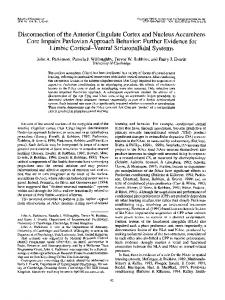
\. ;-^^v;:;'\ Figure 1. a: Schematic representation of lesions to the anterior cingulate cortex. Shaded areas represent the smallest (black) and largest (gray) extent of neuronal damage in a single rat. Coronal sections are from +2.1 to +0.48 mm relative to bregma (Paxinos & Watson, 1998). b-c: Photomicrographs showing cresyl violet-stained coronal sections through the anterior cingulate cortex (approximately +1.7 mm from bregma). b: Sham lesion, c: Anterior cingulate cortex lesion. The lesioned area is indicated by the dotted lines. Cgl = cingulate cortex area 1; Cg2 = cingulate cortex area 2; M2 = secondary motor cortex.
47
CORTICOSTRIATAL LESIONS AND AUTOSHAPING
jm
c
Hi 41
Acbc aca
/
Acbsh
\ - /'
d y
% e V
,/
,
.
I ', I
'v.X,
g Acbc
- 1 r^N i - P^j
o
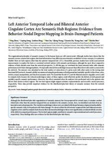
Figure 2. Schematic representations and photomicrographs of excitotoxic lesions to the nucleus accumbens. Shaded areas represent the smallest (black) and largest (gray) extent of neuronal damage in a single rat. Lesions are represented by dotted lines in photomicrographs. Coronal sections are from +2.7 to +0.48 mm relative to bregma (Paxinos & Watson, 1998). a: Nucleus accumbens shell (Acbsh) lesion; b-e: Photomicrographs showing cresyl violet-stained coronal sections through the nucleus accumbens (approximately +1.2 mm from bregma). b: Sham lesion; c: Acbsh lesion; d: high magnification of the sham lesion section shown in b; e: High magnification of the Acbsh lesion section shown in c. f: Schematic representation of excitotoxic lesions to the nucleus accumbens core (Acbc). g-h: Photomicrographs showing cresyl violet-stained coronal sections through the nucleus accumbens (approximately +1.2 mm from bregma). g: Sham lesion; h: Acbc lesion, aca = anterior commissure; icjm = major islands of Calleja; LV = lateral ventricle. presentations) were analyzed for each sham and lesion group and are shown in Figure 3a. Analysis of the number of approaches during the acquisition of autoshaping revealed a main effect of lesion, F(l, 15) = 4.95, p < .05, characterized by an increased number of approaches by the Ant Cinglesioned group relative to sham controls. There was also a main effect of CS, F(l, 15) = 20.16, p < .01, due to a greater number of approaches toward the CS + relative to the CS—. More importantly, there was a Lesion X CS
interaction, F(l, 15) = 8.53, p < .05, and a CS X Block interaction, F(9, 135) = 7.12, p < .01. Post hoc analysis revealed that the former interaction was due to discriminated approach demonstrated by the sham control group (p < .05) but not by the lesioned group. Further, there was no significant difference between the Ant Cing sham CS + approaches and the Ant Cing CS + approaches, but there was a significant difference between the CS — approaches of the two groups, with the Ant Cing group showing a higher
48
PARKINSON, WILLOUGHBY, ROBBINS, AND EVERITT
b
a 10-
10-
9-
9-
8-
8-
7-
7-
6-
6-
5-
5-
4-
4-
3-
3-
2-
2 -
1-
1
0
0
-O-
Ant Cing sham CS+ Ant Cing sham CSAnt Cing CS+ Ant Cing CS-
SED
1 2 3 4 5 6 7 8 9 1 0 1 Blocks of 10 stimulus presentations Figure 3. Acquisition of autoshaping behavior (a) after lesions of the anterior cingulate cortex (Ant Cing and sham control). The performance of these rats was also assessed during an omission session (b). Data points represent the mean number of approach responses made toward the conditioned (CS+) and control (CS—) stimuli per block of 10 trials. SED bar represents 1 standard error of the difference between the means taken from the between-groups component of the Lesion X CS X Block interaction term of the repeated-measures analysis of variance. This is an appropriate index of the variance for computing post hoc tests of significance between the mean values of the groups and is calculated according to the formula provided by Cochran and Cox (1957).
number of CS- approaches (p < .05). The latter interaction revealed a block-dependent development of discriminated approach to the CS + . There were no interactions between lesion and block, F(9, 135) = 0.69, p = .72, or between lesion, CS, and block, F(9,135) = 0.42, p = .78.
Approach Latencies Whereas the number of absolute approaches gives a good indication of learning the association between the CS+ and food reward, measuring the latency to make an approach can offer not only a corroborative indication of the extent of learning, but also an indication of lesion effects on response times and, therefore, an index of locomotor capacity. Thus, the mean latency to approach a stimulus was calculated for all the instances that a rat actually approached (a maximum of 100 approaches per stimulus) during acquisition. There was a significant effect of lesion, F(l, 15) = 5.01, p < .01, on approach latencies (see Figure 4). Specifically, lesioned rats were significantly faster to approach than were sham controls (p < .05). There was also a significant main effect of CS, F(l, 15) = 15.11, p < .01, characterized as faster approaches to the CS+ relative to the CS-. There was no Lesion X CS interaction, F(l, 15) = 0.88,p = .36. Omission Session Analyzing the omission data not only reveals any betweengroup differences, but also can offer insight as to whether learning during the acquisition of autoshaping is due predominantly to Pavlovian mechanisms or other forms of learning (Bussey, Everitt, & Robbins, 1997; Williams & Williams,
1969). Thus, the data were subjected to both a repeatedmeasures ANOVA as well as individual Student's / tests on the difference scores of the lesion and sham groups separately. The difference score measure indicated whether there was discriminated approach behavior (toward the CS+) over the entire omission session. Analysis of omission data revealed a main effect of CS, F(l, 15) = 24.90, p < .001, 6001
500
400
300-
SED 200 t/3
$
CJ
a
3
X in
1 1/3
Figure 4. Mean approach latencies during the acquisition of autoshaping behavior (calculated over the 100 acquisition trials). Open bars represent latencies to approach the conditioned stimulus; filled bars represent latencies to approach the control stimulus. Ant Cing = rats with lesions to the anterior cingulate cortex; Core = rats with lesions to the nucleus accumbens core; Shell = rats with lesions to the nucleus accumbens shell. SED bar represents 1 standard error of the difference of the means.
49
CORTICOSTRIATAL LESIONS AND AUTOSHAPING
and a main effect of block, F(4, 60) = 5.10, p < .01, produced by an overall discriminated approach toward the CS+ during omission, but with a general reduction in approaches over blocks. There were no significant effects of lesion, F(l, 15) = 2.17, p = .16, Lesion X CS, F(l, 15) = 3,62,p = .07; Lesion X Block, F(4, 60) = 0.12, p = .91; or Lesion X CS X Block, F(4, 60) = 0.27, p = .78. Difference scores were obtained, and individual Student's t tests were carried out on each block of approach data to determine exactly when a significant level of discriminated approach behavior was attained by each experimental group. The sham group showed a significant level of discriminated approach throughout the omission session (ps < .05), whereas the Ant Cing group showed a significant level of discriminated approach on Blocks 2 and 3 only (p < .05). In summary, whereas sham controls developed a significant level of discriminated approach toward the CS+ relative to the CS-, rats with Ant Cing lesions were impaired in this behavior. The level of approach to the CS + demonstrated by lesioned rats was no different from that of shams, but the level of CS— approach was significantly higher than that of sham controls. However, toward the end of training, Ant Cing rats did begin to show a greater level of approach toward the CS+ versus the CS-, but this did not reach significance. Both groups approached the CS+ with lower latencies than those for the CS-. Further, the lesioned group responded more quickly in general when approaching either stimuli. Finally, sham controls showed a preserved discriminated level of approach during the entire omission session. Rats with Ant Cing lesions actually exhibited discriminated approach on two of the five blocks even after showing no significant degree of discrimination during the acquisition of
autoshaped responding. Thus, although Ant Cing-lesioned rats were significantly impaired at autoshaping, there was evidence to suggest that they were beginning to learn toward the end of the experiment.
Effects ofNAcc Core Lesions Discriminated Approach As shown in Figure 5a, repeated-measures ANOVAs carried out for NAcc core lesioned rats and sham-lesioned controls revealed a significant main effect of lesion, F(l, 18) = 6.12, p < .05, and CS, F(l, 18) = 47.07, p < .001; aLesion X CS interactionF(l, 18) = 15.03,;? < .001; aCS X Block interaction, F(9,162) = 14.66, p < .001; and a Lesion X CS X Block interaction, F(9, 162) = 5.36, p < .001. There was no main effect of block F(5,87) = 1.43, p = .18, or a Lesion X Block interaction, F(5, 87) = 1.66, p = .12. Newman-Keuls post hoc analysis of simple main effects and interactions revealed that the main effect of lesion was due to significantly lower approach scores in the NAcc core-lesioned group (p < .05), and the main effect of CS revealed lower approach scores to the CS— (p < .05) relative to the CS +. The Lesion X CS interaction revealed a significant difference between CS+ and CS- in the NAcc core shams (p < .05) but not the NAcc core-lesioned group. There was also a significant difference between the CS + scores for the NAcc core shams and NAcc core-lesioned groups (p < .05); but this was not true for the CS(p > .05). Thus, sham subjects showed discriminated approach toward the CS+, relative to the CS-, but corelesioned subjects did not. Post hoc analysis of the CS X Block interaction revealed an increase in discriminated approach over blocks between CS+ and CS— from Block 4
Core sham CS+ Core sham CSCore CS+ Core CS-
SED
1 2 3 4 5
1 7 8 9 10 Blocks of 10 stimulus presentations
Figure 5. Acquisition of autoshaping behavior (a) after lesions of the nucleus accumbens core (and sham control). The performance of these rats was also assessed during an omission session (b). Data points represent the mean number of approach responses made toward the conditioned (CS+) and control (CS—) stimuli per block of 10 trials. SED bar represents 1 standard error of the difference of the means.
50
PARKINSON, WILLOUGHBY, ROBBINS, AND EVERTTT
onward (ps < .05). Because there was a Lesion X CS X Block interaction, separate ANOVAs were calculated for the NAcc core sham and NAcc core-lesioned groups to further analyze their autoshaping behavior. Analysis of the NAcc core sham group alone revealed significant main effects of CS, F(l, 10) = 56.17, p < .001, characterized by a higher level of approach to the CS+, a main effect of block, F(9, 10) = 2.19, p < .05, demonstrating an increase in approaches over blocks, and a CS X Block interaction, F(9, 90) = 24.14, p < .0001, revealing a significant discriminated approach toward the CS+ from Block 4 onward (p < .05) and a significant change in approaches to both the CS+ (increased) and CS- (decreased) over blocks. Analysis of the NAcc core-lesioned group alone revealed no significant effects. However, there was a trend toward a significant main effect of CS (p = .068), characterized by an increased approach toward the CS+.
Approach Latencies An ANOVA revealed a significant main effect of lesion, F(l, 18) = 12.31, p < .005, and a Lesion X CS interaction, F(l, 18) = 13.72, p < .005, but no main effect of CS (see Figure 4). NAcc core shams were significantly faster to approach both the CS+ and CS-. Examination of the Lesion X CS interaction showed that there was a significant difference between approach latencies for the CS+ and CS - in the NAcc core sham group (p < .005) but not in the NAcc core-lesioned group (ns) and that the CS + approach latencies were significantly faster for the NAcc core sham group relative to the NAcc core-lesioned group (p < .005) but with no difference in the CS— approach latencies (p > .05). Thus, NAcc core shams approached the CS4more quickly than the CS- and with shorter latencies than either approach in the NAcc core-lesioned group. There was no significant difference between CS+ and CS— scores in the NAcc core-lesioned group (p > .05). Although rats with sham lesions demonstrated faster approach latencies toward the predictive CS+ than toward the CS—, core-lesioned rats did not show any differences between their CS + and CS — approach latencies.
Omission Session Analysis of approach scores revealed a main effect of CS, F(l, 18) = 35.32, p < .001, and of block, F(3, 46) = 8.81, p < .05, and a Lesion X CS interaction, F(l, 18) = 7.98, p < 0.05, but no main effect of lesion, F(l, 18) = 2.92, p = . 1, or the following interactions: Lesion X Block, F(3,46) = 0.11, p = .98; CS X Block, F(2, 42) = 1.05, p = .39; or Lesion X CS X Block, F(2, 42) = 1.12, p = .35 (see Figure 5b). Newman-Keuls post hoc analysis revealed that the main effect of CS was due to a higher number of approaches to the CS+ (p < .01) relative to the CS-. The main effect of block was due to a general reduction in the number of approaches over blocks, and the Lesion X CS interaction was due to a significant difference between the NAcc core shams' CS+ approaches compared with the NAcc core-
lesioned CS+ approaches (p < .05). Although there was a significant difference between the CS+ and CS- approaches in the NAcc core sham group (p < .01), there was no such difference in the behavior of the NAcc core-lesioned group. Student's t tests of the difference between CS+ and CS— approaches were performed on individual blocks to determine whether NAcc core shams and NAcc corelesioned rats (separately) continued to show a discriminated approach during the omission training. Difference scores were significantly different from zero for all five blocks (ps < .05) for the NAcc core sham group. None of the NAcc core-lesioned groups' scores were significantly different from zero (ps > .05). In summary, NAcc core shamoperated rats continued to show discriminated approach behavior during the omission procedure, although their approach scores did decrease over blocks. NAcc corelesioned rats, in contrast, did not reach a significant level of discrimination.
Food Consumption Test After NAcc Core Lesions Because of the severe impairment in autoshaping after NAcc core lesions, a food consumption test was carried out to determine whether there was a deficit in primary motivation, rather than in Pavlovian approach (see Figure 6). Core-lesioned rats and sham controls were tested under four conditions: They were first given 50 Noyes food pellets (the amount presented in a typical 2-hr autoshaping session), and the time to consume them was recorded. Second, the two groups were given an amount of Rat Chow equivalent to the weight of 50 Noyes pellets (i.e. 2.3 g), and again the time to consume was measured. The third and fourth conditions involved giving rats' free access to either pellets or chow for a 30-min period, and the amount consumed was measured in grams. A one-way ANOVA comparing the time to consume 50 Noyes food pellets by the core-lesioned and sham control groups revealed no significant main effect of lesion, F(l, 18) = 2.05, p = .18. Analysis of the time to consume 2.3 g of laboratory chow revealed slightly (though not significantly) longer times for NAcc core-lesioned rats relative to sham controls, as revealed by the lack of a main effect of lesion, F(l, 18) = 3.16, p = .1. Thus, there was no significant difference in the time it took NAcc core-lesioned subjects to consume the amount of food available in an autoshaping session, relative to sham controls. Analysis of the amount of pellets consumed over a 30-min period revealed no significant effect of lesion, F(l, 18) = 0.02, p = .9. However, core-lesioned rats ate relatively less laboratory chow over a 30-min period, F(l, 18) = 43.36, p < .001. Thus, core-lesioned subjects appeared to have a selective impairment in the consumption, over time, of laboratory chow but showed no significant deficit when consuming Noyes food pellets.
Effects of NAcc Shell Lesions Discriminated Approach As shown Figure 7a, there were no significant effects of lesion on autoshaping performance, F(l, 23) = 1.07, p =
51
CORTICOSTRIATAL LESIONS AND AUTOSHAPING
a 700 -, 600 500 400 300 -
SED
200 100 -
0 sham
core
sham
sham
core
sham
core
Figure 6. A test of food consumption after nucleus accumbens core lesions (core) and sham controls (sham), in a food-deprived state, a: Mean latency to consume 50 reinforcer pellets. Fifty pellets is the total number of reinforcers given in a standard 2-hr autoshaping session (50 trials), b: Mean latency to consume 2.3 g of laboratory chow. This amount of chow is equivalent in weight to 50 pellets, c: Mean weight of reinforcer pellets consumed in 30 min. d: Mean weight of laboratory chow consumed in 30 min. Asterisk indicates a significant main effect of lesion (p < .05). SED bar represents 1 standard error of the difference of the means. .31, nor any interactions between Lesion X CS, F(l, 23) = 2.06,p = .17; Lesion X Block, F(9,207) = 1.04, p = .41; or Lesion X CS X Block, F(9,207) = 0.87, p = .56. There was a main effect of CS, F(l, 23) = 76.66, p < .0001, manifest by increased approach toward the CS+ relative to the CS— (p < .01). There was also a significant CS X Block interaction, F(9, 207) = 27.89, p < .001, but no main effect of block, F(9, 23) = 1.64, p = .12, showing that, although the overall number of approaches made did not significantly change, the nature of those approaches did. Thus, approaches to the CS+ increased over blocks and became significantly higher than approaches to the CS- (which reduced over blocks) from Block 4 onward (p < .001). In summary, NAcc shell shams and NAcc shell-lesioned groups did not significantly differ in their acquisition of autoshaping; both groups developed a significant discriminated approach to the CS + relative to the CS -.
Approach Latencies Analysis of the approach latencies revealed a main effect of CS, F(l, 23) = 19.87, p < .0005, demonstrating that approaches to the CS+ were made with shorter latencies than approaches to the CS— (p < .01; see Figure 4). There were no effects of lesion on approach latency, F(l, 23) = 1.15, p = .29, or Lesion X CS interaction, F(l, 23) = 0.40, p = .53. Thus, both NAcc shell and sham-lesioned rats approached the CS+ with shorter latencies than those for theCS-. Omission Session Figure 7b shows that, consistent with the lack of any effects of the shell lesion in the acquisition of autoshaping, there were no significant effects of lesion for the omission
52
PARKINSON, WILLOUGHBY, ROBBINS, AND EVERTTT
-•-•-V-V
Shell sham CS+ Shell sham CSShell CS+ Shell CS-
;•-•--». SED 1
2
3
4
5
6 7 8 9 1 0 1 2 Blocks of 10 stimulus presentations
3
4
5
Figure 7. Acquisition of autoshaping behavior (a) after lesions of the nucleus accumbens shell (and sham control). The performance of these rats was also assessed during an omission session (b). Data points represent the mean number of approach responses made toward the conditioned (CS+) and control (CS—) stimuli per block of 10 trials. SED bar represents 1 standard error of the difference of the means. session, F(l, 23) = 0.32, p = .58. There also were no significant interactions of Lesion X CS, F(l, 23) = 0.1, p = .99; Lesion X Block, F(4, 92) = 0.28, p = .89; or Lesion X CS X Block, F(4,92) = 0.39, p = .82. However, there was a main effect of CS, F(l, 23) = 35.33,p < .0001, showing the continuing discriminated approach to the CS+ over the omission session. There was also a main effect of block, F(4, 92) = 6.09, p < .001, revealed as a reduction in overall approaches over blocks. There was no significant interaction between CS X Block, F(4,92) = 1.15, p = .34. In summary, both the NAcc shell sham and NAcc shell-lesioned groups continued to show a discriminated approach toward the CS+, and there were no significant differences between these groups. This was further emphasised by the individual t tests on the difference scores, which were significantly different from zero for the NAcc shell shams over all blocks (ps < .05) whereas those for the NAcc shell-lesioned group were significant on all but Block 4 (p < .01 for Blocks 1-3, p