Function, Washington University, Saint Louis, MO 63110. Communicated by Michael I. Posner, August 25, 1989 (receivedfor review June 8, 1989). ABSTRACT.
Proc. Natl. Acad. Sci. USA Vol. 87, pp. 256-259, January 1990 Neurobiology
The anterior cingulate cortex mediates processing selection in the Stroop attentional conflict paradigm (attention/positron emission tomography)
Jose V. PARDO*tt, PATRICIA J. PARDO*t§¶, KEVIN W. JANER§, AND MARCUS E. RAICHLEt¶1I Departments of *Psychiatry, §Psychology, and ¶Neurology and Neurosurgery, IlMallinckrodt Institute of Radiology, and tMcDonnell Center for Higher Brain Function, Washington University, Saint Louis, MO 63110
Communicated by Michael I. Posner, August 25, 1989 (received for review June 8, 1989)
Regional cerebral blood flow, an index of ABSTRACT local neuronal activity, was measured using positron emission tomography (PET) during the performance of the classic Stroop color/word task in eight healthy right-handed subjects. In the first condition of this paradigm, subjects name the color of the words presented on a video monitor. All the words are the color names congruent to the color presented (e.g., the noun "red" displayed in red color). In the second condition, subjects also name the color of the words presented on the monitor. However, during these trials all words are color names incongruent to the color presented (e.g., the noun "red" displayed in green color). The difference in brain activity between these two conditions (i.e., incongruent minus congruent) could reveal brain systems involved in the attentionally mediated resolution of the conflict between the habitual response of reading words vs. the task demands of naming the color of the words-i.e., the Stroop interference effect. The most robust responses occurred in the anterior cingulate cortex. Other responses noted were in the left premotor cortex, left postcentral cortex, left putamen, supplementary motor area, right superior temporal gyrus, and bilateral peristriate cortices. These data provide support for the role of the anterior cingulate cortex in attentional processing through the selection and recruitment of processing centers appropriate for task execution. Furthermore, the extensive distributed network of activated regions suggests that the Stroop interference effect cannot be explained simply in terms of stimulus encoding or response interference.
interference but also help define the brain systems mediating such phenomena. What are the likely anatomical loci and associated cognitive operations involved in attentional conflict paradigms such as the Stroop task? Two general areas are suggested on the basis of data from several sources: the prefrontal cortex and the anterior cingulate. The prefrontal cortex, based on experimental evidence from both monkeys as well as humans (4, 5), has been demonstrated to be important not only for memory buffering to permit "on-line" processing but also for inhibition of "prepotent" habitual, albeit sometimes inappropriate, responses. Lesions of the left prefrontal cortex result in impaired performance in the Stroop task (6). Linguistic tasks requiring the inhibition of reading a noun and the generation of a verb appropriate to the noun result not only in activation of the left prefrontal cortex but also the anterior cingulate (7, 8). Some human diseases, such as schizophrenia, involving prefrontal (9) and anterior cingulate abnormalities (10) result in impairments in attentional conflict paradigms (11). Recent technological advances in nuclear medicine brain imaging permit the visualization of neuronal activity engaged in elementary cognitive operations in the human brain with considerable accuracy (12). This is quite effectively done by measuring regional cerebral blood flow (rCBF) using positron emission tomography (PET; refs. 12 and 13). An active-task condition can be selected for its potential to engage specific cognitive operations over a control reference state. The difference in regional neuronal activity between the active and reference state, measured as a change in rCBF, then isolates neural components associated with the cognitive operations under investigation. In this study, such a strategy is employed to identify-those regions of the human brain concerned with the attentionally mediated resolution of processing conflicts, as defined by the Stroop paradigm.
The Stroop task (1, 2) is a classic experimental paradigm used in behavioral neuroscience in both clinical and research settings. Subjects note the strong interference of word reading upon color naming, called the Stroop interference effect, when a noun presented is a color name displayed visually using a different color (see Fig. 1). The interference is quantitated frequently in terms of the increase in reaction time to color naming when both noun and presentation color are incongruent compared to the condition when they are congruent. The task demands resolution of a conflict between two competing tendencies, that of reading vs. naming. This paradigm demonstrates elegantly the requirement for a limited-capacity attentional system in the selection of processing centers appropriate for job execution. Dissection of the Stroop task into particular cognitive operations as well as the definition of the locus of interference of the Stroop effect have undergone extensive study (for review, see ref. 3). Whether the interference results from stimulus encoding or response competition has been debated. Nevertheless, there is scant information concerning the brain centers engaged during the Stroop paradigm. Such data could identify not only which cognitive operations are the sites of
METHODS Subjects. Subjects for PET studies were right-handed healthy volunteers recruited from the university community and were native English speakers. Seven were male and one was female. The average age and age range were, respectively, 26 and 18-40. All subjects gave informed consent according to the guidelines established by the Washington University Human Studies Committee and the Food and Drug Administration-approved Radioactive Drug Research Committee of Washington University. Task Paradigm. To isolate and visualize with PET neural systems active during the incongruent condition of the Stroop task (see Fig. 1), individual words were presented with a video monitor (AOC, model CM-312). These word stimuli
The publication costs of this article were defrayed in part by page charge payment. This article must therefore be hereby marked "advertisement" in accordance with 18 U.S.C. §1734 solely to indicate this fact.
Abbreviations: PET, positron emission tomography; rCBF, regional cerebral blood flow. tTo whom reprint requests should be addressed. 256
Neurobiology: Pardo et al.
A. red B. red
green
yellorw blue
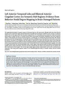
FIG. 1. The Stroop task. (A) Color words as presented visually in the congruent condition. (B) Color words as presented visually in an incongruent condition. In both conditions subjects are asked to name as quickly as possible the color of the letters presented and not to read the word itself. Words were presented individually and at a constant rate on a video monitor during these experiments.
consisted of a random presentation of four color names (red, yellow, or blue) presented in one of these four colors. The words were approximately 6 mm x 15 mm and were presented individually against a black background with a video monitor placed '40 cm directly in front of the subject's face. A fixation mark was present at all times -3 mm below the words. The words were displayed for 1300 msec with an interstimulus interval of 350 msec (1300 msec on, 350 msec off). In the congruent condition, the color in which the word was presented matched the color name (e.g., the word red displayed in red color-see Fig. 1A). In the incongruent condition, the color of the word presented did not match the color name (e.g., the word red displayed in green color-see Fig. 1B). No color names or presentation colors were ever repeated consecutively. During both conditions, subjects were instructed to name as quickly as possible the color ofthe word presented on the video monitor. They were explicitly asked not to read the word presented. To acquaint the subject with the task, the easier congruent condition task was presented first, followed 8 min later by the incongruent condition task. No practice was permitted. PET Studies. Subjects were prepared for PET scanning as described (14). Room lights were dimmed. Low-level noise consisted of that from cooling fans and the PETT VI tomograph (15). The tasks were started about 5 sec before injection of the isotope and concluded 90 sec later. Approximately 70 mCi (1 Ci = 37 GBq) of H2[150] in about 8 ml of saline were injected as an intravenous bolus, and tissue activity was acquired with PETT VI in the low-resolution mode by using an acquisition time of 40 sec after arrival of radioactivity to the head (16). No arterial catheters were used, as tissue activity (PET counts) has been demonstrated to be an excellent measure of rCBF change (16). green,
Proc. Natl. Acad. Sci. USA 87 (1990)
257
Data Analysis. The global (whole slice) average tissue activity with all seven PETT VI slices was calculated for each scan. All scans were then scaled linearly to produce an average global activity of 1000 PET counts to correct for global changes across scans (14). All intrasubject scan pairs (i.e., incongruent vs. congruent) were checked for motion artifact (14), and none was detected. The differences in tissue activity between conditions (i.e., incongruent minus congruent-a total of eight scan pairs, one such subtraction for each subject) were then averaged in stereotactic space as described (17). The location of all local minima and maxima were determined with a center of mass algorithm that used a sphere of 14-mm diameter (18); the magnitude of the responses was calculated as the average tissue activity within a sphere of 14-mm diameter placed at the center of mass of the response (18). All response locations are referenced to brain HD6 of the Talairach stereotactic atlas (19). Response outliers in the averaged stereotactic image were assessed to be significant at P < 0.01 by using the y-2 statistic (17). For post hoc analysis, the principal responses with Z scores >2 (P < 0.01) are reported.
RESULTS The largest reponses map to the right anterior cingulate cortex. Fig. 2 displays a transverse section through the averaged stereotactic image of cerebral activation during the Stroop task and demonstrates the robust anterior cingulate activity. Despite the apparent simplicity of the task, a complex distributed array of additional foci of activity were present (see Table 1 and Fig. 3). These included bilateral peristriate responses, left premotor and left postcentral foci, a left putamen response, supplementary motor area, inferior anterior cingulate cortex, and a right temporal response.
DISCUSSION The Stroop color/word task is a prototypic paradigm in cognitive neuroscience to probe attentional phenomena, such as the selection of processing and the inhibition of habitual responses (20). This task is, in fact, an archetype of human intentional behavior for many cognitive neuropsychologists (3). Numerous chronometric studies have sought the mental locus of the powerful Stroop effect. Perceptual conflict (21), conceptual encoding (22), response interference (23), or combinations of these (24) have been posited as the source of the effect. However, these sensitive reaction time studies
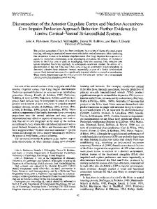
A
FIG. 2. Anterior cingulate activation during the Stroop task. (A) Diagram of the human brain demonstrating the transverse section under study. (B) Transverse section (see A) from averaged stereotactic image of resting rCBF; anterior is at the top, and the subjects' right is at right. (C) Transverse section (see A and B) from averaged stereotactic image of rCBF change between incongruent and congruent Stroop task conditions. Note activation of anterior cingulate cortex (arrow). Scale maximum of 55 PET counts corresponds approximately to a rCBF change of 5 ml/min per 100 g.
258
Neurobiology: Pardo et al.
Proc. Natl. Acad. Sci. USA 87 (1990)
Table 1. Response locations and magnitudes during the Stroop task Magnitude, Coordinate, mm z y PET count x Region 29 48 30 -10 1. Anterior cingulate (R) 27 47 30 -7 2. Anterior cingulate (R) 35 46 3. Anterior cingulate (R) 28 -17 11 45 0 27 4. Putamen (L) -3 42 32 25 5. Buried postcentral (L) 40 6. Postcentral sulcus (L) 36 41 -11 7. Peristriate (L) 40 -2 17 -71 34 11 40 8. Promoter (L) 25 9. Anterior inferior 20 13 53 40 cingulate sulcus (L) 21 39 10. Supplementary motor area 46 7 11. Superior temporal 10 -71 -1 39 gyrus (R) 2 17 -69 39 12. Peristriate (L) 8 -20 -63 38 13. Peristriate (R) All responses have magnitudes with Z scores of 2 or greater, P < 0.01. Coordinates are for brain HD6 of the Talairach atlas (19). L, left; R, right.
have not provided totally conclusive cognitive dissection of the operations involved in the Stroop task. Reaction time studies obviously have intrinsic limitations in providing an anatomical analysis of this popular neuropsychological instrument. No direct visualization has been available of the neural systems representing the cognitive components that mediate the high-level operations required by the Stroop task. This study utilized the Stroop paradigm and PET technology to isolate and visualize the functional anatomy of the neural systems involved with attentionally mediated resolution of processing conflicts (12). The Stroop task had the advantage that under both congruent and incongruent con-
ditions the subject was exposed, on the average, to the same color words and color features. Correspondingly, no differences were observed in the left medial extrastriate cortex, an area uniquely concerned with the visual word form (see below). Also, the subject produced, on the average, the same set of vocalizations under identical task instructions. There was no difference in activity between conditions in the mouth primary somatosensory cortex (see ref. 7, regions 12 and 13) during simple repetition of words. Therefore, much of the cerebral activity associated with low-level stimulus encoding, instructional set, and response output was controlled. Differences between the incongruent and congruent conditions must, therefore, be related directly to the attentionally mediated selection of cognitive processing between color naming and reading. Such conflict underscores the intentional nature of this paradigm and the attentional demands on the processing system required for task execution-thus, the well-known increase in reaction time during the incongruent as compared to the congruent condition-i.e., the Stroop effect. Such features made the Stroop task ideally suited for PET subtraction methodology, wherein the PET scans obtained during two tasks sharing all but isolable cognitive operations are subtracted from each other to reveal the anatomy of the isolated components (12). The robust activation of the anterior cingulate cortex (regions 1-3, see Table 1 and Fig. 3) during the incongruent condition suggests that this cortical region may be a component of an anterior attentional system. Support for involvement of this part of the anterior cingulate cortex in attention comes from a converging experiment reported recently from this laboratory (7, 8). That experiment was designed to study language processing of single nouns presented visually on a video monitor. During the highest level linguistic stage of that study, PET scans from subjects while generating verbs appropriate to the nouns were compared with PET scans obtained while reading aloud the nouns. This comparison was expected to reveal neural components associated with the
STROOP INTERFERENCE PARADIGM
10mm
FIG. 3. Cortical activation foci during the Stroop task. Diagram of medial (at top) and lateral (at bottom) views of the right and left hemispheres demonstrating the cortical responses in Table 1. The axes shown correspond to the transverse fiduciary through the anterior and posterior commissures (x axis) and the vertical fiduciary through the midpoint of the line segment connecting the commissures (z axis). The response in the left putamen (Table 1) is not shown. (Bar = 10 mm.)
Neurobiology: Pardo et al. cognitive operations of semantic processing and selection for action. Although the Stroop task is not in detail similar to such a language study, some analogies can be appreciated. On the average, the subjects during both scans were exposed to similar sensory input and produced similar amounts of speech. Both experiments probed for processing of single words. The generation task required the intentional inhibition of the habitual response (reading the nouns) as well as the selection and recruitment of processing centers appropriate for task execution. The major (P < 0.01) cortical responses common to both the generation and Stroop task occurred in the same region of the anterior cingulate. In contrast, as might be expected from the markedly different processing requirements of the generation and Stroop tasks, most of the other activated brain centers were different between the two tasks. For example, in comparison to the respective control reference states, none of the peristriate responses recruited during the incongruent condition of the Stroop paradigm were recruited during the generation task of the language study. Also, the left anterior frontal cortex (which was suggested to be involved in the semantic processing of the noun so as to produce appropriate verbs) recruited in the generation task was not activated during the Stroop paradigm. The many other smaller responses noted in this study suggest that the Stroop task requires an extensive and distributed network of processing centers for stimulus encoding and verbal output. Some of these responses fit well with emerging functional neuroanatomy of human higher cognitive systems developed in this laboratory using PET. For example, the left and right peristriate regions (regions 7, 12, and 13) most likely correspond to left and right hemispheric processing of the visual features of the stimulus (7, 8). A distinction between the primary visual features of the stimulus and word form can be made on the basis of converging experiments from this laboratory (S. E. Petersen, A. Snyder, P. T. Fox, and M.E.R., unpublished work), which show left medial extrastriate activation when words are compared to false fonts and consonant letter strings. The absence of activation in this area in the present experiment supports the hypothesis that analysis of visual word form was similar in congruent (control) and incongruent conditions. Additionally, passive viewing of single words is known to activate not only striate and extrastriate regions but also the left putamen (region 4; refs. 7 and 8). The Stroop task appears, thus, to entail some alterations in the high-level sensory encoding of the visual information. The roles of the left premotor, supplementary motor area, and postcentral responses are involved presumably in task-modulated verbal output, as these regions correspond closely with some of those seen during simple repetition of visually presented words (7, 8). There is too little information yet available to even speculate as to the function of the right superior temporal response. In conclusion, two converging experiments each with different processing requirements but with analogous attentional demands have now resulted in anterior cingulate activation in association with the recruitment of very different cortical areas sustaining the task-specific demands of each job. These data suggest that the anterior cingulate is involved
Proc. Natl. Acad. Sci. USA 87 (1990)
259
in the selection process between competing processing alternatives on the basis of some preexisting internal, conscious plan. Note Added in Proof. Since this manuscript was submitted, a report identifying a color processing center in the human cerebral cortex has been published (25). Of interest, no changes were observed in this region between congruent and incongruent conditions of the Stroop paradigm within the range of outliers examined (P < 0.01).
We thank the experimental subjects for their patience and efforts. Funding for this work was provided by The McDonnell Center for Higher Brain Function, The National Alliance for Research on Schizophrenia and Affective Disorders, and National Institutes of Health Center Grants HL 13851 and NS 06833. P.J.P. is a predoctoral trainee (MH 17104 to Lee N. Robins). J.V.P. is a Pfizer Fellow. 1. Stroop, J. R. (1935) J. Exp. Psychol. 18, 643-662. 2. Dyer, F. H. (1973) Mem. Cognit. 1, 106-120. 3. Posner, M. 1. (1986) Chronometric Explorations of the Mind (Oxford Univ. Press, New York), pp. 91-97. 4. Fuster, J. M. (1989) The Prefrontal Cortex (Raven, New York). 5. Goldman-Rakic, P. S. (1987) in Handbook of Physiology: Higher Cortical Function of the Brain, eds. Plum, F. & Mountcastle, V. B. (Am. Physiolog. Soc., Washington, DC), Vol. 5, pp. 373-417. 6. Perret, E. (1974) Neuropsychologia 12, 323-330. 7. Petersen, S. E., Fox, P. T., Posner, M. I., Mintun, M. A. & Raichle, M. E. (1988) Nature (London) 331, 585-589. 8. Petersen, S. E., Fox, P. T., Posner, M. 1. & Raichle, M. E. (1989) J. Cognit. Neurosci. 1, 153-170. 9. Weinberger, D. R., Berman, K. F. & Zee, R. F. (1986) Arch. Gen. Psychiatry 43, 114-125. 10. Benes, F. M. & Bird, E. D. (1988) Arch. Gen. Psychiatry 44, 608-616. 11. Posner, M. I., Early, T. S., Reiman, E. M., Pardo, P. J. & Dhawan, M. (1988) Arch. Gen. Psychiatry 45, 814-821. 12. Posner, M. I., Peterson, S. E., Fox, P. T. & Raichle, M. E. (1988) Science 240, 1627-1631. 13. Raichle, M. E. (1987) in Handbook of Physiology: Higher Cortical Function of the Brain, eds. Plum, F. & Mountcastle, V. B. (Am. Physiolog. Soc., Washington, DC), Vol. 5, pp. 643-674. 14. Fox, P. T., Miezin, F. M., Allman, J. M., Van Essen, D. C. & Raichle, M. E. (1987) J. Neurosci. 7, 913-922. 15. Yamamoto, M., Ficke, D. C. & Ter-Pogossian, M. M. (1982) I.E.E.E. Trans. Nucl. Sci. 29, 529-533. 16. Herscovitch, P., Markham, J. & Raichle, M. E. (1983) J. Nucl. Med. 14, 782-789. 17. Fox, P. T., Mintun, M. A., Reiman, E. M. & Raichle, M. E. (1988) J. Cereb. Blood Flow Metab. 8, 642-653. 18. Mintun, M. A., Fox, P. T. & Raichle, M. E. (1989) J. Cereb. Blood Flow Metab. 9, 96-107. 19. Talairach, J. & Szikla, G. (1967) Atlas d'Anatomie Stereotaxique du Telencephale (Mason, Paris). 20. Mesulam, M. M. (1985) Principles of Behavioral Neurology (Davis, Philadelphia), pp. 125-163. 21. Hock, H. S. & Egeth, H. E. (1970) J. Exp. Psychol. 83, 299-303. 22. Seymour, P. H. K. (1977) Q. J. Exp. Psychol. 29, 245-265. 23. Proctor, R. W. (1978) Percept. Psychophys. 23, 413-419. 24. Stirling, N. (1979) Q. J. Exp. Psychol. 31, 121-132. 25. Lueck, C. J., Zeki, S., Friston, K. J., Deiber, M.-P., Cope, P., Cunningham, V. J., Lammertsma, A. A., Kennard, C. & Frackowiak, R. S. J. (1989) Nature (London) 340, 386-389.