Nov 5, 2006 - a bead-based chemiluminescent ELISA procedure conducted under ... a mixture of Luminol and hydrogen peroxide is transported over the ...
DISK-BASED PARALLEL CHEMILUMINESCENT DETECTION OF DIAGNOSTIC MARKERS FOR ACUTE MYOCARDIAL INFARCTION L. Riegger1, J. Steigert1, M. Grumann1, S. Lutz1, G. Olofsson2, M. Khayyami2, W. Bessler3, K. Mittenbuehler3, R. Zengerle1,4, and J. Ducrée1,4 1
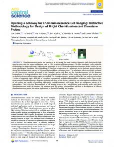
Lutz Riegger, Laboratory for MEMS Applications, Department of Microsystems Engineering (IMTEK), University of Freiburg, Georges-Koehler-Allee 106, 79110 Freiburg, Germany 2 Prolight Diagnostics AB, Sweden, 3IMMZ, Germany, 4HSG-IMIT, Germany Abstract We present a novel concept for the parallel detection of cardiac markers on a rotating lab-on-a-disk. Antigens indicating an acute myocardial infarction (AMI) are captured by a bead-based chemiluminescent ELISA procedure conducted under rotation on a polymer disk and detected by a photomultiplier (PMT). Parallel channel structures allow the quasi-simultaneous screening for different markers. To proof this concept, diluted, HRP-labeled myoglobin antibodies are captured with cardiac myoglobin coated beads. We obtain a CV of 12.4% and a cmin=12.2 ng/ml which is well below the myoglobin cutoff for the diagnosis of AMI [1]. Furthermore, two different antibody concentrations are read out simultaneously during spinning to demonstrate the capability of parallelization. Keywords: cardiac marker, ELISA, parallel readout, centrifugal microfluidics 1. Introduction So-called “lab-on-a-chip” systems for diagnostic point-of-care technologies attract a growing interest due to their immanent benefits of reduced turn-around times, their amenability for full process integration and automation, increased reliability, and minimized costs [2]. Among various lab-on-a-chip concepts, “lab-on-a-disk” [3,4] systems exploit centrifugal forces for metering, accelerated sedimentation of particles in biological suspensions, and mixing in order to run fully process integrated assays. In our approach, we implement a disk-based test to screen the relevant cardiac markers in parallel. This enables the early diagnosis of acute myocardial infarction in a single run. 2. Assay Principle Polystyrene beads (rbead = 75 μm) are coated off-disk with capture antibodies specific to the respective antigen M and then aggregated in the disk-based detection chamber. On-disk, the plasma sample is incubated with the beads N followed by horseradish peroxidase (HRP) labeled detection antibodies O in a flow-through procedure. Finally, a mixture of Luminol and hydrogen peroxide is transported over the beads P. In this step, the HRP enzyme catalyzes the oxidation of Luminol to excite 3-aminophtalate which emits a photon upon relaxation [4] (Fig. 1-A). The photon count is hence a direct measure of the target antigen concentration in the sample.
The 10th International Conference on Miniaturized Systems for Chemistry and Life Sciences (μTAS2006) November 5-9, 2006, Tokyo, Japan 4-9903269-0-3-C3043 © 2006 Society for Chemistry and Micro-Nano Systems 819
3. Experimental Setup and Read-out Procedure Our modular setup (Fig. 1-B) comprises a photomultiplier tube (PMT) and a centrifuge which spins a disposable polymer disk in the format of a standard CD.
Figure 1. Assay principle and experimental setup. (A) Beads are first functionalized with capture antibodies off-disk M. After bead aggregation, target antigens are transported into the detection chamber to form the specific antigen-antibody complexes N which are coupled with HRP-labeled detection antibodies subsequently O. The chemiluminescent reaction [5] is catalyzed by the HRP enzyme under constant flow of Luminol/H2O2 P and read out under rotation. (B) The measurement setup comprises a centrifuge with a disk holder, the PMT module, an electronics box for adjustment of the gain voltage, and an x-y drive for the positioning of the PMT relative to the disk.
Figure 2. Microfluidic disk and read-out concept. (A) The chip with the format of a conventional CD features passive microfluidic elements, only: an inlet, a detection chamber, a geometrical barrier for bead aggregation, and an outlet. (B) For the parallel read-out, the disk is spun at a frequency ν and the chemiluminescent signal for each chamber is detected by a high-bandwidth PMT once per revolution. Reagents are transported through the fluidic structures by the centrifugal force which is controlled by a designated frequency protocol ν(t). Each measurement channel on the disk (Fig. 2-A) features a set of passive microfluidic structures, i.e. an inlet, a detection chamber, an outlet (h = 200 μm), and a geometrical barrier for the bead aggregation The 10th International Conference on Miniaturized Systems for Chemistry and Life Sciences (μTAS2006) November 5-9, 2006, Tokyo, Japan 820
(h = 100 μm). The chemiluminescent signal for each detection chamber is read out in parallel during rotation by the PMT (Fig. 2-B). 4. Results For the validation of the concept, diluted HRP-labeled myoglobin antibodies are captured with human cardiac myoglobin coated beads. Our calibration curve (Fig. 3-A) features a CV of 12.4 % which represents 4 disks with 8 channels each processed on different days and a cmin=12.2 ng/ml as the lower limit of detection which is far below the myoglobin threshold at 100 ng/ml for the diagnosis of an AMI [1]. The capability for parallelization is demonstrated by the quasi-simultaneous read-out of two different antibody concentrations (500 ng/ml and 160 ng/ml, respectively) in Fig. 3-B.
Figure 3. (A) Calibration curve for the myoglobin assay featuring a CV = 12.4% and a cmin = 12.2 ng/ml which is far below the AMI cut-off at 100 ng/ml [1]. (B) Spectrum of the parallel read-out of two concentrations, 500 ng/ml and 160 ng/ml, respectively. The signals can easily be discriminated and are in agreement with the calibration curve. 5. Conclusion We realized a versatile concept for the automated processing and high-speed (< 1 s) read-out of parallel chemiluminescent ELISAs. It is particularly suited for the early determination of AMI where three distinct cardiac markers, e.g. Myoglobin, Troponin-I, and CK-MB, have to be detected simultaneously. The simple and rugged concept is implemented in a modular setup comprising a PMT, a standard centrifuge, and an exchangeable polymer substrate containing passive fluidic structures which can thus readily be fabricated by standard polymer micromachining. References [1] F. Apple et al., Clinical Chemistry, 45:2, pp. 199-205, (1999). [2] A. van den Berg, E. Oosterbroek, Micro Total Analysis Systems, Elsevier Science, Amsterdam, NL, (2003). [3] M. Madou and G. Kellogg, Proceedings of SPIE, SPIE publisher, San Jose, 3259, pp 80–93, (1998). [4] J. Steigert et al., J. Assoc. for Laboratory Autom.,10, pp. 331-341, (2005). [5] P. Whitehead, K. Kricka, J. Thorpe, Clinical Chemistry, 25, pp. 1531-1546, (1979). The 10th International Conference on Miniaturized Systems for Chemistry and Life Sciences (μTAS2006) November 5-9, 2006, Tokyo, Japan 821