Downloaded By: [Brooke, Charlene][informa internal users] At: 17:28 9 May 2011
Aging, Neuropsychology, and Cognition, 2011, 18 (3), 362–384 http://www.psypress.com/anc ISSN: 1382-5585/05 print; 1744-4128 online DOI: 10.1080/13825585.2011.560243
Dissociated deficits of visuo-spatial memory in near space and navigational space: Evidence from brain-damaged patients and healthy older participants L. Piccardi1,2 , G. Iaria3 , F. Bianchini4 , L. Zompanti4 , and C. Guariglia2,4 1
Dipartimento di Scienze della Salute, Università degli Studi di L’Aquila, Coppito, Italy Sezione di Neuropsicologia, I.R.C.C.S. Fondazione Santa Lucia, Rome, Italy 3 Neurolab, Departments of Psychology, Hotchkiss Brain Institute, and Department of Clinical Neurosciences, University of Calgary, Calgary, AB, Canada 4 Dipartimento Psicologia 39, Sapienza Università di Roma, Rome, Italy 2
ABSTRACT Defects confined to spatial memory can severely affect a variety of daily life activities, such as remembering the location of objects or navigating the environment, until now the skills involved have been mostly assessed with regard to the visual domain using traditional pencil and paper tests. Our aim was to test the efficacy of a recently developed psychometric instrument (Walking Corsi Test: WalCT) to assess the specific contribution of spatial memory to the complex task of retrieving route knowledge. The WalCT is a 3×2.5-m version of the well-known Corsi Block-tapping Test (CBT), in which patients are required to memorize (and replicate) a sequence of body displacements. We assessed the ability of left and right brain-damaged patients, as well as healthy young and senior controls, to perform both the CBT and the WalCT. Results showed differences related to age in the healthy individuals and specific functional dissociations in the brain-damaged patients. The double dissociations found in this study demonstrate the importance of having a task able to detect navigational disorders, because virtual reality tasks are often much too difficult for aged brain-damaged patients to perform. Keywords: Cerebrovascular patients; Memory for routes; Memory for object location; Corsi Block-Tapping Test; Topographical disorientation; Spatial learning; Aging. This research was supported by a grant to C.G. from the European Community (FP6-NEST: Wayfinding 12959). L.P. thanks Dr. Raffaella Nori for her useful suggestions during the reviewing process. Address correspondence to: Dr Laura Piccardi, Ph.D., Centro Ricerche di Neuropsicologia, I.R.C.C.S. Fondazione Santa Lucia, Via Ardeatina, 306, 00179 Rome, Italy. E-mail:
[email protected] © 2011 Psychology Press, an imprint of the Taylor & Francis Group, an Informa business
Downloaded By: [Brooke, Charlene][informa internal users] At: 17:28 9 May 2011
WALKING MEMORY IN BRAIN-DAMAGED PATIENTS
363
By the 1960s, it had become clear that human memory was not a unitary concept. Strong evidence of a dissociation between short- and long-term memory was found in studies on two types of neurological patients: those with a classical form of amnesia as a consequence of lesions in the temporal lobe and hippocampi and those with lesions in the perisylvian region of the left hemisphere. The first type of patients has normal short-term memory and a pronounced deficit in learning and remembering new verbal and visual items (Milner, 1966). On the contrary, the second type of patients reveals the opposite pattern (Shallice & Warrington, 1970). Although it is now generally accepted that there are at least two independent systems for retaining information for few seconds or for an entire lifetime and that verbal and visuo-spatial information is stored by means of separate processes, different types of visuo-spatial memory systems and their dissociations are not still well studied. Many studies investigated nature, characteristics and anatomical basis of verbal and non-verbal memory in the past century, but the two domains were not studied equally. Indeed, verbal memory was investigated in most of the studies and detailed experimental paradigms were developed considering also structural and semantic characteristics of stimuli. Differently, visuo-spatial memory, often defined as ‘non-verbal memory’, received less attention. Only in the last few decades, attempts have been made to disentangle the ‘visual’ and ‘spatial’ aspects of memory (i.e., Della Sala, Gray, Baddeley, Allamano, & Wilson, 1999; Grossi, Becker, Smith, & Trojano, 1993). Another neglected point refers to the type of visuo-spatial material to be stored. Little attention has been paid to the nature of visuo-spatial stimuli and the idea that non-verbal stimuli do not need to be differentiated because of their nature (objects, faces, buildings, geometric shapes, etc.) has dominated for a very long time. One distinction, that needs to be made, is that between memory for locations in reaching space and memory for locations in navigational space (route memory). Recent studies (Ekstrom et al., 2003; Hartley & Burgess 2005; Hartley, Maguire, Spiers, & Burgess, 2003; Maguire et al. 1998; Piccardi, Iaria, et al., 2008; White & McDonald, 2002) describe the existence of at least two distinctive spatial memories for topographical orientation and object locations in both humans and animals. Postma, Jager, Kessels, Koppeschaar, and van Honk (1998) found sex differences in object location memory, specifically in their experiment women outperformed men, an atypical result, since men show better capability than women in performing several visuo-spatial tasks. Authors hypothesize a type of visuo-spatial memory devoted in memorizing object location and in a following study they observed selective impairments in object-location binding, metric encoding and their integration after ischemic stroke (Kessels, Postma, Kappelle, & de Haan, 2002). The distinction between a memory for locations in reaching space and a memory for locations in navigational space (route memory) is suggested
Downloaded By: [Brooke, Charlene][informa internal users] At: 17:28 9 May 2011
364 L. PICCARDI ET AL. by the absence of reports of associations between deficits in near space and navigational space; differently, most patients are affected by memory disorders confined to just one type of space (De Renzi, 1982). Even studies on the neural basis of navigational processes (Bird & Burgess, 2008) and neural models of visuo-spatial memory (Byrne, Becker, & Burgess, 2007) underline involvement of different parts of the hippocampal and retrosplenial areas (Ekstrom et al., 2003; Hartley & Burgess, 2005; Hartley et al., 2003; Iaria, Petrides, Dagher, Pike, & Bohbot, 2003; Maguire et al., 1998; Voermans et al., 2004; White & MacDonald, 2002). Ghaem et al. (1997) reported that a mental ‘replay’ of navigation seems to be subserved by two distinct networks, one for long-term memory reactivation that involves the posterior and middle hippocampal regions, the dorsolateral prefrontal cortex and the posterior cingulate gyrus, and the other for dynamic spatial mental imagery that involves the medial part of left hippocampal regions, visuo-spatial and sensorimotor areas. Considering the complexity of the environment and our flexibility to navigate, it is reasonable to hypothesize that different memory processes are involved in coding and storing different aspects. To assure the possibility to move through the environment also after a brain lesion, it is possible that there are two systems for memorizing individual movements and environmental features. Furthermore, considering Wang and Spelke’s (2002) theoretical distinction between the cognitive processes subserving topographical orientation, path integration, which is a process that updates subject location according his movements, represents the first step of environmental knowledge acquired by an individual followed by the view-dependent place recognition, a process based on place and landmark recognition. Therefore, the existence of different memory processes seems to be supported also from theoretical model of environmental knowledge (Montello, 1998; Siegel & White, 1975; Wang & Spelke, 2002). Neuropsychological studies have shown that a specific memory deficit for environmental features may be present in patients affected by topographical disorientation. Indeed, these patients have selectively lost their ability to navigate in large-scale environments (Aguirre & D’Esposito, 1999; Farrell, 1996). Generally, topographical disorientation tends to affect the ability to learn new environments more than to move in familiar ones (Farrell, 1996). Two different mechanisms are considered responsible for this disorder: topographical agnosia or/and topographical amnesia (Peterson & Zangwill, 1945). The former concerns the impaired ability to recognize and identify environmental features and the latter the loss of stored topographical representations built up through experience (Farrell, 1996). Studies on topographical disorientation suggest that memory disorders for environmental features (i.e., shapes/colours of landmarks) play an important role. Nevertheless, it is still unknown whether a specific memory for movements in the environment, coding sequences of locations, exists and
Downloaded By: [Brooke, Charlene][informa internal users] At: 17:28 9 May 2011
WALKING MEMORY IN BRAIN-DAMAGED PATIENTS
365
whether its damage leads to topographical disorders. Disorders of routememory could be responsible for the amnesic component of topographical disorientation, that is, memory disorders for distances and directions between different spatial locations. Previous studies have demonstrated that memory for landmarks is subserved by a specific system not involved in other types of non-verbal memory (Incisa della Rocchetta, Cipolotti, & Warrington, 1996). It is, therefore, possible that the route-memory system is dissociated from the system that memorizes sequences of locations in near space. This hypothesis is supported by clinical observations of patients who fail to store and recall sequences on the Corsi Block-tapping Test (CBT) never affected by navigational disorders, as well as patients affected by topographical disorientation that perform flawlessly on the Corsi Test. The first aim of this study was to verify the existence of a system devoted to memorize sequences of environmental locations in navigational space. The second aim was to verify whether patients affected by topographical disorientation have a damage to the route-memory system. To answer these questions, we adopted a recently developed large-scale version of the CBT, that is, the Walking Corsi Test (WalCT: Piccardi, Iaria, et al., 2008), which requires memorizing a sequence of places by following a path. The advantages of the large-scale test are the following: (a) it mimics memory for route knowledge in a controlled setting and (b) it allows investigating the ability to memorize sequences in navigational space and in near space by comparing performances on WalCT and on CBT. Also if, CBT has not traditionally been used as a ‘reaching space’ memory task, and it is considered a visual spatial memory task, it measures the capability to immediately and delayed recall a sequence in the reaching space requiring to learn small locations with minor variations in location. Differently, the WalCT permits evaluating route-memory ability by measuring a pathway span and testing acquisition and long-term recall of a pathway supra-span. Two groups of right and left brain-damaged patients as well as healthy young and senior controls performed short- and long-term memory tasks on the WalCT and on the CBT. We expected to find no differences in performances if the two tests actually measure the same sequential memory system. Differently, we expected to find dissociated performances on the CBT and the WalCT in at least some patients if the two tests assess different types of visuo-spatial memory. In general, we expect poor performances in both tests in right braindamaged patients as already demonstrated by De Renzi, Faglioni, and Previdi (1977) that found a dominant role played by the posterior region of the right hemisphere in subserving spatial memory mechanisms. However, patients assessed by De Renzi et al. (1977) have also perceptual disorders that could partially explain their results. Moreover, we assessed young and old healthy participants to observe if CBT and WalCT are sensitive to measure age
366 L. PICCARDI ET AL. differences. Once again, if they actually measure different types of visuospatial memory we should observe differences related to age in performing them. In the present study, we also included two patients suffering from topographical disorders to verify whether they would show a specific performance defect on the WalCT and if WalCT is really predictive of route memory.
Downloaded By: [Brooke, Charlene][informa internal users] At: 17:28 9 May 2011
SUBJECTS The study was designed in accordance with the ethical principles for human experimentation stated in the Declaration of Helsinki and was approved by the local ethics committee. All participants provided their informed written consent after the procedures had been fully explained to them. The research has been carried out in I.R.C.C.S. Fondazione Santa Lucia of Rome. Healthy senior participants were relatives of inpatients, while healthy young subjects were college students. Following the preliminary clinical neuropsychological examinations reported below, the subjects were included in one of the following four groups and then participated in the experiments: ●
●
●
●
Right brain-damaged patients (RBD): 10 patients were recruited (eight males). Their mean age was 62.50 years (SD = 12.76) and their mean educational level was 11.10 years (SD = 3.14). Left brain-damaged patients (LBD): 15 patients were recruited (eight males). Their mean age was 57.53 years (SD = 16.22) and their mean educational level was 12.47 years (SD = 3.66). Both fluent and non-fluent aphasic patients were included in this group if their comprehension deficits allowed them to understand the easy verbal instructions required to perform the tests. Healthy senior subjects (HSS): 25 subjects (15 males) whose mean age was 62.68 years (SD = 6.42) and mean educational level was 10.84 years (SD = 3.80), were recruited. Both age (one-way ANOVA: F(2, 44) = 1; p = ns) and education (one-way ANOVA: F(2, 44) = 1.030; p = ns) were comparable to those of the two groups of brain-damaged participants. Healthy young subjects (HYS): 59 subjects (30 males) whose mean age was 23.1 years (SD = 2.20) and mean educational level was 13.08 years (SD = 0.65) were included in this group.
NEUROPSYCHOLOGICAL TESTING Before being enrolled in the study, each healthy senior subject was administered the Milan Overall Deterioration Assessment (M.O.D.A.: Brazzelli, Capitani, Della Sala, Spinnler, & Zuffi, 1994) to be sure they were not
Downloaded By: [Brooke, Charlene][informa internal users] At: 17:28 9 May 2011
WALKING MEMORY IN BRAIN-DAMAGED PATIENTS
367
suffering from mental decay. To exclude left-handers, all participants were requested to complete a handedness questionnaire (Salmaso & Longoni, 1983). Brain-damaged patients were submitted to an extensive neuropsychological assessment evaluating language (Ciurli, Marangolo, & Basso, 1996; Miceli, Laudanna, Burani, & Capasso, 1994), short- and long-term verbal memory (Spinnler & Tognoni, 1987) and abstract reasoning (Basso, Capitani, & Laiacona, 1987; Spinnler & Tognoni, 1987). The presence of hemi-neglect has been tested by administering Star Cancellation task (Wilson, Cockburn, & Halligan, 1987). Right brain-damaged patients also underwent a standard battery of tests to assess the presence of hemi-neglect including peri-personal and personal tests (Pizzamiglio et al., 1990; Zoccolotti, Antonucci, & Judica, 1992). Representational neglect deficits were assessed by asking the patients to describe from memory a familiar place as it would appear from two different perspectives (Bisiach & Luzzatti, 1978). A laterality quotient was calculated on the basis of the number of elements reported for each side in the two descriptions (Bartolomeo, D’Erme, & Gainotti, 1994); a score equal or inferior to –20 was considered a sign of representational neglect (Guariglia, Piccardi, Iaria, Nico, & Pizzamiglio, 2005). Exclusion criteria were: mental deterioration, short-/long-term memory deficits, perceptual neglect, severe aphasia. Only one right brain-damaged patient was affected by pure representational neglect. The language assessment of the left brain-damaged patients showed that their comprehension skills were adequate for understanding the easy instructions for performing the experiments. The patients’ clinical data are provided in Table 1. Only patients who could walk were included in the study. All brain-damaged patients were submitted to a neurological examination and a neuro-radiological scan (TC/RMI) (Figure 1a, b). METHODS The Corsi Block-Tapping Test (CBT) Short- and long-term spatial memory was tested using the CBT (Spinnler & Tognoni, 1987). This test is composed of nine wooden blocks (4.5×4.5 cm) fixed on a baseboard (30×25 cm) in a scattered array. On the experimenter’s side, the blocks are numbered for easy identification (Figure 2a). Both short- and long-term memory were tested. In the Visual Shortterm Memory Test (VSTM), the examiner tapped a sequence of blocks at the rate of one block per two seconds and the subject had to reproduce the same sequence in the same order. Sequences of increasing length (i.e., number of blocks starting from a 2-block sequence) were presented until the subject failed to reproduce two out of three trials of a given length.
14 10 9 8 12 4 7 14 11 1
52 52 51 51 53 32 53 np 53 53
51 51 47 51 51 29 51 np 51 51
11 11 11 11 11 10 11 np 11 11
10 10 10 10 10 10 10 np 10 10
0 0 0 0 0 0 0 np 0 0
0 0 0 0 0 1 0 np 0 0
6 6 6 6 6 6 6 np 6 6
0 0 0 0 0 0 0 np 0 np
23 41 1390 159 61 67 56 6495 84 4887
Onset (days)
FTP/I I F/cr/th/wm FTP cr F/wm/cr cr wm Ic/cr FT
Lesion’s site
1
np, not performed; UR∗ , unexpected responses; F, frontal; T, temporal; P, parietal; th, thalamus; cr, corona radiata; I, insulae; wm, white matter. Basso et al. (1987); 2 Carlesimo et al. (1996); 3 Diller et al. (1974); 4 Albert et al. (1973); 5 Massironi et al. (1988); 6 Pizzamiglio et al. (1990); 7 Zoccolotti et al. (1992).
53 40 41 40 49 39 38 51 44 24
RBD RBD RBD RBD RBD RBD RBD RBD RBD RBD
AMC DO EP SS LZ RR LT OC GC CS
19 25 17 23 29 28 26 Np 28 34
(Err = 9)
No case
Wundt-Jastrow5
Raven Delayed Coloured Imm. Recall Left Right Left Right Left PM1 Recall Right Group (Hits = 36) (Hits = 75) (Hits = 15) (Hits = 53) (Hits = 51) (Hits = 11) (Hits = 10) (UR∗ = 20) (UR∗ = 20) (Hits = 6)
Barrage4
Personal neglect7
Letter cancellation3
Screening Neglect
Reading6
Rey’s 15 words learning task2
TABLE 1. (a) Clinical data of right brain-damaged patients
Downloaded By: [Brooke, Charlene][informa internal users] At: 17:28 9 May 2011
368 L. PICCARDI ET AL.
LBD
LBD LBD LBD LBD LBD LBD LBD LBD
LBD LBD
LBD LBD LBD LBD
GM
US RM VP OV VB GC PS LT
VI GG
MB FM BS VDO
preserved preserved preserved preserved
preserved preserved
preserved preserved mild preserved preserved mild preserved preserved
mild
Oral comprehension11 Actions
3,33% 17,8% 80% 0%
14,29% 6,6% 78% 6%
40% 50% Naming objects10 (Hits = 32) 29 33,33% 39.28% 40% 27,28% 3.33% 10,71% 31% 18% np 25% 0% np Standard Language Standard Language Examination11 + Examination11 + Naming objects11 (Hits = 32) 32 0% 0%
Objects
25 27 27 np 27 26 np 27
27
Right (Hits = 27)
27 27 27 27 Letter Cancellation3 Left (Hits = 53) Right (Hits = 51) 53 51 np np 27 27 np np
27 27 27 np 27 27 np 27
27
Left (Hits = 27)
Star Cancellation9
19 21 35 26
30 31
18 32 34 26 29 33 29 29
17
Raven1 (Hits = 36)
96 36 181 70
5324 75
952 180 35 671 46 53 28 38
241
Onset (days)
np, not performed; F, frontal; T, temporal; P, parietal; bg, basal ganglia; ic, internal capsule; cr, corona radiata; I, insulae; wm, white matter; bs, brain stem. 1 Basso et al. (1987); 3 Diller et al. (1974); 8 Miceli et al. (1994); 9 Wilson et al. (1987); 10 Basso et al. (1987); 11 Ciurli et al. (1996).
Group
No case
BADAs Naming
TABLE 1. (b) Clinical data of left brain-damaged patients
Downloaded By: [Brooke, Charlene][informa internal users] At: 17:28 9 May 2011
FPT/wm ic/wm/cr T/wm FP/wm
T/ic lc
wm;bg F/cr/wm TP FTP/wm/I FT/wm TP/wm bg/wm bs
TP/wm
Lesion’s site
WALKING MEMORY IN BRAIN-DAMAGED PATIENTS
369
370 L. PICCARDI ET AL.
Downloaded By: [Brooke, Charlene][informa internal users] At: 17:28 9 May 2011
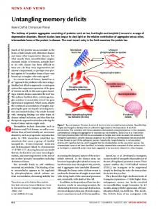
F IGURE 1. (a) CT and MRI performed by right brain-damaged patients. (b) CT and MRI performed by left brain-damaged patients.
WALKING MEMORY IN BRAIN-DAMAGED PATIENTS
371
Downloaded By: [Brooke, Charlene][informa internal users] At: 17:28 9 May 2011
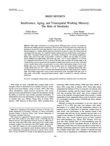
F IGURE 2. (a) Apparatus used for administering the stimuli in the CBT. (b) Modified large-scale version of the CBT. The scale was 1:10 and measured 3×2.50 m; black squares were 30×30 cm.
Two aspects of long-term memory were assessed: learning (VSL) and delayed recall (VSDR). In VSL, the subjects were required to learn an 8block sequence (Capitani, Grossi, Lucca, Orsini, & Spinnler, 1980; Spinnler & Tognoni, 1987) presented by the examiner. The learning criterion was reached if the subject reproduced the correct sequence three times in a row (max number of trials: 18). The learning score was calculated as follows: one point was attributed to each cube/square correctly tapped/stepped until the criterion was reached; then it was added to the score corresponding to correct performance of the remaining trials (up to the 18th). For example, if the subjects reached the learning criterion by the third repetition, they obtained a score of 8 cubes/squares × 3 = 24 plus 8 cubes/squares × 15 = 120 for the remaining trials. Thus, they obtained a total score of 144, the maximum score for both the CBT and WalCT. Five minutes later (Capitani et al., 1980; Spinnler & Tognoni, 1987), the VSDR was administered. The examiner asked the subject to reproduce the previously learned 8-block sequence. Scores were calculated on the basis of the number of cubes correctly reproduced. In each task, the subjects were tested individually in a quiet laboratory room with artificial lighting. They were seated facing the examiner on a height-adjustable office chair in front of the CBT baseboard. The Walking Corsi Test (WalCT) In an empty room, a larger version (3×2.5 m; scale 1:10 of the CBT) of the CBT was set up. In this test, the subject has to actually walk and reach
Downloaded By: [Brooke, Charlene][informa internal users] At: 17:28 9 May 2011
372 L. PICCARDI ET AL. different locations. The WalCT (Piccardi, Iaria, et al., 2008) is composed of nine black squares placed on a light grey carpet in a scaled position of the standard CBT (see Figure 2b). Although the same experimental conditions were adopted in the WalCT as in the CBT, the starting position was different; that is, in the WalCT, but not in the CBT, both the examiner and the participants started from the same point. The examiner illustrated the sequence by walking on the carpet and stopping on each square for two seconds. Then, the subject had to repeat the same sequence as the examiner by walking and stopping on the squares included in the sequence. The administration order of the CBT and WalCT was counterbalanced across the subjects in each group. RESULTS Figure 3 shows mean and standard deviations (SD) of the span reached in the CBT and WalCT, as well as the number of cubes/squares necessary to reach the learning criterion plus the overall cubes/squares in the remaining trials and the number of cubes/squares recalled after a 5-minute delay. Separate statistics analyses were performed for short-term memory (VSTM), learning (VSL) and delayed recall (VSDR). Visual Short-term Memory (VSTM) A 4 (group: RBD vs. LBD vs. HSS vs. HYS) × 2 (type of test: CBT vs. WalCT) mixed ANOVA on span scores revealed significant main effects for group, F(3, 104) = 7.754; p < .0001; d = 1.185. A Duncan post-hoc analysis revealed a significantly (p < .001) larger span on both tests in Healthy Young Subjects than in the other groups (HSS, RBD, and LBD), which showed no difference between one another. The span on the CBT was significantly larger than that on the WalCT, F(1, 104) = 33.077; p < .000; d = 0.241. There were no significant differences for the interaction group × test, F(3, 104) = 0.794; p = ns; d = 0.022. Visual Long-term Memory: Learning (VSL) A two-way ANOVA revealed main effects for group, F(3, 104) = 9.314; p < .00; d = 0.212, and for type of test, F(1, 104) = 12.38; p < .00; d = 0.111, as well as a significant group × test, F(3, 104) = 3.743; p < .01; d = 0.097, interaction. A Duncan post-hoc analysis showed that left brain-damaged patients differed (p < .01) significantly from right braindamaged patients on WalCT but not on the CBT, learning WalCT sequence significantly faster than those of CBT. Right brain-damaged patients also differed significantly from healthy subjects on both tests; specifically, they had
WALKING MEMORY IN BRAIN-DAMAGED PATIENTS
Downloaded By: [Brooke, Charlene][informa internal users] At: 17:28 9 May 2011
F IGURE 3. Mean and standard deviations (SD) of the span reached in the CBT and WalCT (a), as well as the number of cubes/squares necessary to reach the learning criterion plus the overall cubes/squares in the remaining trials (b) and the number of cubes/squares recalled after a 5-minute delay (c).
373
374 L. PICCARDI ET AL. difficulty in learning both the 8-cube and the 8-square sequences. Healthy Young subjects learned faster than Healthy Senior subjects only in the WalCT (see Figure 3).
Downloaded By: [Brooke, Charlene][informa internal users] At: 17:28 9 May 2011
Visual Long-term Memory: Delayed recall (VSDR) A 2×2 (Group×Test) ANOVA on the number of cubes/squares the subjects were able to recall after a delay yielded a significant main effect for Group, F(3, 104) = 3.829; p < .01; d = 0.100. A post-hoc analysis (Duncan’s test) showed that the HYS had better recall than the two groups of brain-damaged patients, which did not differ from each other. The HSS performances were comparable to those of the HYS and better than those of the two groups of brain-damaged patients. The WalCT sequence was recalled better than the CBT sequence, F(1, 104) = 14.31; p < .00; d = 0.122. The interaction group × test, F(3, 104) = 1.460; p = ns; d = 0.041, did not reach significance. INDIVIDUAL DIFFERENCES To better explore the hypothesis that WalCT and CBT assess different types of visuo-spatial memory, we analyzed individual performances in detail using Crawford’s analysis (Crawford & Howell, 1998). This method permits estimating the abnormality of individual scores compared to the control sample detecting dissociations by statistically analyzing the performance differences for the two different tests (see Crawford & Garthwaite, 2005). Visual Short-term Memory (VSTM) No dissociated performances were found in the right brain-damaged patients and none showed deficits on the CBT or the WalCT. Differently, in the left brain-damaged group 2 out of 15 patients performed significantly worse (G.M. and M.B.: t-test = –2.525; p < .005) than the healthy subjects group on the CBT, but their performance on the WalCT was comparable to that of the healthy subjects. Both G.M. and M.B. have a lesion involving temporo-parietal regions as well as white matter. No left brain-damaged patient showed a deficit on the WalCT. Visual Long-term Memory: Visual learning (VSL) As regards learning, we found a double dissociation in the group of right brain-damaged patients. Indeed, patient A.M.C. performed significantly worse than the healthy subjects on the CBT (t-test = –3.059; p < .01) but not on the WalCT (t-test = –1.45; p = ns) and patient S.C. showed the opposite pattern of performance (CBT: t-test = 0.63; p = ns; WalCT: t-test = –2.563;
WALKING MEMORY IN BRAIN-DAMAGED PATIENTS
375
p < .01; for details, see section below: Two case reports). Both patients have lesions involving fronto-temporal regions.
Downloaded By: [Brooke, Charlene][informa internal users] At: 17:28 9 May 2011
Visual Long-term Memory: Delayed recall (VSDR) With regard to delayed recall, we observed one right brain-damaged patient (O.C.) who failed on the WalCT (t-test = –9.806; p < .01) but not on the CBT, revealing a pattern of performance corresponding to a classical dissociation. We also observed four left brain-damaged patients (G.M.; U.S.; F.M.; V.D.) who failed on the WalCT (p < .01) but not the CBT. According to Crawford’s analysis, three (G.M.; U.S.; and V.D.) of them showed a pattern of performance that fulfilled the criteria for a strong dissociation, whereas the fourth (F.M.) revealed a classical dissociation. Both the right brain-damaged patient and the four left brain-damaged patients show lesions involving also white matter.
TWO CASE REPORTS While running the experiment, we observed that two patients were affected by navigational disorders. As we hypothesized a close connection between poor performance in the WalCT and navigational impairment, we administered both the CBT and the WalCT to these patients to see whether their performances were dissociated. Clinical and demographic data of these two patients are provided in Table 2. S.C., a 40-year-old right-handed male (Salmaso & Longoni, 1983) with 13 years of education, was employed in a family business. In December 1995, he suffered from a hemorrhage in the right temporal lobe due to the rupture of an angioma during physical exertion. He was admitted to the I.R.C.C.S. Fondazione Santa Lucia Hospital suffering from severe topographical disorientation. This disorder has been present since the onset of his illness, and limits his social life and his possibility of changing jobs. Thus, we performed a complete neuropsychological examination and found that he had divided attention deficits (TEA 10.2 Italian version: for details see Zimmermann & Fimm, 1992, 1995), severe topographical disorientation (he even gets lost at home) and pervasive pure representational neglect (Familiar Square description Test: LQ = –69: Bisiach & Luzzatti, 1978). To test our first hypothesis, that is, that the WalCT is sensitive to navigational disorders, we administered this test to S.C. He was unable to learn the 8-cube sequence in the WalCT but was able to learn a different 8-cube sequence in the CBT. When we compared his performance to that of healthy subjects by means of Crawford’s analysis, we found that S.C.’s pattern of performance fulfilled the criteria for a putatively classical dissociation. The patient’s score (t-test = –2.563; p < .01) was significantly below the controls’ average for the WalCT, while
376 L. PICCARDI ET AL.
Downloaded By: [Brooke, Charlene][informa internal users] At: 17:28 9 May 2011
TABLE 2. Clinical and demographic data of S.C. and C.G. Test
S.C.’ score
G.C.’ score
Temporal orientationa Spatial orientationa Raven coloured PMb Verbal judgmentsa Digit spana Corsi Block-tapping Testa Rey 15 words learning taskc Immediate recall 15 min delayed recall Episodic memorya Visual searcha TEA 10.2d Street Completion Testa Constructional apraxiaa Picture naming Verbal fluency (semantic)a Verbal fluency (phonetic)a Token teste Rey figuref Copy Immediate recall Delayed recall Forward digit spana Rivermeadg
99/100 100/100 34/36 (cut-off: 18) 46/60 (SS∗ = 1) 7 (SS∗ = 4) 5 (SS∗ = 3)
100/100 100/100 32/36 (cut-off: 18) 52/60 (SS∗ = 2) 6 (SS∗ = 4) 5 (SS∗ = 3)
24 (SS∗ = 0) 1 (SS∗ = 0) 7 (cut-off: 8)
33(SS∗ = 1) 6 (SS∗ = 1) 6.9 (cut-off: 8) 56 (SS∗ = 3)
Go/nogo = median percentile 79 8/14 (SS∗ = 2) 14/14 (cut-off: 8) 31/32 (SS∗ = 3) 22 (SS∗ = 4) 57 (SS∗ = 4) 36/36 (cut-off: 29) 36 (SS∗ = 4) 16 (SS∗ = 2) 13.5 (SS∗ = 1) Screening 2/12 (SS∗ = 0)
14/14 (cut-off: 8)
36/36 (cut-off: 29) 31 (SS∗ = 1) 6.5 (SS∗ = 0) 2 (SS∗ = 0) 7 (PP = 14) Screening 8/12 (SS∗ = 0)
The neuropsychological assessment included only tests standardized for use with Italian-speaking patients. ∗ SS = standard score from 1 to 4 is in the normal range; 0 is a pathological score. a Spinnler and Tognoni (1987); b Basso, Capitani, and Laiacona (1987); c Carlesimo, Caltagirone, and Gainotti (1996); d Zimmerman and Fimm (1992); e De Renzi and Faglioni (1978); f Carlesimo et al. (2002); g Wilson et al. (1985).
his performance on the CBT did not differ from that of the healthy subjects (t-test = 0.63; p = ns). He was quite deficient in learning a sequence in a macro space but showed no deficit in learning a similar one in a micro space. He was surprisingly capable of remembering both sequences in visual long-term memory. C.G. is the second patient with topographical disorientation. He was excluded from the group study because of a bilateral hippocampal lesion resulting from herpetic encephalitis he had suffered 2 years before. C.G. is a 59-year-old right-handed male (Salmaso & Longoni, 1983) with 18 years of education. He also has severe topographical disorientation; in particular, he has great difficulty in trying to learn new environments. The patient suffers from a number of memory and writing deficits but, above all, from a form of topographical disorientation that limits his social and working life. We performed a complete neuropsychological examination, which revealed that the patient had a deficit in long-term verbal memory (Rey’s Auditory
Downloaded By: [Brooke, Charlene][informa internal users] At: 17:28 9 May 2011
WALKING MEMORY IN BRAIN-DAMAGED PATIENTS
377
Verbal Learning: Carlesimo et al., 1996) and in visuo-spatial memory (Rey’s figure: Carlesimo et al., 2002); he presented a mild deficit on the Rivermead test because he was unable to compensate for his memory deficit (score 8). He suffers from a severe form of topographical disorientation when he is in a new environment (e.g., he was unable to learn a simple route in the hospital or use a map to navigate a new environment). Compared to the healthy subjects tested, C.G. showed a clear learning deficit on both the WalCT and the CBT (WalCT: t-test = –5.314; p < .01 CBT: t-test = –1.813; p < .05), but his performance was significantly worse on the former than on the latter test (Crawford: p < .01). Indeed, the patient’s pattern of performance fulfils Crawford’s criteria for a strong dissociation. Unlike C.S., C.G. was unable to remember the sequences in the delayed recall task. However, there was a significant difference between his recall of the CBT sequence and the WalCT sequence. On the CBT, he was able to repeat at least 4 of the 8-cube sequences and his performance did not differ from that of healthy senior subjects (t-test = –1.337; p = ns). On the WalCT, instead, he remembered only 1 of the 8-square sequences and, thus, showed a clear deficit in the macro-space context (t-test = –17.16; p < .001). According to Crawford’s analysis C.G.’s pattern of performance indicated a classical dissociation. Neither C.G. nor C.S. showed a deficit in visual short-term memory. DISCUSSION We used the Walking Corsi test, an enlarged version of the well-known CBT, to assess route-memory ability by measuring a pathway span, learning and long-term recall of a pathway supra-span. By using this test, we were able to detect visual-memory deficits in macro-space contexts without requiring subjects to learn and reproduce pathways in the hospital. Indeed, a recent paper by Davis, Therrien, and West (2009) studying place learning and working memory performances in a group of older women, found that the place learning in a virtual environment was affected by the type of cues present and from the working memory capability. However, they did not find any significant effect with spatial working memory (i.e., performance on CBT, Corsi, 1972), in particular, they observed that CBT was not a significant predictor of performance for either time to find the target or heading error. Davis et al. (2009) explained this result stressing the fact that CBT is a test of small-scale space and may not be related to search strategies in large-scale environments, like the CG-Arena, a virtual variant of the Morris Water Maze (Morris, 1981) a well-known paradigm to test place learning human and animals’ literature. In order to investigate whether there is a route-memory system associated with navigational disorders and segregated from the memory system for the near space, we examined right and left brain-damaged patients and
Downloaded By: [Brooke, Charlene][informa internal users] At: 17:28 9 May 2011
378 L. PICCARDI ET AL. two large groups of healthy subjects (young and old) and compared their performances on the WalCT and the CBT. Data collected from the healthy participants allowed us to demonstrate that the two tests are of comparable difficulty. Indeed, our results showed a significant difference in short-term memory and learning performance due to age in the healthy subjects, while no difference emerged for delayed recall. As to short-term memory, healthy young subjects had a larger span than healthy senior subjects on both tests. Differently, learning differences between healthy young and senior subjects emerged only in the WalCT, because young subjects learn significantly faster than senior subjects. Moreover, it is noteworthy that there was no significant difference in the ability of healthy senior subjects to learn supra-span sequences in CBT or in WalCT, whereas for the healthy young subjects it was easier to learn a supra-span sequence in WalCT than in CBT. In the delayed recall test, both healthy-subject groups remembered steps more readily than touched cubes. It is generally assumed that normal aging is associated with a decline in numerous cognitive processes including episodic memory, attention, working memory, and spatial learning (Driscoll, Derek, Yeo, Brooks, & Sutherland, 2005; Evans, Brennan, Skorpanich, & Held, 1984; Kausler, 1994; Rutledge, Hancock, & Walker, 1997; Sharps & Gollin, 1987). This decline seems to depend on structural and biochemical changes in the hippocampus and may underlie the cognitive decays observed in performance (Driscoll et al., 2003). It is interesting to note that in learning a supra-span sequence the healthy senior-subject group examined here showed no difference between the reaching and the navigational space and a significant difference compared to the healthy young subjects’ group only for route-memory performance. Turcotte, Gagnon, and Poirier (2005) showed a dissociation in age-related learning deficits depending on the nature of the to-be-remembered material. The effect of repetition is similar for younger and older adults with familiar and unfamiliar verbal material (words and pseudowords) but is significantly reduced in older adults when learning is assessed with a visuospatial version of Hebb’s supra-span learning task (Corsi, 1972). Results found by Turcotte et al. (2005) are in line with our data. Indeed, while healthy senior participants did not show significant differences in performing CBT from young participants, they performed significant worse the WalCT in respect to healthy young group. Again, this finding could stress the presence of different types of visuospatial memory, one of which may be more age-dependent than the other. This may depend on changes in the hippocampus, a structure demonstrated to be crucial for place learning (Baddeley, Bressi, Della Sala, Logie, & Spinnler, 1986; Driscoll et al., 2005; Hartley & Burgess, 2005). Furthermore, our results not only indicate a general difference due to age, they also underline that the CBT and WalCT measure different types of visuo-spatial memory, as
Downloaded By: [Brooke, Charlene][informa internal users] At: 17:28 9 May 2011
WALKING MEMORY IN BRAIN-DAMAGED PATIENTS
379
already found by Piccardi, Iaria, et al. (2008) in a previous study on sex difference studying which men outperformed women in both tests, but women showed a better performance in WalCT in respect to CBT. With regard to short-term memory, right and left brain-damaged patients did not differ from healthy senior subjects but differed significantly from healthy young subjects. This confirms an age-dependent trend towards a reduction of visuo-spatial span that is unrelated to the presence of unilateral brain damage. This result should not seem surprising because in the literature a decay due to aging has already been reported in both spatial working memory and spatial short-term memory (Andiel & Liu, 1995; Baddeley et al., 1986; Belleville, Peretz, & Malefant, 1996; Golomb, Kluger, George, Tarshish, & Ferris, 1993). Thus, we can confirm that on both tests our patients reached performance levels within the normal range for aged people. However, the span in the CBT was greater than that in the WalCT for the healthy senior subjects as well as the patients, though not for the healthy young subjects. One explanation might be related to the rate of stimuli presentation differentiating the two tests. The WalCT has a longer inter-stimulus interval than the CBT and there are reports in the literature that the interstimulus interval has little or no effect on performance in the non-elderly population (Neils, Newman, Hill, & Weiler, 1991; Skottun, 2004). In fact, in our previous research we found a completely opposite performance pattern: a larger span on the WalCT than the CBT. Differently, in this study we found no differences between performances on the CBT and WalCT in the healthy young subjects, but we observed differences only in the senior groups (both healthy subjects and patients), which were probably due to the inter-stimulus interval. Another more likely interpretation could be linked with the independence of the two types of system, that is, that route-memory has a smaller short-term memory store. In visual learning, left brain-damaged patients learned faster than right brain-damaged patients on the WalCT. Moreover, they learned significantly faster on the WalCT than the CBT. In fact, performance of the right braindamaged patients was significantly worse than that of the healthy senior controls on both tests. To our knowledge, this result is in line with a classical paper by De Renzi et al. (1977) in which the CBT was administered to 40 controls and 80 brain-damaged patients to assess the relationship between different aspects of spatial memory and locus of hemispheric lesion. The authors found that patients with right lesions performed worse than controls a delayed reproduction of a 3-cube sequence (which was within the span of every patient). Moreover, they observed that about 65% of right braindamaged patients with a visual-field defect failed to learn the supra-span even if it was shown in up to a maximum of 50 trials. This percentage was significantly higher than that found not only in the control group but also in the left brain-damaged patients. According to De Renzi et al. (1977), their
Downloaded By: [Brooke, Charlene][informa internal users] At: 17:28 9 May 2011
380 L. PICCARDI ET AL. results point to the dominant role played by the posterior region of the right hemisphere in subserving spatial memory mechanisms, especially when the acquisition of stable traces is required. Our results also highlight the poor performance of right brain-damaged patients compared to the other groups on both the WalCT and the CBT. However, in delayed recall both groups of patients (left and right) performed significantly worse than healthy subjects only on the WalCT, not the CBT. Moreover, on average they were able to recall about 6 cubes out of the 8-cube sequence on the CBT and about 7 squares out of the 8-square sequence on the WalCT. Observing anatomical localization of their lesion, left brain-damaged patients that fail in delayed recall of WalCT showed an impairment in white matter. It is difficult to interpret a damage like that because an impairment in white matter could interrupt the communication between subcortical and cortical areas. Unfortunately, in the present study we are unable to draw conclusions about the anatomo functional basis of route memory. In fact, most of our left and right brain-damaged patients suffered from large lesions due to an extensive vascular accident in the territory of the middle cerebral artery. Thus, a more sophisticated, goal-directed, neuroradiological study is needed to clarify the neural mechanisms subserving route memory and near space memory. However, the present data confirm the hypothesis that posterior region of the right hemisphere was crucial in subserving spatial memory mechanisms (De Renzi et al., 1977). Further neuroimaging studies will be useful to clarify which specific posterior regions of right hemisphere are responsible for route memory as well as to define the role of left hemisphere. Healthy subjects’ performance was at ceiling in the WalCT and corresponded to about 7 cubes out of the 8-cube sequence in the CBT. Our group of right brain-damaged patients reached a better performance level than the group described by De Renzi et al. (1977). This may be due to differences in the inclusion criteria. In fact, contrary to De Renzi et al.’s sample, our sample included no patient suffering from visual-field defects. To support our hypothesis that the WalCT and the CBT assess different types of visuo-spatial memory, we made a detailed analysis of individual visual short- and long-term memory performances and found some dissociated performances in both tests. In particular, we observed a double dissociation in visual learning. One right brain-damaged patient failed specifically on the CBT but not on the WalCT and another right brain-damaged patient failed on the WalCT but not on the CBT. Our hypothesis about a close connection between the WalCT, which assesses route-memory, and navigation is confirmed by the fact that patient S.C., who failed on the WalCT, also suffered specifically from severe topographical disorientation and pure pervasive representational neglect. Recent reports have demonstrated an intimate link between representational neglect and navigation (Guariglia et al.,
Downloaded By: [Brooke, Charlene][informa internal users] At: 17:28 9 May 2011
WALKING MEMORY IN BRAIN-DAMAGED PATIENTS
381
2005; Nico et al., 2008; Piccardi, Bianchini, Zompanti, & Guariglia, 2008). The strong connection between representational neglect and human navigation is also supported by the recent BBB model (Byrne et al., 2007), which addresses the relationship between long-term memory, short-term memory and imagery, egocentric and allocentric and visual and idiothetic representations. In S.C., both topographical disorientation and representational neglect prevented successful navigation. His poor performance on the WalCT attests to the sensitivity of this recently developed test in detecting navigational memory deficits in contrast with the CBT on which C.S. performed flawlessly. In the same period, we also assessed another patient (C.G.) suffering from bilateral hippocampal damage who was affected by a severe form of topographical disorientation and performed deficiently in the WalCT but not in the CBT. Taken together, our results demonstrate the usefulness of the WalCT not only for assessing topographical memory deficits but also for evaluating the effects of age in a large-scale environment, distinguishing between young adulthood and senescence. In conclusion, the present findings support the hypothesis that several types of visuo-spatial memory exist and that they can be selectively damaged. At least two different types of visuo-spatial memory can be hypothesized on the basis of the present data: route-memory for paths and environments and sequential memory for arrays of locations in near space. The next step in this line of research is to investigate the importance of the neural mechanisms underlying these two different memory systems using equivalent but clearly dissociated instruments like the CBT and the WalCT. Original manuscript received 24 June 2010 Revised manuscript accepted 25 January 2011
REFERENCES Aguirre, G. K., & D’Esposito, M. (1999). Topographical disorientation: A synthesis and taxonomy. Brain, 122, 1613–1628. Andiel, C., & Liu, L. (1995). Working memory and older adults: Implications for occupational therapy. American Occupational Therapy Association, 49, 681–686. Baddeley, A. D., Bressi, S., Della Sala, S., Logie, R., & Spinnler, H. (1986). Dementia and working memory. Quarterly Journal of Experimental Psychology A, 38, 603–618. Bartolomeo, P., D’Erme, P., & Gainotti, G. (1994). The relationship between visuospatial and representational neglect. Neurology, 44, 1710–1714. Basso, A., Capitani, E., & Laiacona, M. (1987). Raven’s Coloured Progressive Matrices: Normative values on 305 adults normal controls. Functional Neurology, 2, 189–194. Belleville, S., Peretz, I., & Malefant, D. (1996). Examination of the working memory components in normal aging and in dementia of the Alzheimer type. Neuropsychologia, 34, 195–207.
Downloaded By: [Brooke, Charlene][informa internal users] At: 17:28 9 May 2011
382 L. PICCARDI ET AL. Bisiach, E., & Luzzatti, C. (1978). Unilateral neglect of representational space. Cortex, 14, 129–133. Brazzelli, M., Capitani, E., Della Sala, S., Spinnler, H., & Zuffi, M. (1994). M.O.D.A. – Milan overall dementia assessment. Firenze, Italy: O.S. Organizzazioni Speciali Firenze. Byrne, P., Becker, S., & Burgess, N. (2007). Remembering the past and imagining the future: A neural model of spatial memory and imagery. Psychological Review, 114, 340–375. Capitani, E., Grossi, D., Lucca, M., Orsini, A., & Spinnler, H. (1980). Spatial and colour-cues in a spatial learning task. Acta Neurologica (Napoli), 35, 305–314. Carlesimo, G. A., Caltagirone, C., Gainotti, G., Fadda, L., Gallassi, R., Lorusso, S., Marfia, G., Marra, C., Nocentini, U., & Parnetti, L. (1996). The mental deterioration battery: Normative data, diagnostic reliability and qualitative analyses of cognitive impairment. The group for the standardization of the mental deterioration battery. European Neurology, 36, 378–384. Carlesimo, G. A., Buccione, I., Fadda, L., Graceffa, A., Mauri, M., Lorusso, S., Bevilacqua, G., & Caltagirone, C. (2002). Standardizzazione di due test di memoria per uso clinico: Breve racconto e Figura di Rey. Nuova Rivista di Neurologia, 12, 3–13. Ciurli, P., Marangolo, P., & Basso, A. (1996). Esame del Linguaggio II. Firenze, Italy: O.S. Organizzazioni Speciali Firenze. Corsi, P. M. (1972). Human memory and the medial temporal region of the brain. Unpublished doctoral dissertation, McGill University, Montreal. Crawford, J. R., & Garthwaite, P. H. (2007). Comparison of a single case to a control or normative sample in neuropsychology: Development of a Bayesan approach. Cognitive Neuropsychology, 24(4), 343–372. Crawford, J. R., & Howell, D. C. (1998). Comparing an individual’s test score against norms derived from small samples. The Clinical Neuropsychologist, 12, 482–486. Davis, R. L., Therrien, B. A., & West, B. T. (2009). Working memory, cues, and wayfinding in older women. Journal of Applied Gerontology, 28(6), 743–767. De Renzi, E. (1982). Disorders of space exploration and cognition. New York, NY: Wiley and Son. De Renzi, E., Faglioni, P., & Previdi, P. (1977). Spatial memory and hemispheric locus of lesion. Cortex, 13, 424–434. Della Sala, S., Gray, C., Baddeley, A., Allamano, N., & Wilson, L. (1999). Pattern span: A tool for unwelding visuo-spatial memory. Neuropsychologia, 37, 1189–1199. Driscoll, I., Hamilton, D. A., Petropoulos, H., Yeo, R. A., Brooks, W. M., Baumgartner, R. N., & Sutherland, R. J. (2003). The aging hippocampus: Cognitive, biochemical and structural findings. Cerebral Cortex, 13, 1344–1351. Driscoll, I., Derek, A. H., Yeo, R. A., Brooks, W. M., & Sutherland, R. I. (2005). Virtual navigation in humans: The impact of age, sex, and hormones on place learning. Hormones and Behavior, 47, 326–355. Ekstrom, A. D., Kahana, M.J., Caplan, J.B., Fields, T.A., Isham, E.A., Newman, E. L., & Fried, I. (2003). Cellular networks underlying human spatial navigation. Nature, 425, 184–188. Evans, G. W., Brennan, P. L., Skorpanich, M. A., & Held, D. (1984). Cognitive mapping and elderly adults: Verbal and location memory for urban landmarks. Journal of Gerontology, 39, 452–457. Farrell, M. J. (1996). Topographical disorientation. Neurocase, 2, 509–520. Ghaem, O., Mellet, E., Crivello, F., Tzourio, N., Mazoyer, B., Berthoz A., & Denis, M. (1997). Mental navigation along memorized routes activates the hippocampus, precuneus, and insula. NeuroReport, 8, 739–744.
Downloaded By: [Brooke, Charlene][informa internal users] At: 17:28 9 May 2011
WALKING MEMORY IN BRAIN-DAMAGED PATIENTS
383
Golomb, J., Kluger, A., George, A. E., Tarshish, C., & Ferris, S. H. (1993). Hippocampal atrophy in normal aging. An association with recent memory impairment. Archives of Neurology, 50, 967–973. Grossi, D., Becker, J. T., Smith, C., & Trojano, L. (1993). Memory for visuospatial patterns in Alzheimer’s disease. Psychological Medicine, 23(1), 65–70. Guariglia, C., Piccardi, L., Iaria, G., Nico, D., & Pizzamiglio, L. (2005). Representational neglect and navigation in real space. Neuropsychologia, 43, 1138–1143. Hartley, T, & Burgess, N. (2005). Complementary memory systems: Competition, cooperation and compensation. Trends in Neuroscience, 28(4), 169–170. Hartley, T., Maguire, E. A., Spiers, H. J., & Burgess, N. (2003). The well-worn route and the path less travelled: Distinct neural bases of route following and wayfinding in humans. Neuron, 37(5), 877–888. Iaria, G., Petrides, M., Dagher, A., Pike, B., & Bohbot, V. D. (2003). Cognitive strategies dependent on the hippocampus and caudate nucleus in human navigation: Variability and change with practice. Journal of Neuroscience, 23, 5945–5952. Incisa della Rocchetta, A., Cipolotti, L., & Warrington, E. K. (1996). Topographical disorientation: Selective impairment of locomotor space? Cortex, 32(4), 727–735. Kausler, D. H. (1994). Learning and memory in normal aging. San Diego, CA: Academic Press. Kessels, R. P. C., Postma, A., Kappelle, L. J., & de Haan, E. H. F. (2002). Selective impairments in object-location binding, metric encoding and their integration after ischemic stroke. Journal of Clinical and Experimental Neuropsychology, 24, 115–129. Maguire, E. A., Burgess, N., Donnett, J. G., Frackowiak, R. S., Frith, C. D., & O’Keefe, J. (1998). Knowing where and getting there: A human navigation network. Science, 280, 921–924. Miceli, G., Laudanna, A., Burani, C., & Capasso, R. (1994). Batteria per l’Analisi dei Deficit Afasici, Roma: B.A.D.A. CEPSAG, Università Cattolica del Sacro Cuore. Milner, B. (1966). Amnesia following operation on the temporal lobes. In C. W. M. Whitty & O. L. Zangwill (Eds.), Amnesia. London, UK: Butterworth. Montello, D. R. (1998). A new framework for understanding the acquisition of spatial knowledge in large-scale environments. In M. J. Egenhofer & R. G. Golledge (Eds.), Spatial and temporal reasoning in geographic information systems (pp. 143–154). New York, NY: Oxford University Press. Morris, R. G. (1981). Spatial localization does not require the presence of local cues. Learning and Motivation, 12, 239–260. Neils, J., Newman, C. W., Hill, M., & Weiler, E. (1991). The effects of rate, sequencing, and memory on auditory processing in the elderly. Journal of Gerontology, 46(2), 71–75. Nico, D., Piccardi, L., Iaria, G., Bianchini, F., Zompanti, L., & Guariglia, C. (2008). Landmark based navigation in braindamaged patients with neglect. Neuropsychologia, 46(7), 1898–1907. Peterson, A., & Zangwill, O. L. (1945). A case of topographical disorientation associated with a unilateral brain lesion. Brain, 68, 188–212. Piccardi, L., Iaria, G., Ricci, M., Bianchini, F., Zompanti, L., & Guariglia, C. (2008). Walking in the Corsi test: Which type of memory do you need? Neuroscience Letters, 432, 127–131. Piccardi, L., Bianchini, F., Zompanti, L., & Guariglia, C. (2008). Pure representational neglect and navigational deficits in a case with preserved visuo-spatial working memory. Neurocase, 14(4), 329–342. Pizzamiglio, L., Antonucci, G., Guariglia, C., Judica, A., Montenero, P., Razzano, C., & Zoccolotti, P. (1990). La rieducazione neurocognitiva dell’eminattenzione in pazienti con lesione emisferica unilaterale. Milano: Masson. Postma, A., Jager, G., Kessels, R. P. C., Koppeschaar, H. P. F., & van Honk, J. (2004). Sex differences for selective forms of spatial memory, Brain Cognition, 54, 24–34.
Downloaded By: [Brooke, Charlene][informa internal users] At: 17:28 9 May 2011
384 L. PICCARDI ET AL. Rutledge, P. C., Hancock, R. A., & Walker, L. (1997). Effects of retention interval length on young and elderly adults’ memory for spatial information. Experimental Aging Research, 23, 163–177. Salmaso, D., & Longoni, A. M. (1983). Hand preference in an Italian sample. Perceptual and Motor skills, 461(57), 1039–1042. Shallice, T., & Warrington, E. K. (1970). Independent functioning of verbal memory stores: A neuropsychological study. Quarterly Journal of Experimental Psychology, 22, 261–273. Sharps, M. J., & Gollin, E. S. (1987). Memory for object locations in young and elderly adults. Journal of Gerontology, 42, 336–341. Siegel, A. W., & White, S. H. (1975). The development of spatial representations of largescale environments. In H. W. Reese (Ed.), Advances in child development (pp. 37–55). New York, NY: Academic Press. Skottun, B. C. (2004). On the use of discrimination to assess memory. Perception and Psychophysics, 66(7), 1202–1205. Spinnler, H., & Tognoni, G. (1987). Standardizzazione e taratura italiana di test neuropsicologici, The Italian Journal of Neurological Sciences, (Suppl. 8), 1–120. Turcotte, J., Gagnon, S., & Poirier, M. (2005). The effect of old age on the learning of supraspan sequences. Psychological Aging, 20(2), 251–260. Voermans, N. C., Petersson, K. M., Daudey, L., Weber, B., van Spaendonck, K. P., Kremer, H. P. H., & Fernández, G. (2004). Interaction between the human hippocampus and the caudate nucleus during route recognition. Neuron, 43, 427–435. Wang, R. F., & Spelke, E. S. (2002).Human spatial representation: Insight from animal. Trends in Cognitive Sciences, 6, 376–381. White, N. M., & McDonald, R. J. (2002). Multiple parallel memory systems in the brain of the rat. Neurobiology of Learning and Memory, 77, 125–184. Wilson, B., Cockburn, J., & Halligan, P. W. (1987). The Behavioural Inattention Test. Bury St. Edmunds, UK: Thames Valley Test Company. Zimmermann, P., & Fimm, B. (1992). Testbatterie zur Aufmerksamkeitsprüfung (TAP). Würselen, Germany: Psytest. Zoccolotti, P., Antonucci, G., & Judica, A. (1992). Psychometric characteristics of two semi-structured scales for functional evaluation of hemi-inattention in extra personal and personal space. Neuropsychological Rehabilitation, 2, 179–191.