MOLECULAR AND CELLULAR BIOLOGY, Aug. 1996, p. 4163–4171 0270-7306/96/$04.0010 Copyright q 1996, American Society for Microbiology
Vol. 16, No. 8
DNA Binding Domain and Subunit Interactions of Transcription Factor IIIC Revealed by Dissection with Poliovirus 3C Protease YUHONG SHEN,1 MEGAN IGO,2 PADMAJA YALAMANCHILI,2 ARNOLD J. BERK,1,3* AND ASIM DASGUPTA1,2 Molecular Biology Institute, University of California, Los Angeles, Los Angeles, California 90095-15701; Department of Microbiology and Immunology, UCLA School of Medicine, Los Angeles, California 90095-17472; and Department of Microbiology and Molecular Genetics, University of California, Los Angeles, Los Angeles, California 90095-14893 Received 29 February 1996/Returned for modification 24 April 1996/Accepted 14 May 1996
Transcription factor IIIC (TFIIIC) is a general RNA polymerase III transcription factor that binds the B-box internal promotor element of tRNA genes and the complex of TFIIIA with a 5S rRNA gene. TFIIIC then directs the binding of TFIIIB to DNA upstream of the transcription start site. TFIIIB in turn directs RNA polymerase III binding and initiation. Human TFIIIC contains five different subunits. The 243-kDa a subunit can be specifically cross-linked to B-box DNA, but its sequence does not reveal a known DNA binding domain. During poliovirus infection, TFIIIC is cleaved and inactivated by the poliovirus-encoded 3C protease (3Cpro). Here we analyzed the cleavage of TFIIIC subunits by 3Cpro in vitro and during poliovirus infection of HeLa cells. Analyses of the DNA binding activities of the resulting subcomplexes indicated that an N-terminal 83-kDa domain of the a subunit associates with the b subunit to generate the TFIIIC DNA binding domain. Cleavage with 3Cpro also generated an ;125-kDa C-terminal fragment of the a subunit which remained associated with the g and « subunits. mobility shift assay (EMSA), transcriptionally inactive forms of B-box binding activity that had mobilities much greater than that of the TFIIIC-DNA complex were detected (6). B-box DNA-protein complexes with similar mobilities could be generated in vitro by treating partially purified TFIIIC with recombinant poliovirus 3C protease (3Cpro), indicating that the more rapidly migrating B-box DNA-protein complexes are generated by the cleavage of TFIIIC by this poliovirus protease (7). In the studies reported here, we used antisera generated against specific fragments of the a subunit to analyze the cleavage of TFIIIC subunits by poliovirus 3Cpro in vitro and during poliovirus infection of cultured cells. Characterization of the subcomplexes generated indicated that an N-terminal 83-kDa domain of the a subunit associates with the b subunit to generate the TFIIIC DNA binding domain, while an ;125-kDa C-terminal domain of the a subunit associates with the g and ε subunits.
RNA polymerase III (pol III) transcribes a variety of small, stable RNA molecules. One subclass of these includes tRNA and certain viral RNAs whose promotor regions are located within transcribed DNA in two separate regions of ;11 bp called the A- and B-boxes (20, 34, 35). The process of tRNA transcription is best understood in the yeast system (17) in which the first protein to interact with a tRNA gene is the six-subunit transcription factor IIIC (TFIIIC) (9) that binds the B-box with high affinity. TFIIIC then directs the binding of TFIIIB to a region centered ;50 bp upstream of the A-box. TFIIIB is a three-subunit protein in the yeast system, one of which is the TATA-box-binding protein. TFIIIB then positions pol III at the start site through interactions between the BRF subunit of TFIIIB and the unique 34-kDa subunit of pol III (33). During purification from human cells, TFIIIC activity separates into two protein fractions, one of which binds specifically to B-box DNA (24, 36). We use the term TFIIIC to refer to the B-box binding activity and the term TFIIIC1 to refer to the additional protein fraction required for transcription. Purified human TFIIIC contains polypeptides of 240, 110, 100, 80, and 60 kDa, which we refer to hereafter as the a, b, g, d, and ε subunits, respectively (23, 37). The a subunit of both yeast TFIIIC and human TFIIIC can be specifically cross-linked to B-box DNA (16, 23, 37). However, no recognizable, conserved DNA binding domain is apparent in the primary sequence of the yeast or mammalian subunits (24, 26, 27). Poliovirus infection inhibits pol III transcription in HeLa cells (1, 10, 19). pol III nonspecific polymerase activity is not altered (10, 15, 31). Instead, the activities of both TFIIIB and TFIIIC are reduced (15). When TFIIIC DNA binding activity was assayed in extracts of infected cells by an electrophoretic
MATERIALS AND METHODS Cells and viruses. HeLa cells were grown in spinner culture in SMEM (GIBCO-BRL) or in monolayer culture in Dulbecco modified Eagle medium (DMEM), both supplemented with 10% newborn calf serum. Cells were infected with poliovirus type I (Mahoney strain) at a multiplicity of infection of 25, as previously described (12). Antibodies, immunoprecipitation, and immunoblot analysis. The regions corresponding to amino acids 302 to 585, 753 to 1082, and 1745 to 2110 of TFIIICa were subcloned into the 6-His (Qiagen; Chatsworth, Calif.) expression vector pQE8 to generate pQEIIICa2, pQEIIICa4, and pQEIIICa7, respectively. The 6-His fusion polypeptides were expressed in Escherichia coli, extracted, and purified through nitrilotriacetic acid-nickel columns under denaturing conditions by descending pH gradient elution, as previously described (30a). The 302-585 and 1745-2110 polypeptides were extracted from induced E. coli cells with buffer A containing 6 M guanidine-HCl, whereas the 753-1082 polypeptide was extracted with buffer Ap (5 M guanidine thiocyanate, 1 M NaCl, 0.1 M NaH2PO4, 0.1 M Tris [pH 8.0], 1% Triton X-100, 10 mM b-mercaptoethanol). Extract in buffer Ap was diluted 1:1 with dH2O before binding to the nickel column. Fractions from the nitrilotriacetic acid-nickel column were pooled and subjected to preparative sodium dodecyl sulfate-polyacrylamide gel electrophoresis (SDSPAGE). Gel slices containing the polypeptides were cut from the gel after
* Corresponding author. Phone: (310) 825-9370. Fax: (310) 2067286. Electronic mail address:
[email protected]. 4163
4164
SHEN ET AL.
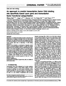
FIG. 1. Fragments of TFIIICa used to generate antisera. The TFIIICa amino acid sequence is diagrammed; the fragments shown below were expressed as His-tagged polypeptides in E. coli and used to generate antisera. The positions of glutamine-glycine bonds, preferred cleavage sites for poliovirus 3Cpro, are indicated above the diagram.
staining with Coomassie blue, pulverized by forcing through a syringe, and used to immunize rabbits twice every month with 50 to 100 mg of protein. Preimmune and immune sera were collected. For immunoprecipitation, 10 to 30 ml of phosphocellulose C (PC-C) fraction was diluted to 300 ml with IP buffer (0.7 M KCl, 1% Triton X-100, 2 mg of bovine serum albumin [BSA] per ml in buffer A [20 mM HEPES {N-2-hydroxyethylpiperazine-N9-2-ethanesulfonic acid}, pH 7.9, 20% glycerol, 0.2 mM EDTA, 1 mM dithiothreitol, 0.5 mM phenylmethylsulfonyl fluoride]) or 0.8-ml column fractions from the experiment shown in Fig. 7 were brought to 1.2 ml with the same buffer. These samples were precleared by incubation with 5 ml of nonrelated rabbit serum and 25 ml of protein A-Sepharose beads at room temperature for 45 min followed by centrifugation. Five microliters of preimmune or immune serum was added to the supernatant and incubated with mixing for 2 h at room temperature before the addition of 25 ml of protein A-Sepharose beads. After 1 additional h of incubation, immunoprecipitates were recovered by centrifugation and the pellets were washed five times each with 1 ml of IP buffer and phosphate-buffered saline (PBS). Immunoprecipitated proteins were recovered by elution with 30 ml of 1.53 SDS-PAGE sample buffer, and proteins were resolved by SDS–8% PAGE followed by 1 M sodium salicylate treatment and autoradiography. Immunoblot assays using a 1:1,000 dilution of antiserum in 3% nonfat dried milk–500 mM NaCl–20 mM Tris (pH 7.5)–0.1% Tween 20 were performed. Bound antibody was detected by using an Amersham ECL Western Kit. In vivo labeling conditions. For 35S labeling, HeLa cells on 150-mm-diameter plates at 80% confluency were washed with PBS three times. Cells were washed with DMEM-Met-Cys (Gibco BRL), and then 1.2 mCi of [35S] Protein Labeling Mix (catalog no. NEG-072; NEN) was added to 6 ml of DMEM-Met-Cys supplemented with 5% dialyzed fetal calf serum (Gibco BRL) per plate. Labeling was for 5 to 6 h. For 32P labeling, cells were washed three times with DMEM without phosphate (Gibco BRL). 32Pi (1.5 mCi) (ICN) was added to 6 ml of DMEM without phosphate supplemented with 5% dialyzed fetal calf serum per 150-mm-diameter plate. Labeling was for 6 h. For 35S or 32P labeling during poliovirus infections, poliovirus was added to each plate at a multiplicity of infection of 25 in 10 ml of DMEM-Met-Cys or DMEM without phosphate. Label was added at the same time as was virus. Virus absorption was carried out for 1 h, with occasional gentle shaking, after which newborn calf serum was added to a final concentration of 8%. Infection was continued for 5 to 5.5 h. Mock infections were performed identically, except that virus was omitted. In vitro transcription and translation. Template plasmids pTMIIICa1-732 and pTMIIICa1-740, containing the sequences of TFIIICa from the N terminus to amino acids 732 and 740, respectively, in the pTM-1 expression vector (14), were transcribed with T7 RNA polymerase according to the manufacturer’s instructions (Promega). The resultant cRNAs were translated by incubation in the rabbit reticulocyte lysate according to the manufacturer’s instructions (Promega). Two microliters of the in vitro-translated proteins was used in the immunoblot analysis shown in Fig. 4. Purification of and cleavage with recombinant 3Cpro. 3Cpro amplified by PCR from pT7PV1 was cloned into pQE30 (Qiagen) and transformed into E. coli M15. For protein induction, the E. coli culture at an optical density at 600 nm of 0.7 was induced with 2 mM IPTG (isopropyl-b-D-thiogalactopyranoside) for 4 h. 3Cpro was purified from E. coli lysate under nondenaturing conditions by nitrilotriacetic acid-nickel affinity chromatography. All 3Cpro digestions were carried out at 308C for 6 h, except the experiment shown in Fig. 7A, which was performed at 308C for 12 h. For the 3Cpro treatment of immunoprecipitated TFIIIC, 7 mg of 35S-labeled PC-C fraction or 11 mg of 32P-labeled PC-C fraction was immunoprecipitated with the individual sera. Each immunoprecipitated sample on 25 ml of protein A-Sepharose beads was resuspended in 15 ml of PBS with 6.8 or 8 mg of 3Cpro and digested at 308C for 12 h, after which 9 ml of 43 SDS-PAGE sample buffer was added to each sample. Extract preparation and fractionation. Nuclear extracts were prepared as previously described (13). For small-scale nuclear extract preparations from 35Sor 32P-labeled cells, cells were lysed as previously described (25). For 32P-labeled samples, phosphotase inhibitors (final concentrations: 20 mM NaF, 1 mM Na
MOL. CELL. BIOL. PPi, and 0.5 mM Na3VO4) were added to cells before they were lysed. Nuclear extract was fractionated by chromatography on phosphocellulose (P11; Whatman) in buffer A. Columns were loaded at 0.1 M KCl in buffer A and washed with 5 column volumes of 0.35 M KCl in buffer A, and the PC-C fraction was eluted with 0.7 M KCl in buffer A. The gradient elution of poliovirus-infected cell nuclear extracts was performed as previously described (6), except that 10 mg of poliovirus-infected nuclear extract was loaded at 0.1 M KCl onto a 1-ml phosphocellulose column, washed with 5 ml of 0.1 M KCl in buffer A, and eluted with 20 ml of a 0.1 to 0.7 M KCl gradient in buffer A and subsequently with 5 ml of 0.7 M KCl in buffer A. For the gradient elution of in vitro-cleaved TFIIIC, 4 mg of HeLa cell PC-C fraction was incubated with 280 mg of 3Cpro at 308C for 12 h, loaded on a 1-ml phosphocellulose column at 0.1 M KCl, washed with 5 column volumes of 0.1 M KCl, and eluted with 20 ml of a 0.1 to 0.7 M KCl gradient, all in buffer A. Four microliters (each) of fractions 3 through 16 was analyzed by EMSA. For the experiment shown in Fig. 7B, 400 mg of 35S-labeled HeLa cell PC-C fraction was digested with 3Cpro, precleared with nonrelated rabbit serum and protein A-Sepharose beads, and adjusted to 90 mM KCl in buffer Z (20 mM HEPES [pH 7.9], 10% glycerol, 0.1 mM ZnCl2, 5 mM dithiothreitol, 5 mM MgCl2, 0.1% Nonidet P-40, 0.5 mM phenylmethylsulfonyl fluoride) before binding to 100 ml of B-box oligonucleotide affinity resin as previously described (27). The column was washed with 0.6 ml of 0.1 M KCl–buffer Z and subsequently with 0.4 ml of 0.2 M KCl–buffer Z. The TFIIIC activity was eluted with 0.5 ml of 0.7 M KCl–buffer Z. The 0.7 M KCl eluate was diluted with buffer A containing 1 mg of BSA per ml to 0.1 M KCl before being loaded on a 1-ml phosphocellulose column and subjected to gradient elution. To determine the stoichiometry of the a and b subunits, the 0.7 M KCl eluate of the B-box column was diluted to 0.09 M KCl with buffer Z and incubated with another 100 ml of B-box oligonucleotide affinity resin. The second round of B-box affinity chromatography was performed as described for the first round. Protein eluted with 0.7 M KCl–buffer Z was subjected to SDS-PAGE, and the dried gel was analyzed with a Molecular Dynamics PhosphorImager. EMSA. The EMSA probe was prepared by labeling the 39 ends of the 129-bp XbaI-BstEII fragment of pVA1A containing the B-box. Protein fractions were incubated for 30 min at 308C with ;10 fmol of probe in 70 mM KCl–3.5 mM MgCl2–20 mM HEPES (pH 7.9)–8% glycerol–5 mM dithiothreitol–0.04 mM EDTA in 15 ml, with 1.8 mg of poly(dI-dC) and 0.3 mg of pBluescriptII SK1 as nonspecific competitors. Samples were resolved on a prerun 15-cm 4% polyacrylamide (acrylamide/bisacrylamide ratio, 30:0.8) gel in 0.253 TBE (22.5 mM Tris, 22.5 mM boric acid, 0.5 mM EDTA) at room temperature for 2 h at 150 V. For EMSA experiments with the addition of anti-TFIIICa antibodies, preimmune or immune immunoglobulin G was purified in batch on protein A-Sepharose as previously described (18). The final volumes were one-third of the starting volumes. Protein fractions were incubated with 1 ml of purified antibodies on ice for 30 min in EMSA buffer, the EMSA probe was added, and the incubations were continued at 308C for 30 min. The reaction mixtures were loaded onto a 1.6% agarose gel in 0.53 TBE and subjected to electrophoresis at room temperature for 3 h at 125 V. For the EMSA experiment with the antiTFIIICb antiserum, 0.3 ml of the control nonrelated rabbit serum or antiTFIIICb serum was preincubated on ice with protein fractions for 15 min in EMSA buffer. Then the EMSA was performed as described above.
RESULTS Specific antisera raised against fragments of TFIIICa immunoprecipitate the multimeric TFIIIC complex. Previous results showed that TFIIIC was cleaved by 3Cpro both in vivo and in vitro to generate two types of complexes with specific DNA binding activities. These complexes were distinguished from undigested TFIIIC by EMSA using a DNA probe containing the adenovirus type 2 VAI gene B-box (6, 7). We hereafter call the complex with the highest mobility TFIIICpolio and the more slowly migrating complex TFIIICpolio*. To analyze which subunit(s) of TFIIIC is cleaved by 3Cpro, we raised specific antisera against different fragments of TFIIICa expressed in E. coli. Ab2, Ab4, and Ab7 were raised against portions of TFIIICa corresponding to amino acid residues 302 to 585, 753 to 1082, and 1745 to 2110, respectively (Fig. 1). These three antisera specifically immunoprecipitated the five TFIIIC subunits from partially purified fractions of TFIIIC prepared from HeLa cells metabolically labeled with [35S]methionine and cysteine or 32Pi (Fig. 2). Less 35S label was observed in the b and d subunits compared with the other subunits. This was not caused by a significant difference in the half-lives of these TFIIIC subunits, since the relative amounts of label in the subunits were similar when cells were labeled continuously for either 4 or 36 h and similarly slow rates of
VOL. 16, 1996
DNA BINDING DOMAIN AND SUBUNIT INTERACTIONS OF TFIIIC
FIG. 2. Immunoprecipitation of the TFIIIC complex. Shown are autoradiograms of SDS-PAGE gels of immunoprecipitates of the PC-C fraction (0.35 to 0.7 M KCl eluate) prepared from labeled HeLa cells. Lanes 1 and 2, immunoprecipitates from 35S-labeled HeLa cells with preimmune (PI) serum and Ab4 immune serum, respectively; lanes 3 and 4, immunoprecipitates from 32P-labeled HeLa cells with PI serum and Ab2 immune serum, respectively. The positions of molecular mass standards (M; in kilodaltons) are indicated.
decay were observed for all five subunits in pulse-chase experiments (31a). Since cDNAs encoding the a and b subunits have been isolated and sequenced, the amino acid compositions of these subunits are known. On the basis of this information and the relative number of counts per minute in the a and b subunits after metabolic labeling with methionine and cysteine, we estimate that the molar ratio of the a and b subunits in TFIIIC is 2:1 (calculated molar ratios of 2.0 and 2.1 in two independent experiments). All five subunits were labeled with 32 P. Comparing the intensities of TFIIIC subunits from 35Sand 32P-labeled cells, the a, b, and ε subunits showed higher 32 35 P/ S ratios than did the g and d subunits, indicating that they were more extensively phosphorylated. In subsequent experiments, the 110-kDa b subunit was observed most clearly with 32 P-labeled protein. TFIIIC a and d subunits are cleaved by poliovirus 3Cpro in vitro. Purified bacterially expressed poliovirus 3Cpro cleaves TFIIIC in vitro, as shown by an increase in the mobility of the B-box binding activity assayed by EMSA (7). To determine which subunits are cleaved by 3Cpro, 35S-labeled TFIIIC (Fig. 3B, lanes 11 to 16) and 32P-labeled TFIIIC (Fig. 3C, lanes 9 to 12) were purified by phosphocellulose chromatography and immunoprecipitation and the immune complexes bound to protein A-Sepharose beads were incubated with purified recombinant 3Cpro. The digestion products were analyzed by SDS-PAGE and autoradiography. Similar results were observed with antibodies directed against three different portions of the a subunit (Fig. 3B, lanes 9 to 16) (data not shown for 32 P-labeled protein), indicating that bound antibody did not block the access of this proteinase to the TFIIIC complex. The b (110-kDa), g (100-kDa), and ε (60-kDa) subunits did not appear to be cleaved by 3Cpro. The a subunit was cleaved, and
4165
two major cleavage products with molecular masses of ;125 and ;90 kDa were generated. The 32P-labeled sample showed that the d (80-kDa) subunit was also cleaved by 3Cpro (Fig. 3C, lane 12). Cleavage of the d subunit was also apparent in a longer exposure of the 35S-labeled protein (data not shown). Several minor cleavage products, including a doublet of ;150 kDa and an ;78-kDa fragment, were also observed. To specifically analyze the cleavage products of the a subunit, partially purified labeled TFIIIC was cleaved with 3Cpro prior to immunoprecipitation and the resulting subcomplexes were then subjected to immunoprecipitation using the antisera raised against specific regions of the a subunit. Under these conditions, TFIIIC was completely cleaved by 3Cpro to generate TFIIICpolio and TFIIICpolio* complexes, as analyzed by EMSA (Fig. 3A). When Ab2 was used to immunoprecipitate cleaved TFIIIC, it brought down much more of the ;90kDa cleavage product than the ;125-kDa cleavage product (Fig. 3B, lanes 4 and 16, and C, lanes 4 and 12). The 110-kDa b subunit coprecipitated with the 90-kDa cleavage product (Fig. 3C, lane 4). In contrast, when Ab7 was used, it brought down the ;125-kDa cleavage product and the 100- and 60-kDa g and ε subunits but very little of the ;90-kDa cleavage product or the 110-kDa b subunit (Fig. 3B, lanes 8 and 12, and C, lanes 8 and 12). Immunoprecipitation of 3Cpro-cleaved TFIIIC with Ab4 produced results similar to those with Ab7 (data not shown). These immunoprecipitation experiments suggested that the ;90-kDa cleavage product was derived from the N-terminal region of the a subunit, while the ;125kDa cleavage product was derived from the C-terminal region. This was confirmed by immunoblot analyses. Ab2 recognized the ;90-kDa cleavage product, while both Ab4 and Ab7 recognized the ;125-kDa cleavage product and minor cleavage products of ;150 kDa (Fig. 3D). The intensities of the ;150kDa doublet bands were variable but were always less than that of the ;125-kDa cleavage product, suggesting that the ;150kDa doublets are digestion intermediates which are further cleaved to the ;125-kDa fragment. The data in Fig. 3 indicate that the 110-kDa b subunit of TFIIIC is associated with an ;90-kDa domain from the N-terminal region of the a subunit, while the 100-kDa g and 60-kDa ε subunits are associated with an ;125-kDa domain from the C-terminal region of the a subunit. One 3Cpro cleavage site maps near or at Q-732G-733. Poliovirus 3Cpro preferentially cleaves glutamine-glycine bonds (30). There are seven glutamine-glycine bonds in the TFIIICa sequence (Fig. 1). The results shown in Fig. 3 suggested that either the Q-732G-733 or Q-740G-741 site was cleaved to generate an ;85-kDa cleavage product from the N terminus which migrated as ;90 kDa on the SDS-PAGE gel. To assess whether the Q-732G-733 or Q-740G-741 site was cleaved by 3Cpro, PCR was used to introduce stop codons into cDNA clones of the a subunit after Q-732 or Q-740. These mutant cDNAs were transcribed and translated in vitro. Then the translation products were analyzed by immunoblotting, using Ab2 and TFIIIC cleavage products generated by 3Cpro digestion as markers. Fragment 1-732 of TFIIICa (lane 3) comigrated with the 3Cpro cleavage product (Fig. 4), strongly suggesting that 3Cpro cleaves TFIIIC between Q-732 and G-733 to generate an 83-kDa N-terminal fragment. Poliovirus infection generates the same TFIIIC cleavage products as does 3Cpro in vitro digestion. We analyzed the cleavage of TFIIIC subunits in vivo during poliovirus infection to determine if it could be explained by the 3Cpro cleavage sites observed in vitro. HeLa cells were metabolically labeled with [35S]methionine (Fig. 5A) or 32Pi (Fig. 5B) during poliovirus infection. Extracts prepared from cells 5 h after infection
4166
SHEN ET AL.
MOL. CELL. BIOL.
FIG. 3. Cleavage of TFIIIC with purified 3Cpro in vitro. (A) 35S-labeled PC-C fraction was cleaved with 3Cpro (lane 3) or was incubated under identical conditions without the addition of 3Cpro (lane 2). The protein fractions were then used in an EMSA with a 32P-labeled B-box DNA probe. An autoradiogram of the EMSA gel under conditions that detect only 32P is shown. Lane 1, no protein added. The positions of uncleaved and cleaved TFIIIC complexes are indicated on the right. FP, free probe; NS, nonspecific DNA binding activity. (B) Lanes 1 to 8, 35S-labeled PC-C fraction cleaved with 3Cpro (1) or incubated under identical conditions without the addition of 3Cpro (2) was immunoprecipitated (IP) after digestion with the indicated preimmune (PI) or immune serum; lanes 11 to 16, TFIIIC digested with 3Cpro after immunoprecipitation with the indicated serum before dissociation from protein A-Sepharose beads; lanes 9 and 10, immunoprecipitations of 35S-labeled PC-C fraction incubated without 3Cpro; lane M, molecular mass markers. The numbers on the left refer to the molecular masses (in kilodaltons) of the markers; the numbers on the right refer to specific protein bands with the indicated estimated molecular masses. Numbers without parentheses indicate the positions of uncleaved TFIIIC subunits; numbers in parentheses refer to new bands generated by 3Cpro cleavage. (C) The same as described for panel B, except that the PC-C fraction was prepared from HeLa cells labeled with 32Pi. (D) Immunoblots of HeLa PC-C fraction digested with 3Cpro (1) or incubated under identical conditions without the addition of 3Cpro (2). The antisera used for immunoblotting are indicated above the lanes, and the major bands detected are indicated on the right.
were fractionated by chromatography on phosphocellulose, and equal amounts of the PC-C fraction from mock-infected and poliovirus-infected cells were subjected to immunoprecipitation using the three anti-TFIIICa sera. Less label was incorporated into TFIIIC polypeptides from poliovirus-infected
cells compared with that from mock-infected cells because of the increasing inhibition of host mRNA translation during the course of poliovirus infection (1, 19). The same cleavage products of ;90 and ;125 kDa and a doublet of ;150 kDa observed after TFIIIC cleavage with purified recombinant 3Cpro
VOL. 16, 1996
DNA BINDING DOMAIN AND SUBUNIT INTERACTIONS OF TFIIIC
4167
TFIIICpolio*, along with uncleaved TFIIIC, eluted at 380 to 470 mM KCl (data not shown) (see Fig. 7A). Fractions containing TFIIICpolio were pooled, as were fractions containing uncleaved TFIIIC and TFIIICpolio*, and the pooled fractions were incubated with preimmune or specific anti-TFIIICa serum before an EMSA was performed (Fig. 6). Ab2 specifically inhibited complex formation by TFIIIC (Fig. 6, lane 11) as well
FIG. 4. Mapping of the N-terminal 3Cpro cleavage site in TFIIICa. Immunoblots were performed with Ab2 on uncleaved PC-C (lane 1), 3Cpro-cleaved PC-C (lane 2), and in vitro-transcribed and -translated pTMIIICa1-732 (lane 3) and pTMIIICa1-740 (lane 4). aa., amino acids.
in vitro (Fig. 3) were observed during poliovirus infection in vivo (Fig. 5), confirming that poliovirus 3Cpro is responsible for TFIIIC cleavage during poliovirus infection. However, cleavage was not as complete as in our in vitro cleavage reactions, since the full-length a subunit persisted and the intensities of the ;150-kDa partial digestion products from the Cterminal region were similar to that of the C-terminal ;125kDa final digestion product. An ;200-kDa polypeptide migrating just faster than the uncleaved a subunit was also observed. A fragment of similar size was observed after short periods of cleavage with 3Cpro in vitro (data not shown), suggesting that it is also a product of partial 3Cpro digestion. The 80-kDa d subunit was probably also cleaved during poliovirus infection since it was observed at a much lower ratio to the other subunits in immunoprecipitates from poliovirus-infected cells than in those from mock-infected cells. 3Cpro cleavage identifies a TFIIIC DNA binding domain with portions of both the a and b subunits. TFIIICa is the only human TFIIIC subunit which is specifically UV crosslinked to bromouracil-containing B-box DNA (23, 37). However, no obvious known DNA binding motif can be identified in the a subunit sequence (24, 27). To determine which portion of TFIIICa forms the B-box DNA binding domain, we analyzed the specific DNA-binding subcomplexes generated from TFIIIC by 3Cpro digestion. The different TFIIIC complexes generated during poliovirus infection were separated by salt gradient elution of nuclear extract protein absorbed to a phosphocellulose column. As assayed by EMSA, TFIIICpolio eluted first from the column at 310 to 380 mM KCl, while
FIG. 5. Analysis of TFIIIC cleavage during poliovirus infection in vivo. Shown are immunoprecipitations of PC-C fractions prepared from poliovirusinfected, in vivo-labeled cells. (A) Equal amounts of 35S-labeled PC-C prepared from mock-infected (lanes 1, 2, 5, 6, and 9) and poliovirus-infected (lanes 3, 4, 7, 8, and 10) cells were subjected to immunoprecipitation with the indicated sera (Ab). Major bands are indicated on the right, with bands specific for poliovirusinfected cells in parentheses. (B) Equal amounts of 32P-labeled PC-C prepared from mock-infected (lanes 1 to 6) and poliovirus-infected (lanes 7 to 12) cells were subjected to immunoprecipitation with the indicated sera (Ab). The bands on the right are the same as described for panel A. Lanes M, molecular mass markers (in kilodaltons) whose positions are shown on the left.
4168
SHEN ET AL.
FIG. 6. The TFIIICa DNA binding domain is within the N-terminal 732 residues. EMSA was used to analyze the effects of anti-TFIIICa antibodies (Ab) on the specific DNA binding activities of TFIIIC and poliovirus-specific TFIIIC complexes. Poliovirus-infected HeLa cell nuclear extract was fractionated on a phosphocellulose column by linear salt gradient elution. Fractions (Fr) containing mainly TFIIICpolio (lanes 2 to 8) or containing uncleaved TFIIIC with small amounts of TFIIICpolio* and TFIIICpolio (lanes 9 to 15) were used in gel shift assays. Protein fractions were either untreated (no Ab; lanes 2 and 9) or preincubated with the indicated protein A-Sepharose-purified antibodies before incubation with the B-box DNA probe. No protein was added in lane 1. The positions of DNA-protein complexes are indicated on the right. NS, nonspecific DNA binding activity; FP, free probe; pp, supershifted complex.
as by TFIIICpolio (lane 4) and TFIIICpolio* (lane 11). However, while Ab4 and Ab7 specifically inhibited complex formation by TFIIIC (Fig. 6, lanes 13 and 15), they did not inhibit complex formation by TFIIICpolio (lanes 6 and 8) or TF II ICpolio* (lanes 13 and 15). These results indicate that TF II ICpolio and TFIIICpolio* contain the N-terminal 83-kDa fragment of the a subunit generated by 3Cpro cleavage but not larger fragments of the a subunit that would interact with Ab4 or Ab7. Consistent with this hypothesis, we found that the 83-kDa N-terminal fragment was preferentially retained on a B-box DNA affinity column compared with the ;125- and ;150-kDa fragments (data not shown). To directly analyze the polypeptide compositions of TF II ICpolio and TFIIICpolio* complexes, complexes generated by 3Cpro in vitro cleavage of partially purified TFIIIC were separated by phosphocellulose chromatography and immunoprecipitated. Under the conditions used, all of the TFIIIC was cleaved into either TFIIICpolio or TFIIICpolio*, which was then resolved by salt gradient elution from phosphocellulose (Fig. 7A). TFIIIC polypeptides were analyzed by immunoprecipitation with Ab2 (Fig. 7B). The a subunit N-terminal fragment (indicated as 90 kDa) was found to fractionate into two peaks, one coincident with the peak of TFIIICpolio activity and the other coincident with TFIIICpolio* activity. An immunoprecipitated polypeptide with the mobility of TFIIICb (110 kDa) also coeluted with the peak of TFIIICpolio* activity. Light bands corresponding to an immunoprecipitated polypeptide of ;78 kDa coeluted with the TFIIICpolio activity peak. This ;78-kDa cleavage product was observed earlier (Fig. 3B and C), but it was not possible to distinguish from which TFIIIC subunit it was derived. While other polypeptides were observed on the autoradiogram, they did not correspond to the
MOL. CELL. BIOL.
molecular weights of the known TFIIIC subunits or 3Cpro cleavage products. To test if the 110-kDa polypeptide coprecipitating with the TFIIICa N-terminal fragment is indeed the b subunit, EMSAs using these protein fractions and specific antiserum raised against the b subunit (32) were performed. TFIIICpolio* complex formation was specifically inhibited by the anti-b subunit antiserum, demonstrating that the B-box-binding subcomplex includes the b subunit (Fig. 8, lanes 6 and 9). Of particular interest, complex formation by TFIIICpolio in fraction 6 was also blocked by the anti-b subunit serum and a fraction of the probe was observed in a supershifted complex (Fig. 8, lane 3). The results of immunoprecipitations and EMSAs together demonstrate that the TFIIICpolio* B-box-specific DNA binding activity generated from TFIIIC by digestion with 3Cpro results from a complex of the N-terminal 83-kDa fragment of the a subunit with the intact 110-kDa b subunit. TFIIICpolio activity appears to be due to the N-terminal 83-kDa fragment of the a subunit complexed with an ;78-kDa fragment of the b subunit. Taken as a whole, the results of these studies demonstrate that the DNA binding domain of TFIIICa lies within amino acid residues 1 to 732. Furthermore, the specific DNAbinding subcomplexes of TFIIIC generated by digestion with 3Cpro also contain TFIIICb or a large fragment of it. DISCUSSION The inactivation of TFIIIC during poliovirus infection was first detected by a decrease in TFIIIC transcriptional activity in extracts of infected cells, although no decrease in B-box-specific DNA binding activity was apparent when assayed by DNase I footprinting (15). The cleavage of TFIIIC during the course of infection became evident when EMSA was used to assay B-box binding activity. Two new B-box binding activities were generated at the expense of the TFIIIC complex with a much slower mobility (6). The mobilities of the B-box binding activities generated during poliovirus infection were similar to those of complexes formed by TFIIIC after partial digestion with chymotrypsin or V8 protease (4). Digestion of partially purified TFIIIC with the poliovirus 3Cpro in vitro produced DNA-protein complexes with the same rapid mobilities as those in extracts of infected cells. This demonstrated that 3Cpro, the protease that makes most of the cleavages in the poliovirus polyprotein, was responsible for the cleavage of TFIIIC during poliovirus infection (7). TFIIIC is an unusual cellular protein in this regard. Most cellular proteins are not cleaved during viral infection (30). Indeed, TFIIIC (7) and the TATA-box-binding polypeptide (8) are the only cellular proteins known to be cleaved by this viral protease. The development of techniques for the purification of TFIIIC (37), the cloning of a cDNA encoding the 243-kDa TFIIICa (24, 27), and the availability of recombinant poliovirus 3Cpro (29) allowed us to characterize the 3Cpro cleavages made in the a subunit of TFIIIC. This allowed us to localize the B-box DNA binding domain in the a subunit by analyzing the polypeptide composition of the B-box binding activity dissected from TFIIIC by 3Cpro. The cleavage of TFIIIC by recombinant 3Cpro in vitro was more complete than it was in poliovirus-infected HeLa cells isolated just before cell lysis (Fig. 3 and 5). 3Cpro has a preference for cleavage at accessible glutamine-glycine bonds in the poliovirus polyprotein (6, 11, 30). One site of TFIIICa cleavage appears to be between Q-732 and G-733, since an N-terminal 3Cpro cleavage product comigrated during SDS-PAGE with an in vitro-translated fragment of TFIIICa extending from residues 1 to 732 (Fig. 3 and 4). A second major cleavage product had a mobility on the
VOL. 16, 1996
DNA BINDING DOMAIN AND SUBUNIT INTERACTIONS OF TFIIIC
4169
FIG. 7. Resolution of TFIIICpolio and TFIIICpolio* complexes by phosphocellulose column gradient elution. (A) PC-C fraction was digested with 3Cpro in vitro and subjected to chromatography on phosphocellulose using a linear gradient elution from 0.1 to 0.7 M KCl in buffer A. Fractions (Fr) were assayed for specific B-box DNA binding activity by EMSA. FP, free probe. (B) Immunoprecipitation of phosphocellulose gradient-eluted fractions with Ab2. 35S-labeled HeLa cell PC-C fraction was cleaved with 3Cpro in vitro. The resultant TFIIICpolio and TFIIICpolio* were purified on a B-box DNA oligonucleotide affinity column and subjected to chromatography on phosphocellulose, as described for panel A. Column fractions were immunoprecipitated with Ab2, and the immunoprecipitates were resolved by SDS-PAGE. An autoradiogram of the gel is shown. The numbers on the left refer to the molecular masses (in kilodaltons) of marker proteins (M); the numbers on the right indicate protein bands discussed in the text.
SDS-PAGE gel that predicted a molecular mass of ;125 kDa. Since it reacted with antibodies raised to TFIIICa fragments from amino acid residues 753 to 1082 (Ab4) and 1745 to 2110, it must extend in the N-terminal direction to approximately at least residue 1075 and in the C-terminal direction to approximately at least residue 1750. A likely candidate for this fragment generated by 3Cpro extends from the glutamine-glycine bond at G-1067 to near the C terminus, where there are two glutamine-glycine bonds (Fig. 1). Further work will be required to precisely delineate the a subunit residues present in this 3Cpro digestion product. Two B-box binding activities, called TFIIICpolio and TF II ICpolio*, were generated by TFIIIC cleavage with recombinant 3Cpro. The mobilities of both were significantly greater than that of the B-box complex with native TFIIIC (Fig. 3A).
Both of these complexes contained the N-terminal 83-kDa fragment of TFIIICa and lacked other fragments of the a subunit generated by 3Cpro (Fig. 7). This conclusion was supported by the effects of antibodies raised to specific regions of the a subunit primary sequence on the DNA binding activities of TFIIICpolio and TFIIICpolio*. Antibodies raised against TFIIICa fragments C terminal to Q-732 inhibited B-box DNA binding by undigested TFIIIC but did not inhibit binding by TFIIICpolio or TFIIICpolio*, whereas antibodies raised to an N-terminal fragment inhibited binding by native TFIIIC as well as by TFIIICpolio and TFIIICpolio*. TFIIICpolio* also contained the 110-kDa b subunit of TFIIIC (Fig. 7B and 8). TFIIICpolio contained an ;78-kDa polypeptide generated by 3Cpro cleavage. This was probably a fragment of the b subunit, since antiserum to the b subunit inhibited B-box binding by
4170
SHEN ET AL.
FIG. 8. The 3Cpro fragments of TFIIIC that bind DNA specifically contain the b subunit. PC-C fraction (lanes 10 to 12) or the indicated fractions (Fr) from the phosphocellulose column analyzed in Fig. 7A containing TFIIICpolio and/or TFIIICpolio* (lanes 1 to 9) were incubated with antiserum (Ab) raised against TFIIICb (anti-TFIIICb) or a control antiserum (control) or were untreated (none). The protein fractions were then subjected to EMSA using a B-box DNA probe. FP, free probe; NS, nonspecific DNA binding activity; pp, supershifted complex; Gel ori., gel origin.
both TFIIICpolio and TFIIICpolio* and generated supershifted complexes (Fig. 8). These results indicate that the DNA binding domain of the a subunit lies within the N-terminal 83-kDa fragment generated by 3Cpro. We assayed for specific B-box binding activity by both the full-length a subunit and the N-terminal fragment extending to Q-732 generated by in vitro translation. However, we failed to observe this activity even when we used several times the mass of protein required to detect specific B-box binding with TFIIICpolio or TFIIICpolio*. These results suggest that the portion of the b subunit included in the ;78-kDa fragment is required for specific B-box DNA binding activity. 3Cpro cleavage of TFIIIC inactivates its transcriptional activity (7), although the DNA binding activities of the cleavage products were unchanged (Fig. 3A). We found that cleaved TFIIIC inhibited transcription when added to an in vitro reaction mixture reconstituted with partially purified factors and polymerase (31a). Inhibition was as expected for a simple competition for template binding between active TFIIIC and the two transcriptionally inactive forms of cleaved TFIIIC. Since the cleaved forms of TFIIIC that bind the B-box promoter element appear to lack the 100-kDa g, 80-kDa d, and 60-kDa ε subunits and are defective for transcription, it is likely that at least one of these subunits is required for TFIIIC’s function in transcription. Much of the uncleaved TFIIIC in extracts of poliovirusinfected cells may be inactivated by dephosphorylation (6). For this model to be correct, active TFIIIC must be phosphorylated. Consistent with this, we found that all five subunits of TFIIIC can be metabolically labeled with 32Pi (Fig. 2). Full-
MOL. CELL. BIOL.
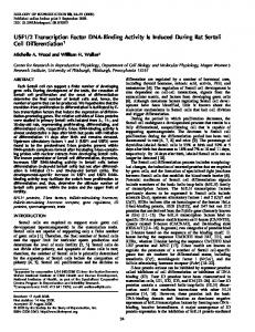
length a, b, g, and ε subunits immunoprecipitated from extracts of 32P-labeled poliovirus-infected HeLa cells were also labeled, indicating that none are fully dephosphorylated during poliovirus infection. The lower overall level of 32P label observed in TFIIIC subunits from poliovirus-infected cells compared with that from mock-infected cells (Fig. 5B) may be due to the inhibition of protein synthesis during poliovirus infection (1, 19), which also resulted in a decrease in TFIIIC subunit label in [35S]methionine-labeled cells (Fig. 5A). Further studies will be required to determine if phosphate is removed from any specific sites in any of the TFIIIC subunits during poliovirus infection. Antisera raised against fragments of TFIIICa specifically immunoprecipitated the five polypeptides identified as TFIIIC subunits by purification of transcriptional and B-box binding activities (Fig. 2) (23, 37). In proteins labeled metabolically with [35S]methionine and cysteine, much more label was observed in the immunoprecipitated a (240-kDa), g (100-kDa), and ε (60-kDa) subunits than in the immunoprecipitated b (110-kDa) and d (80-kDa) subunits (Fig. 2). By taking into account the methionine and cysteine contents of the a and b subunits predicted from the sequences of their cDNA clones (24, 27, 32), the molar ratio of a to b subunits in human TFIIIC purified by B-box affinity chromatography was ;2 to 1. In contrast, studies of yeast TFIIIC indicated that the two largest subunits are each present in one copy in the yeast protein (26, 28). It seems unlikely that the human and yeast proteins differ from each other in the stoichiometry of subunits with equivalent functions. One possible explanation for this discrepancy is that the b subunit is present only in a subset of human TFIIIC complexes, a hypothesis proposed by Sinn et al. (32). This seems unlikely, however, since all the DNA binding activities of our TFIIIC preparations were blocked by antiserum to the b subunit (Fig. 8, lane 12). The amino acid sequence of the human b subunit is not homologous to that of any of the three yeast subunits so far cloned. Consequently, it is not clear that the second-largest subunits of human TFIIIC and yeast TFIIIC have equivalent functions. Further studies will be required to resolve the apparent discrepancy in the stoichiometry of the subunits of yeast TFIIIC and human TFIIIC. Associations of TFIIIC subunits with the two major 3Cpro fragments of the a subunit were revealed by immunoprecipitating the a fragments separately. The b subunit preferentially coprecipitated with the B-box-binding N-terminal 83-kDa fragment, while the g and ε subunits coprecipitated preferentially with the C-terminal ;125-kDa fragment (Fig. 3B and C). The d subunit was eliminated by 3Cpro digestion and consequently could not be identified in immunoprecipitates of TFIIIC subcomplexes. Figure 9 summarizes the associations of TFIIIC subunits with domains of the a subunit. We propose that the b subunit associated with the DNA binding N-terminal domain of the a subunit lies near the B-box, while the C-terminal region of the a subunit associated with the g and ε subunits extends toward the transcription start site, where it can direct the binding of TFIIIB upstream of the transcription start (17, 21). In yeast TFIIIC, the second-largest subunit, TFC120, is positioned over the 59 end of a tRNA gene (2, 3), where it interacts with the BRF subunit of TFIIIB to position TFIIIB upstream of the transcription start site (3, 5, 22). As mentioned above, the sequence of the human TFIIIC 110-kDa b subunit is not recognizably related to yeast TFC120 (28, 32). Our finding that the human b subunit is bound to the B-box binding domain of the a subunit suggests that the human b subunit is not functionally equivalent to yeast TFC120. At present, it is
VOL. 16, 1996
DNA BINDING DOMAIN AND SUBUNIT INTERACTIONS OF TFIIIC
FIG. 9. Summary of the associations of TFIIIC subunits with domains of the a subunit. Digestion of TFIIICa by poliovirus 3Cpro revealed N- and C-terminal protease-resistant domains connected by a protease-sensitive region. The 83-kDa domain at the N terminus interacts with TFIIICb and binds to the B-box promotor element. The 125-kDa domain at the C terminus associates with the 100-kDa g subunit and the 60-kDa ε subunit. No conclusions concerning the 80-kDa d subunit could be drawn since it was also cleaved by 3Cpro. The arrow labeled Tx indicates the direction of transcription.
not clear which of the TFIIIC subunits is functionally equivalent to the mechanistically critical yeast TFC120 subunit or whether this polypeptide is a component of the TFIIIC1 protein fraction required for transcription in addition to TFIIIC, pol III, and TFIIIB (24, 27, 36). ACKNOWLEDGMENTS We thank Robert Roeder for providing the anti-TFIIICb antiserum. Y.S. thanks Fanping Wang for encouragement and helpful discussions. This work was supported by National Institutes of Health grants to A.J.B. (CA25235) and A.D. (AI-27451). P.Y. was supported by Public Health Service Awards AI-07323 and GM-07104 from the National Institutes of Health. REFERENCES 1. Bablanian, R. 1975. Structural and functional alterations in cultured cells infected with cytocidal viruses. Prog. Med. Virol. 19:40–83. 2. Bartholomew, B., B. R. Braun, G. A. Kassavetis, and E. P. Geiduschek. 1994. Probing close DNA contacts of RNA polymerase III transcription complexes with the photoactive nucleoside 4-thiodeoxythymidine. J. Biol. Chem. 269: 18090–18095. 3. Bartholomew, B., G. A. Kassavetis, and E. P. Geiduschek. 1991. Two components of Saccharomyces cerevisiae transcription factor IIIB (TFIIIB) are stereospecifically located upstream of a tRNA gene and interact with the second-largest subunit of TFIIIC. Mol. Cell. Biol. 11:5181–5189. 4. Boulanger, P. A., N. D. L’Etoile, and A. J. Berk. 1989. A DNA-binding domain of human transcription factor IIIC2. Nucleic Acids Res. 17:7761– 7770. 5. Chaussivert, N., C. Conesa, S. Shaaban, and A. Sentenac. 1995. Complex interactions between yeast TFIIIB and TFIIIC. J. Biol. Chem. 270:15353– 15358. 6. Clark, M. E., and A. Dasgupta. 1990. A transcriptionally active form of TFIIIC is modified in poliovirus-infected HeLa cells. Mol. Cell. Biol. 10: 5106–5113. 7. Clark, M. E., T. Hammerle, E. Wimmer, and A. Dasgupta. 1991. Poliovirus proteinase 3C converts an active form of transcription factor IIIC to an inactive form: a mechanism for inhibition of host cell polymerase III transcription by poliovirus. EMBO J. 10:2941–2947. 8. Clark, M. E., P. M. Lieberman, A. J. Berk, and A. Dasgupta. 1993. Direct cleavage of human TATA-binding protein by poliovirus protease 3C in vivo and in vitro. Mol. Cell. Biol. 13:1232–1237. 9. Conesa, C., R. N. Swanson, P. Schultz, P. Oudet, and A. Sentenac. 1993. On the subunit composition, stoichiometry, and phosphorylation of the yeast transcription factor TFIIIC/tau. J. Biol. Chem. 268:18047–18052. 10. Crawford, N., A. Fire, M. Samuels, P. A. Sharp, and D. Baltimore. 1981. Inhibition of transcription factor activity by poliovirus. Cell 27:555–61.
4171
11. Das, S., and A. Dasgupta. 1993. Identification of the cleavage site and determinants required for poliovirus 3CPro-catalyzed cleavage of human TATA-binding transcription factor TBP. J. Virol. 67:3326–3331. 12. Dasgupta, A. 1983. Purification of host factor required for in vitro transcription of poliovirus RNA. Virology 128:245–251. 13. Dignam, J. D., R. M. Lebovitz, and R. G. Roeder. 1983. Accurate transcription initiation by RNA polymerase II in a soluble extract from isolated mammalian nuclei. Nucleic Acids Res. 11:1475–1489. 14. Elroy-Stein, O., T. R. Fuerst, and B. Moss. 1989. Cap-independent translation of mRNA conferred by encephalomyocarditis virus 59 sequence improves the performance of the vaccinia virus/bacteriophage T7 hybrid expression system. Proc. Natl. Acad. Sci. USA 86:6126–6130. 15. Fradkin, L. G., S. K. Yoshinaga, A. J. Berk, and A. Dasgupta. 1987. Inhibition of host cell RNA polymerase III-mediated transcription by poliovirus: inactivation of specific transcription factors. Mol. Cell. Biol. 7:3880–3887. 16. Gabrielsen, O. S., N. Marzouki, A. Ruet, A. Sentenac, and P. Fromageot. 1989. Two polypeptide chains in yeast transcription factor tau interact with DNA. J. Biol. Chem. 264:7505–7511. 17. Geiduschek, E. P., and G. A. Kassavetis. 1995. Comparing transcriptional initiation by RNA polymerases I and III. Curr. Opin. Cell Biol. 7:344–351. 18. Harlow, E., and D. Lane. 1988. Antibodies: a laboratory manual. Cold Spring Harbor Laboratory, Cold Spring Harbor, N.Y. 19. Kaariainen, L., and M. Ranki. 1984. Inhibition of cell functions by RNAvirus infections. Annu. Rev. Microbiol. 38:91–109. 20. Kassavetis, G. A., B. R. Braun, C. A. P. Joazeiro, M. Pisano, and E. P. Geiduschek. 1994. Transcription by RNA polymerase III, p. 107–126. In R. C. Conaway and J. W. Conaway (ed.), Transcription mechanisms and regulation, vol. 3. Raven Press, New York. 21. Kassavetis, G. A., B. R. Braun, L. H. Nguyen, and E. P. Geiduschek. 1990. S. cerevisiae TFIIIB is the transcription initiation factor proper of RNA polymerase III, while TFIIIA and TFIIIC are assembly factors. Cell 60:235–245. 22. Khoo, B., B. Brophy, and S. P. Jackson. 1994. Conserved functional domains of the RNA polymerase III general transcription factor BRF. Genes Dev. 8:2879–2890. 23. Kovelman, R., and R. G. Roeder. 1992. Purification and characterization of two forms of human transcription factor IIIC. J. Biol. Chem. 267:24446– 24456. 24. Lagna, G., R. Kovelman, J. Sukegawa, and R. G. Roeder. 1994. Cloning and characterization of an evolutionarily divergent DNA-binding subunit of mammalian TFIIIC. Mol. Cell. Biol. 14:3053–3064. 25. Lee, K. A., and M. R. Green. 1990. Small-scale preparation of extracts from radiolabeled cells efficient in pre-mRNA splicing. Methods Enzymol. 181: 20–30. 26. Lefebvre, O., C. Carles, C. Conesa, R. N. Swanson, F. Bouet, M. Riva, and A. Sentenac. 1992. TFC3: gene encoding the B-block binding subunit of the yeast transcription factor IIIC. Proc. Natl. Acad. Sci. USA 89:10512–10516. 27. L’Etoile, N. D., M. L. Fahnestock, Y. Shen, R. Aebersold, and A. J. Berk. 1994. Human transcription factor IIIC box B binding subunit. Proc. Natl. Acad. Sci. USA 91:1652–1656. 28. Marck, C., O. Lefebvre, C. Carles, M. Riva, N. Chaussivert, A. Ruet, and A. Sentenac. 1993. The TFIIIB-assembling subunit of yeast transcription factor TFIIIC has both tetratricopeptide repeats and basic helix-loop-helix motifs. Proc. Natl. Acad. Sci. USA 90:4027–4031. 29. Nicklin, M. J. H., K. S. Harris, P. V. Pallai, and E. Wimmer. 1988. Poliovirus proteinase 3C: large-scale expression, purification, and specific cleavage activity on natural and synthetic substrates in vitro. J. Virol. 62:4586–4593. 30. Palmenberg, A. C. 1990. Proteolytic processing of picornaviral polyprotein. Annu. Rev. Microbiol. 44:603–623. 30a.Qiagen. 1992. The QIAexpressionist, 2nd ed. Qiagen, Chatsworth, Calif. 31. Schwartz, L. B., C. Lawrence, R. E. Thach, and R. G. Roeder. 1974. Encephalomyocarditis virus infection of mouse plasmacytoma cells. II. Effect on host RNA synthesis and RNA polymerases. J. Virol. 14:611–619. 31a.Shen, Y., and A. J. Berk. Unpublished data. 32. Sinn, E., Z. Wang, R. Kovelman, and R. G. Roeder. 1995. Cloning and characterization of a TFIIIC2 subunit (TFIIIC beta) whose presence correlates with activation of RNA polymerase III-mediated transcription by adenovirus E1A expression and serum factors. Genes Dev. 9:675–685. 33. Werner, M., N. Chaussivert, I. M. Willis, and A. Sentenac. 1993. Interaction between a complex of RNA polymerase III subunits and the 70-kDa component of transcription factor IIIB. J. Biol. Chem. 268:20721–20724. 34. White, R. J. 1994. RNA polymerase III transcription. CRC Press, Boca Raton, Fla. 35. Willis, I. M. 1993. RNA polymerase III. Genes, factors and transcriptional specificity. Eur. J. Biochem. 212:1–11. 36. Yoshinaga, S. K., P. A. Boulanger, and A. J. Berk. 1987. Resolution of human transcription factor TFIIIC into two functional components. Proc. Natl. Acad. Sci. USA 84:3585–3589. 37. Yoshinaga, S. K., N. D. L’Etoile, and A. J. Berk. 1989. Purification and characterization of transcription factor IIIC2. J. Biol. Chem. 264:10726– 10731.