Journal o f Nematology 29(3):250-254. 1997. © The Society o f Nematologists 1997.
DNA Sequences from Formalin-Fixed Nematodes: Integrating Molecular and Morphological Approaches to Taxonomy 1 W . KELLEY THOMAS, 2 J. T . VIDA, 2 LINDA M . FRISSE, 2 MANUEL MUNDO, 3 AND JAMES G . BALDWIN 3
Abstract: To effectively integrate DNA sequence analysis and classical nematode taxonomy, we must be able to obtain DNA sequences from formalin-fixed specimens. Microdissected sections of nematodes were removed from specimens fixed in formalin, using standard protocols and without destroying morphological features. The fixed sections provided sufficient template for multiple polymerase chain reaction-based DNA sequence analyses. Key words: DNA sequence, formalin, microdissection, molecular biology, nematode, polymerase chain reaction, taxonomy.
mens for linkage between DNA and classical characters, is not a trivial problem. Nevertheless, with new m e t h o d s o f m o l e c u l a r analysis, very small n e m a t o d e tissue samples can provide sufficient DNA for polymerase chain reaction (PCR)-based analysis (Williams et al., 1992). S t a n d a r d p r o t o c o l s for the collection, preservation, and identification o f nematodes typically include fixation in a formalinbased solution. Unfortunately, formalin and other fixatives are known to damage DNA and represent a potential problem for the integration of molecular characters as part of a standard survey of n e m a t o d e species. Previous studies with paraffin-embedded tissues have shown that some methods o f formalin fixation can yield tissues that are reliable sources of DNA for amplification by PCR, even when the tissues were fixed for m o r e than 8 days (Greer et al., 1991). However, De Giorgi et al. (1994) have recently reported that nematodes fixed in formalin are not useful for PCR-based analysis. Alternatives to the use of formalin-fixed tissue c o u l d include culturing individual species for molecular analysis or microdissetting n e m a t o d e s b e f o r e fixation to recover a small tissue sample for DNA amplification. However, culturing nematodes is not economically feasible for large-scale surReceived for publication 2 December 1996. 1 This research was supported in part by National Science veys and excludes n u m e r o u s unculturable Foundation Grant DEB-9318249 (toJGB and WKT). n e m a t o d e species. Furthermore, the microDivision of Molecular Biology and Biochemistry, University of Missouri, Kansas City, MO 64110. dissection of unfixed nematodes, which are 3 Department of Nematology, University of California, Riveru n d e r hydrostatic pressure, is extremely deside, CA 92521. E-mail:
[email protected] structive to the morphological integrity of The authors thank Claudia Dolinski for culturing specimens, the specimen. This is in contrast to fixed and F-a'ystalynneMorris for comments and discussion.
Surveys including nematodes, the most a b u n d a n t metazoans, are a high priority (Freckman, 1994; Systematics Agenda 2000, 1994), but understanding species diversity in nematodes is often limited by the huge size o f the phylum, and by the inadequate taxonomic understanding of many groups. Urgently n e e d e d advances in taxonomy may be e n c u m b e r e d by the relative lack o f easily utilized, diagnostic morphological characters, and a paucity of specialists. Although estimates of the n u m b e r o f species vary by a u t h o r (Andrgssy, 1992; Pearce, 1995; Poinar, 1983; Systematics Agenda 2000, 1994), there is wide a g r e e m e n t that the majority of n e m a t o d e species have not b e e n taxonomically n a m e d and described. To address this problem, new tools for species identification m u s t be i n t e g r a t e d into n e m a t o d e taxonomy. DNA sequences r e p r e s e n t o n e of the most promising sources of new taxonomic characters. While the sequencing of DNA can be a useful approach in taxonomy, a limitation has b e e n the small size o f most nematodes (0.3-2.0 m m in length). T h e isolation o f DNA f r o m such small individuals, while maintaining morphological voucher speci-
250
DNA Sequences from Formalin-fixed Nematodes: Thomas et al. 251 specimens, where it is possible to remove a slice from a carefully selected region of the n e m a t o d e with little or no impact on the diagnostic morphological characters. For any valuable use of molecular information in n e m a t o d e taxonomy, the DNA sequences must be linked to morphological voucher specimens. O u r goal was to establish a m e t h o d of molecular characterization using formalin-fixed nematodes that minimizes the destruction o f the morphological specimen a n d does not c o m p r o m i s e the quality of the molecular data to be obtained. MATERIALSAND METHODS
Nematodes: Five genera from three families were used for these tests: Caenorhabditis elegans Maupas, 1900 ( D o u g h e r t y , 1953) (Rhabditidae), PS1010 (Rhabditidae, unn a m e d species), Zeldia punctata ( T h o r n e , 1925) T h o r n e , 1937 (Cephalobidae), Aduncospiculum halicti Giblin and Kaya, 1984 (Diplogasteridae), and JB100 (Diplogasteridae, u n n a m e d species). T h e lengths o f the intact nematodes used in this study ranged from 0.6-1.2 ram. All species were obtained from cultures on Escherichia coli-seeded agar petri dishes at the University of California, Riverside (UCR). Formalin fixation: Nematodes were killed and fixed at UCR by means o f standard p r e p a r a t i o n t e c h n i q u e s for light microscopy. Specimens were killed and straighte n e d by placing them for approximately 1 minute in 5 ml of tap water heated to 50 °C. An equal volume of 10% formalin was a d d e d to the tap water for a final solution of 5% formalin. Samples were fixed for 48 hours at r o o m t e m p e r a t u r e b e f o r e processing for PCR. T h e region selected for DNA amplification, and considered to be o f the least value for m o r p h o l o g i c a l diagnosis, was a p o r t i o n o f the intestine posterior to the esophagus but excluding the anterior end o f the gonad. After p h o t o g r a p h i n g the specim e n to record overall form and length, the n e m a t o d e specimen, in a d r o p o f fixative, was placed on the lid o f a plastic petri dish and a 50-1am slice was removed with a small scalpel. T h e slice was transferred with a cap-
illary tube to a 0.5-ml plastic microfuge tube containing 10 pl o f digestion buffer. As a control, a similar minute volume of fixative was transferred to buffer without a nematode slice. T h e n e m a t o d e slices in digestion buffer were placed on dry ice and shipped to the senior a u t h o r for PCR analysis. The remaining portions o f each n e m a t o d e were processed to glycerin with standard procedures (Golden, 1990), m o u n t e d on Cobb slides, a n d d e p o s i t e d as vouchers in the UCR N e m a t o d e Collection for future morphological investigations. Template preparation: N e m a t o d e sections are p r e p a r e d for amplification using "single w o r m " digestion buffer (Williams et al., 1992). Each section was placed in 10 Ill o f digestion buffer (10 mM Tris [pH 8.2]; 2.5 mM MgC12; 50 mM KC1; 0.45% TWEEN 20; 0.05% gelatin; 60 ~ g / m l Proteinase K) and frozen at -70 °C for 15 minutes to several days. T h e extracts were thawed, overlaid with a d r o p of mineral oil, and warmed to 60 °C for at least 60 minutes. Proteinase K was d e n a t u r e d by heating to 95 °C for 15 minutes. A 2.0-tll aliquot o f the extract was used in subsequent PCR amplifications. Amplification and sequencing: Amplifications were p e r f o r m e d in 25 tal o f a solution containing 67 mM Tris (pH. 8.8), 6.7 mM MgCI 2, 16.6 mM (NH4)2SO4, l0 mM 2-mercaptoethanol, each dNTP at 1 mM, each p r i m e r at i ]aM, 2-5 units of AmpliTaq (Thermus aquat/cus) polymerase (Perkin-Elmer/ Cetus, Foster City, CA), and 2 Ill of DNA extract (Thomas and Wilson, 1991). For the ribosomal DNA (rDNA) expansion segment the primers D3A (5'-GACCCGTCTTGAAAC A C G G A - 3 ' ) a n d D3B ( 5 ' - T C G G A A G GAACCAGCTACTA-3') were used with an annealing temperature o f 52 °C. PCR products were separated in 2.0% NuSieve agarose (FMC, Rockland, ME), and a small agarose plug was taken from each band and diluted in 100 Ill o f d H 2 0 . T h e plug was subsequently melted at 65 °C for o n e to several minutes. In s u b s e q u e n t asymmetric PCR amplifications, 1.5 tll o f the diluted plug was used as template. Sequencing templates were g e n e r a t e d by asymmetric amplification using the same
252 Journal of Nematology, Volume 29, No. 3, September 1997 primers with one diluted 1:100 relative to the other. Each 25-01 amplification was desalted on 30,000 NMWL filters (UltrafreeMC, Millipore, Bedford, MA) a n d resusp e n d e d in 12 pl of d H 2 0 . A template of 3.5 lal was used in each subsequent sequencing reaction. S e q u e n c i n g reactions were perf o r m e d with Sequenase version 2.0 (U.S. B i o c h e m i c a l , Cleveland, O H ) following m a n u f a c t u r e r ' s instructions, e x c e p t that each reaction was p e r f o r m e d in half the prescribed volume. Primers for s e q u e n c i n g were the same as those used in the original amplification and an internal sequencing p r i m e r ID3B (5'-TAGT/ATCRCCATCTTTCGGGT-3'). RESULTS AND DISCUSSION
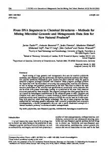
Microdissection of the morphological sample: Diagnostic morphological characters were preserved, even in small, relatively young specimens, with judicious selection of the region from which the tissue was excised for PCR (Fig. 1A,B). A central question concerns the exact section o f the n e m a t o d e that should be sacrificed for molecular analysis. T h e primary c o n c e r n is to minimize the loss of taxonomically important morphological characters. For these selected taxa, the region dissected was a portion of the intestine posterior to the esophagus and excluding the anterior e n d o f the gonad (Fig. 1A,B).
While this region may conserve the most useful morphological characters for these taxa, o t h e r sections may be chosen for o t h e r n e m a t o d e groups. Given the distribution of nuclei within a typical nematode, any section s h o u l d p r o v i d e a d e q u a t e t e m p l a t e quantity and quality. Based on the n u m b e r and distribution o f nuclei in C. elegans, we estimate that a typical 50-t~m section will include approximately 20 nuclei. With this n u m b e r of nuclei and an estimated 55 copies of the target rDNA seq u e n c e p e r h a p l o i d g e n o m e (Files a n d Hirsh, 1981), we expect that a typical C. elegans section would include a few thousand " p o t e n t i a l " template molecules for rDNA loci. However, both the absolute n u m b e r o f target repeats per g e n o m e and the n u m b e r of nuclei in a section are expected to vary from species to species and locus to locus. In addition, the degradation of DNA by the fixation process may vary d e p e n d i n g u p o n factors such as cuticle permeability. For this reason, we included a taxonomically diverse set of nematodes in o u r analysis. A potential problem with the microdissection sample is the existence of other organisms (and potential PCR templates) in the intestine. For the vast majority o f nematodes, these are ingested bacteria and fungi, which can be excluded by use of PCR primers (such as the D3 primers, see below) that do not amplify homologous targets in these
A
st~oma
vulva
I
posterior end of esophagus
anterior end of gonad
4 FIG. 1. Dissection of Zeldiapunctata. A) A Z punctata mature egg-laying female. B) A young Z. punctata female just at the point of reproductive maturity. Arrowhead indicates the region between the esophagus and gonad excised with a scalpel for PCR.
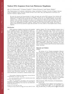
DNA Sequences from Formalin-fixed Nematodes: Thomas et al. 253 organisms. Nevertheless, in some groups o f nematodes, particularly predators o f other nematodes, such as Mononchida, this may represent a greater p r o b l e m and require excised tissue from a m o r e posterior region. In most such cases, it should be possible to eliminate the intestinal contents by starvation or during microdissecfion. Amplifications: PCR amplifications were p e r f o r m e d on each n e m a t o d e extract using the primers D3A and D3B (Nunn, 1992). These primers are known to amplify a region o f the g e n o m e that includes the D3 expansion segment o f the large subunit o f the nuclear rDNA repeat from a wide range of metazoans and all nematodes tested thus far (Litvaitis et al., 1994; N u n n , 1992). The expected size o f the PCR p r o d u c t is 362 bp in C. elegans and does not vary in length significantly in o t h e r n e m a t o d e taxa (Ellis et al., 1986; W. tL Thomas, unpubl.). In 10 attempts to amplify template p r e p a r e d from a n e m a t o d e fixed in formalin, all amplified after 35 cycles. In all cases, extracts from a n e m a t o d e section gave products o f the expected size (Fig. 2A, lanes 1-4).
A 1
O u r results differ f r o m those observed by De Giorgi et al. (1994) with respect to amplification success. In their study, only 20% (9 o f 44) of fixed nematodes amplified even t h o u g h entire n e m a t o d e s or pools of app r o x i m a t e l y 100 n e m a t o d e s were used. By contrast, all (10 of 10) of o u r amplifications were successful. Two distinct differences exist between m e t h o d o l o g y used in our study and that of De Giorgi et al. (1994) that m a y a c c o u n t for the d i f f e r e n c e in amplification success. First, De Giorgi et al. (1994) attempted to amplify a target twice the size o f the o n e in this study. Previously, Greer et al. (1991) clearly d e m o n s t r a t e d that the size of a PCR p r o d u c t that could be amplified is dramatically r e d u c e d with tissue fixation time. A s e c o n d difference is that the DNA isolation from De Giorgi et al. (1994) involved extraction and precipitation steps that could result in substantial loss o f template. By contrast, we have chosen a simple PCR-compatible tissue digestion that does not involve any steps that would remove potential template f r o m the reactions.
B 2
3
4
O
o
1
2
3
4
FIG.2. PCR amplification and sequencing results. A) A 2% agarose gel containing amplification products of the D3 expansion segment from four nematode species fixed in formalin for 2 days (lanes 1-4). ] : Zeldia punctata; 2: PS1010; 3: Aduncospiculum halicti; 4: Diplogastersp. Lane 5 is a lysis extract-PCR negative control. Lane 6 is a HincII digest of qbX174as a size standard. B) A representative region of a DNA sequencing gel demonstrating the quality of results obtainable from formalin-fixed nematodes. Labeled lanes correspond to the same species as in Fig. 2A, lanes 1-4. Nucleotides are loaded in the order G, A, T, C.
254 Journal of Nematology, Volume 29, No. 3, September 1997
Authenticity of sequences from formalin-fixed nematodes: I t is critical t h a t s e q u e n c e s faithfully r e p r e s e n t t h o s e in t h e g e n o m e o f t h e original specimen. Theoretically, although the PCR templates are highly modified and e r r o r s will b e g e n e r a t e d d u r i n g t h e a m p l i f i c a t i o n p r o c e s s , it is n o t p o s s i b l e to g e n e r a t e an incorrect sequence when the sequences are determined for the entire population of amplified molecules (Thomas and Kocher, 1993). N e v e r t h e l e s s , p r o b l e m s s u c h as c o n t a m i n a t i o n c a n l e a d to i n c o r r e c t s e q u e n c e s when the proper controls are not used. T o e v a l u a t e t h e a u t h e n t i c i t y o f t h e seq u e n c e s , we i n c l u d e d several c o n t r o l e x p e r i m e n t s . C o n t r o l s i n c o r p o r a t e d in this a m p l i fication scheme included a negative PCR control, a mock extraction control, and the i n c l u s i o n o f f o u r d i f f e r e n t s p e c i e s with dist i n c t r D N A s e q u e n c e s . I n a d d i t i o n , seq u e n c e s f r o m fresh, u n f i x e d s p e c i m e n s w e r e d e t e r m i n e d a n d c o m p a r e d to t h o s e o f f i x e d tissue. B o t h t h e P C R c o n t r o l a n d t h e e x t r a c t c o n t r o l r e s u l t e d in n o P C R p r o d u c t (Fig. 2A, Cont). T o test t h a t p r o d u c t s a m p l i f i e d f r o m e a c h e x t r a c t w e r e t h o s e o f t h e o r i g i n a l strain, we sequenced each of the amplified products directly after the asymmetric amplifications. Sequences from several nematode taxa c l e a r l y d e m o n s t r a t e d t h a t u n a m b i g u o u s discrete sequence differences can be identified f r o m f o r m a l i n - f i x e d tissue (Fig. 2B, l a n e s 1 4). I n all cases, t h e s e q u e n c e s w e r e i d e n t i c a l to t h o s e p r e v i o u s l y d e t e r m i n e d f r o m t h o s e strains (Ellis e t al., 1986).
Integrating molecular analysis and morphological analysis: T h e g o a l o f this s t u d y was to d e v e l o p a t e c h n i q u e f o r t h e effective use o f n e m a t o d e s c o l l e c t e d with m e t h o d s typical o f s t a n d a r d n e m a t o d e surveys. W e h a v e d e m o n s t r a t e d t h a t n e m a t o d e s f i x e d f o r as l o n g as 2 days b e f o r e e x t r a c t i o n c a n y i e l d suffic i e n t a m o u n t s o f t e m p l a t e D N A to allow acc u r a t e a m p l i f i c a t i o n o f a typical t a r g e t seq u e n c e . T h i s l e n g t h o f f i x i n g t i m e is sufficient for preliminary processing of samples c o l l e c t e d in t h e f i e l d a n d f o r p r e l i m i n a r y morphological sorting of specimens. The size o f t h e s e q u e n c e a m p l i f i e d in this s t u d y
(ca. 360 b p ) is typical o f m a n y P C R - b a s e d analyses. W e also have d e m o n s t r a t e d t h a t sufficient t e m p l a t e is r e c o v e r e d f r o m f o r m a l i n - f i x e d tissue s a m p l e s to allow f o r several a m p l i f i c a t i o n s , m a k i n g it p o s s i b l e to a m p l i f y c o m p l e t e g e n e s a n d (or) s e q u e n c e s f r o m m u l t i p l e d i f f e r e n t loci. F u t u r e i n v e s t i g a t i o n s m i g h t e x p l o r e b o t h t h e m a x i m u m size o f the sequence that can be amplified under various fixation protocols and the maximum t i m e o f f i x a t i o n t h a t c a n still y i e l d a m p l i f i able template. LITERATURE CITED AndrSmy, I. 1992. A short census of free-living nematodes. Fundamental and Applied Nematology 15:187-188. De Giorgi, C. M. F., M. F. Sialer, and F. Lamberti. 1994. Formalin-induced infidelity in PCR-amplified DNA fragments. Molecular and Cellular Probes 8:459-462. Ellis, R. E., J. E. Sulston, and A. R. Coulson. 1986. The rDNA of C. degans: Sequence and structure. Nucleic Acids Research 14:2345-2364. Files, J. G., and D. Hirsh. 1981. Ribosomal DNA of Caenorhabditiselegans.Journal of Molecular Biology 149: 223-240. Freckmma, D. W., ed. 1994. Life in the soil. Soil biodiversity: Its importance to ecosystem processes. Fort Collins, CO: Colorado State University. Golden, A.M. 1990. Preparation and mounting nematodes for microscopic observation. Pp. 197-205 in B.M. Zuckerman, W.F. Mai, and L.R. Krusberg, eds. Plant hematology laboratory manual. Amherst, MA: University of Massachusetts Agricultural Experiment Station. Greer, C. E., J. Y,2Lund, and M. M. Manos. 1991. PCR amplification from paraffin-embedded tissues: Recommendations on fixatives for long-term storage and prospective studies. PCR Methods and Applications 1:46-50. Litvaifis, M.K., G. Nunn, W. K. Thomas, and T. D. Kocher. 1994. A molecular approach for the identification of meiofauna. Marine Biology 120:437-442. Nunn, G.B. 1992. Nematode molecular evolution. Ph.D. thesis. University of Nottingham, UIC Pearce, F. 1995. RockaU mud richer than rainforest. New Scientist 147:8. Poinar, G. O., Jr. 1983. The natural history of nematodes. Englewood Cliffs, NJ: Prentice-Hall. Systemafics Agenda 2000. 1994. Systematics agenda 2000: Charting the biosphere. New York: American Museum of Natural History. Thomas, W. IC, and A. C. Wilson. 1991. Mode and tempo of molecular evolution in the nematode Caenorhabditis. Genetics 128:269-279. Thomas, W. K., and T. D. Kocher. 1993. Sequencing of polymerase chain reaction-amplifiedDNAs. Methods in Enzymology 224:391-399. Williams, B.D., B. Schrank, C. Huynh, R. Shownkeen, and R. H. Waterston. 1992. A genetic mapping system in Caenorhabditiseleg'ansbased on polymorphic sequence-tagged sites. Genetics 131:609--624.