Mar 3, 2014 - especially if the area of decay is at least half way through the enamel or into ... Decay. Advanced Decay. 0-20 Healthy / Sound Tooth Structure.
DentalTeamwork-MAR14-Pediatrics-REV_Layout 1 2014-03-05 9:54 AM Page 46
T T
Do We Really Need Caries Detection Devices?
he detection and treatment of dental caries has not changed radically since the time of G.V. Black. For the past 50 years, detection of caries has depended upon locating mineral loss on bite wing radiographs, examining stain and discoloured areas on the tooth surface, and probing lesions with a sharp explorer looking for soft or sticky areas. But what is caries, and are there any new technologies on the market that aid in its detection? In 2001, the National Institute of Health’s (NIH) Consensus Conference on the Diagnosis and Management of Dental Caries throughout Life concluded:
“Dental caries is an infectious, communicable disease resulting in destruction of tooth structure by acid-forming bacteria found in dental plaque, an intraoral biofilm, in the presence of sugar. The infection results in the loss of tooth minerals that starts with the outer surface of the tooth and can progress through the dentin to the pulp, ultimately compromising the vitality of the tooth.”1
46
Dental TEAMWORK Vol.7 No.3 - March 2014
Dr. Stephen Abrams
If caries is a disease that results in the destruction of the crystal structure of the tooth, shouldn’t we be asking ourselves if we have the right tools (radiographs, explorers and visual exam) to detect the breakdown of tooth structure and monitor the progress of treatment?
How Does Dental Caries Develop?
Dental caries arises from an overgrowth of specific bacteria that can metabolize fermentable carbohydrates and generate acids as waste products of their metabolism. Streptococci mutans and Lactobacillus are the two principal species of bacteria involved in dental caries and are found in the plaque biofilm on the tooth surface.2,3,4 They are not the only bacteria responsible for lesion formation but they are the principal species in this disease process. When these bacteria produce acids, the acids diffuse into enamel, cementum or dentin and dissolve or partially dissolve the mineral from crystals below the surface of the tooth. If the mineral dissolution is not halted or reversed, the early subsurface lesion becomes a “cavity”.
DentalTeamwork-MAR14-Pediatrics-REV_Layout 1 2014-03-05 9:54 AM Page 47
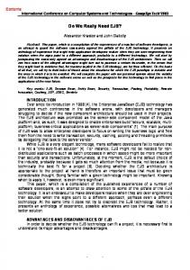
The Anatomy of a White Spot Lesion
Photographic Image of Scanned Area (Spot A)
Polarized Light Microscopy Cross Section Spot A
Spot
Canary Number
DIAGNOdent Peak Value
ICDAS Ranking
PLM Lesion Depth (µm)
A
35 ± 2
2±0
1
533.01
Figure 1
The tooth surface undergoes demineralization and remineralization continuously, with some reversibility. When exposed to acids, the hydroxyapatite crystals dissolve to release calcium and phosphate into the solution between the crystals. These ions diffuse out of the tooth leading to the formation of the initial carious lesion. The reversal of this process is remineralization. Remineralization will occur if the acid in the plaque is buffered by saliva, allowing calcium and phosphate present primarily in saliva to flow back into the tooth and form new mineral on the partially dissolved subsurface crystal remnants.5 The new “veneer” on the surface of the crystal is much more resistant to subsequent acid attack, especially if it is formed in the presence of sufficient fluoride.
White Spots are Caries
The earliest visual clinical sign of dental caries is the “white spot lesion”. When this is first seen, the caries process has been occurring for months. Figure 1 shows a cross-section (using polarized light microscopy, PLM) of a white spot lesion. The tooth surface appears smooth but the PLM image shows a lesion that is at least 533.01 microns in depth (half a millimeter). The findings from a visual examination tell us nothing about the lesion developing beneath the surface. If the lesion were to grow or shrink in response to remineralization therapies, all that would be visible is a white spot. Radiographs may not pick up this lesion until a large area has demineralized beneath the tooth surface. In this
case, scanning with The Canary System™ indicated that a lesion is present while DIAGNODent indicated that there is no lesion present. These early lesions can be treated before cavitation and they are amenable to remineralization.6,7 The key is to find the lesion and use the appropriate technology to monitor the changes in the lesion as it undergoes remineralization.
Can Radiographs and Visual Exams Detect Pit and Fissure Caries?
Radiographic and visual examinations are satisfactory if there is a substantial cavitated lesion, but detecting early pit and fissure caries is a challenge. On occlusal surfaces radiographic imaging is of minimal diagnostic value because of the large amounts of surrounding enamel.8,9 In addition, a number of studies have found the dental explorer inefficient for the diagnosis of occlusal caries.10,11 There are a number of concerns with the use of the explorer in detecting pit and fissure caries: • Since cavitation in pit and fissure caries occurs late in the disease process, using an explorer stick to detect caries only finds larger lesions, • Probing an occlusal pit or fissure could convert a small lesion into a larger one,12 • The probing could produce irreversible traumatic defects in areas that have the potential to remineralize, • Probing can inoculate the fissure with microorganisms from other intraoral sites,13,14 Dental TEAMWORK Vol.7 No.3 - March 2014
47
DentalTeamwork-MAR14-Pediatrics-REV_Layout 1 2014-03-05 9:55 AM Page 48
0
20
Healthy
Canary Scale Decay
70
100
Advanced Decay
0-20
Healthy / Sound Tooth Structure
21-70
Decay
71-100 Advanced Decay Figure 2
Figure 3
Figure 4
Figure 5
• A stick or catch with an explorer may be due to fissure morphology or probe pressure rather than a carious lesion.
for the detection of dental caries is poor for all types of lesion on proximal and occlusal surfaces”. The review found that “it is beneficial only if the intervention is the surgical removal of tooth structure and detrimental if it is used for non-invasive remineralization methods”. Pretty and Maupome in their review of radiographic diagnostic procedures concluded that “for interproximal lesions a clinician using radiographs can be very certain of the lack of disease in apparently sound surfaces (97% specificity) but not as certain that disease is indeed present in suspect interproximal surfaces (54% sensitivity)”.19 Radiographs and visual examination are valid diagnostic tools for the detection of larger lesions20,21 but there is a need for more sensitive methods.
40 Can Radiographs Detect Enamel Lesions in Interproximal Areas?
Radiographs can detect caries in interproximal areas, especially if the area of decay is at least half way through the enamel or into dentin. But in terms of early lesion detection, radiographs are not effective in detecting small lesions in the order of 50 – 100 μ (microns) in the interproximal areas, which could be remineralized if detected early and suitable preventive measures instituted.15 One study, using bitewing radiographs for detection of interproximal caries, found that radiographs, at best bitewing radiographs, could detect deep lesions less than 50% of the time.16 This low sensitivity for detection of enamel lesions in interproximal regions is not unusual and may be due to the irregular shape and low contrast of these small early lesions.17 An extensive literature review by Dove18 found that “overall the strength of the evidence for radiographic methods
48
Dental TEAMWORK Vol.7 No.3 - March 2014
Clinical Example of the Challenges of Early Caries Detection Using Visual Exam and Radiographs
In this clinical situation, a 40 year old female patient with minimal caries risk and only two pre-existing restorations was complaining of pain in the maxillary left first molar. The pain was low grade, not stimulated by chewing or cold.
DentalTeamwork-MAR14-Pediatrics-REV_Layout 1 2014-03-05 9:55 AM Page 49
A routine bitewing radiograph (Figure 2) and visual examination revealed no sign of pathology, and both marginal ridges appeared intact with no signs of any radiolucency. Scanning the mesial contact area with The Canary System, however, indicated that a lesion was present beneath the occlusal aspect of the marginal ridge but towards the buccal surface (Figure 3). Preparation of the tooth for a conventional composite restoration (Figure 4) confirmed caries on the mesial contact area as indicated by The Canary System. This clinical example illustrates situations where radiographs and visual examination may not be able to detect lesions due to their placement beneath a hard intact shell of radiopaque enamel – but treatment was required.
Can Fluorescence Based Technologies Be Used for Caries Detection?
The core technology in most caries detection devices today (e.g., Acteon’s SOPROLIFE (Acteon), Air Techniques’ Spectra and KaVo’s DIAGNODent) is fluorescence.22 Fluorescence is simply the glow from an object that has absorbed light, such as light from LEDs or lasers. Since bacterial porphyrins, stains, tartar, food debris, and prophylaxis paste all fluoresce under the wavelengths used in these devices, whether or not caries is present, 23,24,25,26,27,28 they can lead to false positive readings and unnecessary treatment. In addition, Streptococcus mutans and lactobacilli, the key bacterial initiators of caries, do not have the porphyrins that fluoresce when exposed to the light emitted by these devices.29,30,31,32,33 A number of studies have concluded that measuring fluorescence is not suitable for detecting caries around restoration margins or beneath dental sealants due to false positive readings. 34,35,36,37 The CR Clinicians Report (March 2012) found that restorations interfered with readings from both Spectra and SOPROLIFE.38 Further, fluorescence does not give any information about lesion size or depth, and does not penetrate beneath the tooth surface due to scattering of light from stain, plaque, organic deposits and surface features such as pits and fissures.39,40 One can conclude, therefore, that fluorescence can be used to measure surface changes and a number of common oral factors such as stain, oral bacteria, and tartar, but it should not be relied upon as a means for detecting and monitoring caries.
The Canary System Can Detect Defects in Enamel Crystal Structure
The Canary System, developed by Quantum Dental Technologies, has a very different approach to caries detection. The Canary System looks directly at the status of the enamel crystal by using PTR-LUM technology that measures imperceptible converted heat (PTR) and light (luminescence or LUM) signatures emitted from the tooth surface. Safe pulses of laser light allow one to examine up to 5 mm below the tooth surface. As a lesion grows, there is a corresponding change in the signal; as the heat is confined to the region with crystalline disintegration and LUM decreases. As remineralization progresses and enamel prisms begin to reform their structure, the thermal and luminescence properties begin to revert back in the direction of healthy teeth. The system is directly linked to the status of the tooth crystal structure. Research has demonstrated that the energy conversion technology (PTR-LUM) used in The Canary System can be used to detect and diagnose: • Lesions and defects up to 5 mm. below the enamel surface41,42 • Occlusal pit and fissure caries43,44,45 • Smooth surface caries46,47 • Acid erosion lesions48,49,50 • Root caries51,52 • Interproximal caries lesions53,54,55,56 • Beneath fissure sealants57,58,59 • Caries around the margins of restorations60,61,62 • Caries beneath the intact margins of composite resins63 • Demineralization and remineralization of early lesions64,65,66,67,68,69,70,71,72,73,74
Figure 6
Dental TEAMWORK Vol.7 No.3 - March 2014
49
DentalTeamwork-MAR14-Pediatrics-REV_Layout 1 2014-03-05 9:56 AM Page 50
The Canary System does detect and monitor tooth crystal structure changes and is a valid technique for caries detection. The system measures changes in the tooth crystal structure which is caused by caries and other pathology.
Summary
Caries is a disease that results in the destruction of the crystal structure of the tooth, so the ability to examine the integrity of crystal structure has a direct impact on the effectiveness of the treatment plan. Visual examination, caries risk assessment and radiography all have limitations in terms of their ability to detect, diagnose and monitor caries. Caries risk assessment examines factors that contribute to the disease, but not treatment of the disease. Visual examination looks at the surface of the tooth but typically, early lesions begin and grow beneath the tooth’s surface. In addition, using an explorer to probe lesions is often inaccurate, and it can damage tooth structure and inoculate the pits and fissures with oral bacteria. Dental radiographs can detect interproximal lesions, but only after they’ve demineralized at least 50% of the outer enamel shell. Even at this stage, they do not provide an accurate system to measure and monitor lesion size. Fluorescence-based devices are designed to detect surface changes such as stain and bacterial by-products, but they are not capable of identifying early-stage changes in crystal structure due to caries, cracks or other pathological stimuli. Only the Canary System with its PTR-LUM feature has the ability to quantifiably identify defects in the structure of teeth. With this technology, dental clinicians for the first time have the ability to identify and measure crystal structure defects more accurately than by radiography and visual examination alone.
Disclosure
Dr. Stephen Abrams is the President and Co-Founder of Quantum Dental Technologies which has developed The Canary System mentioned in this article. He has not received any compensation for the preparation of this article. Stephen Abrams is a general dental practitioner with over 33 years of clinical experience. He established a group practice in Toronto Canada which has grown to involve general dentists and dental specialists. Dr. Abrams founded Quantum Dental Technologies, a company developing laser based technology for the early detection and ongoing monitoring of dental caries. He is a fellow of the Pierre Fauchard Academy the Academy of Dentistry International and American College of Dentistry. He is a member of the European Association for Caries Research and International Association of Dental Research. He has published over 90 articles in various international publications. In 2002, Dr. Abrams was awarded the Barnabus Day Award from the Ontario Dental Association for 20 years of distinguished service to the dental profession.
50
Dental TEAMWORK Vol.7 No.3 - March 2014