Review
Does Hemodialysis Increase Protein Breakdown? Dissociation between Whole-Body Amino Acid Turnover and Regional Muscle Kinetics Victoria S. Lim,* T. Alp Ikizler,† Dominic S.C. Raj,‡ and Michael J. Flanigan* *Department of Medicine, University of Iowa College of Medicine, Iowa City, Iowa; †Vanderbilt University School of Medicine, Nashville, Tennessee; and ‡University of New Mexico Health Science Center, Albuquerque, New Mexico Hemodialysis (HD) is a protein catabolic procedure. Whole-body amino acid turnover studies identify dialysate amino acid loss and reduced protein synthesis as the catabolic events; proteolysis is not increased. Regional amino acid kinetics, however, document enhanced muscle protein breakdown as the cause of the catabolism; muscle protein synthesis also increased but to a lesser magnitude than the increment in protein breakdown. This discordance between whole-body and regional kinetics is best explained by the contrasting physiology between the muscle and the liver. During HD, muscle releases amino acids, which then are taken up by the liver for de novo protein synthesis. There seems to be a somatic to visceral recycling of amino acids. Evidence supporting this concept includes the increased fractional synthesis of albumin and fibrinogen during HD. It should be emphasized that region- or organ-specific kinetics vary, and whole-body turnover is a composite of all of the visceral and somatic compartments taken together. Reduced whole-body protein synthesis may be a compensatory adaptation to dialysate amino acid loss with a consequent reduction in plasma amino acid concentration. Notwithstanding the protein catabolic nature of HD, evidence is accumulating that intradialytic nutritional supplementation may blunt its catabolic effect. J Am Soc Nephrol 16: 862-868, 2005. doi: 10.1681/ASN.2004080624
U
ntil recently, uremia was believed to be a protein catabolic state. This view is no longer tenable because numerous whole-body amino acid turnover studies have unequivocally revealed that there is no excess protein catabolism in the chronic renal failure population in the absence of acidosis and/or concomitant illnesses (1). Uremic patients respond to low-protein diets with appropriate downregulation of whole-body proteolysis (2,3). Peritoneal dialysis is protein catabolic primarily because of protein and amino acid losses through the peritoneal effluent (4). The remaining unsettled issue is whether hemodialysis (HD) induces protein catabolism and, if it does, by what mechanism. There are theoretical reasons to believe that HD can augment protein catabolism. These include amino acid loss to the dialysate and cytokinemediated proteolysis as a result of exposure to bio-incompatible membrane and endotoxin-contaminated dialysate, but to date, investigation of protein metabolism using different techniques has yielded conflicting results.
Protein Metabolism during HD: A Review of the Literature This article reviews the available literature of protein metabolism during HD and proposes a unifying hypothesis to explain
Published online ahead of print. Publication date available at www.jasn.org. Address correspondence to: Dr. Victoria S. Lim, Department of Medicine, University of Iowa College of Medicine, Room T310, General Hospital, University of Iowa Hospitals and Clinics, 200 Hawkins Drive, Iowa City, IA 52242. Phone: 319-356-3415; Fax: 319-384-8220; E-mail:
[email protected] Copyright © 2005 by the American Society of Nephrology
the discordant results and diverging conclusions. The work cited in this review is categorized according to the technique used.
Nitrogen Balance Combined with Urea Kinetics The seminal findings of Borah et al. (5) that nitrogen balance is always more negative or less positive, depending on the intake, on dialysis days compared with nondialysis days greatly influenced the belief that HD is a protein catabolic procedure. Lim et al. (6) also reported that nitrogen output, expressed as milligrams per minute, is greater when the interdialytic interval is 2 d as compared with 3 d, suggesting that each HD generates a certain degree of protein catabolism, the effect of which is minimized when the interdialytic interval is lengthened. The classical technique of nitrogen balance is intake minus output. Output measurement is tedious for the study subjects as well as for the investigators because it requires urine, dialysate, and fecal collection, plus calculation of change in body urea nitrogen pool and unmeasured nitrogen loss. In the above-cited work of Borah and Lim, urea generation rate was probably overestimated by the use of unequilibrated postdialysis blood urea nitrogen. This determinate error likely accounts for the lesser nitrogen balance on dialysis days. Despite the laborious nature of the work, nitrogen balance does not give insight into the various components that made up net nitrogen balance, and these include protein intake, breakdown, oxidation, synthesis, and others. The global nitrogen balance also does not furnish information regarding amino acid turnover in the various region- and organ-specific protein pools. ISSN: 1046-6673/1604-0862
C
C
ESRD EAA
Non-EAA ESRD
Raj (9)
C Total AA ESRD Ikizler (8)
Inc Total AA Normal
a Total AA, total amino acids; EAA, essential amino acids; non-EAA, nonessential amino acids; C, biocompatible dialyzer; Inc, bioincompatible dialyzer; HD, hemodialysis.
—
— 2121%
2517%
1123% 2414%
23%
2109%
0.74 (ml/100 g per min) 0.75 (ml/100 g per min) 3.6 (ml/100 ml per min) 3.3 (ml/100 ml per min) 3.3 (ml/100 ml per min) 25% 113%
⫺116 ⫺140 (nmol /100 g per min) (nmol /100 g per min) ⫺152 ⫺309 (nmol /100 g per min) (nmol /100 g per min) ⫺334 15 (nmol/100 ml per min) (nmol/100 ml per min) ⫺290 (nmol/100 ml per min) ⫺2607 (nmol/100 ml per min) ⫺133 (nmol /100 g per min) Leg ⫺148 (nmol /100 g per min) Forearm ⫺65 (nmol/100 ml per min) Leg ⫺131 (nmol/100 ml per min) Leg ⫺335 (nmol/100 ml per min) Leg
HD Pre-HD Subject Dialyzer Location Type
C
Post-HD
Fractional Change
HD/ Post-HD/ Pre-HD Pre-HD
Pre-HD
HD
Plasma Flow
Author (Reference)
Modern amino acid turnover kinetics generally uses stable isotopes. These studies are accomplished by primed-continuous infusion of labeled amino acids into the free amino acid pool, where, under isotope and substrate steady-state condition, dilution of the labeled amino acid by either intake or endogenous protein degradation is allowed to occur. By measuring this dilution in the plasma and by quantifying tracer loss as labeled CO2, one determines whole-body protein degradation and amino acid oxidation rates and indirectly estimates protein synthesis by mass balance method (10). Table 2 summarizes the results of whole-body flux studies during HD. Lim et al. (11) were some of the earliest investigators to quantify whole-body protein turnover during HD in ESRD patients using primed-constant infusion of 13C leucine. Because Gutierrez’s paper identified peak protein catabolism ⬎3 h after dialysis, they studied leucine flux 2 h before and 4 h after dialysis and shortened the dialysis session to 2.5 hours to avoid isotope recycling. To enhance the probability of finding increased protein degradation, bio-incompatible dialyzers were used, and to the surprise of all, whole-body leucine flux, an index of protein degradation in the postabsorptive state, did not increase either during or after dialysis. Plasma 13C leucine and 13C ␣-keto-isocaproic acid enrichment stayed relatively constant during the entire study period. By contrast, protein synthesis and amino acid oxidation rates varied. During HD,
Net Amino Acid Balance
In Vivo Whole-Body Amino Acid Turnover Kinetics
Table 1. Amino acid balance across the limb during hemodialysis in normal subjects and ESRD patientsa
As shown in Table 1, Gutierrez et al. (7) measured amino acid exchange across the leg (nmol/100 g per min) in normal subjects who underwent sham HD with a bio-incompatible dialyzer and reported 13 and 109% increments in amino acid release during and after dialysis, respectively. When dialyzed with a biocompatible dialyzer, the same subjects did not show increased amino acid release. They also showed that the increase in amino acid release could be blocked by concurrent administration of indomethacin, a nonselective cyclo-oxygenase inhibitor. In sharp contrast, Ikizler et al. (8) and Raj et al. (9) found a marked increase in amino acid release (nmol/100 ml per min) using a biocompatible dialyzer. In Ikizler’s paper, forearm total amino acid balance in 11 patients was ⫺65 predialysis, and the negative balance widened to ⫺334 during dialysis, representing a fractional decrement in balance of 414%. The value returned toward normal 2 h after HD. In Raj’s work, net essential amino acid balances across the leg were ⫺131 and ⫺290, and nonessential amino acid balances were ⫺335 and ⫺2067 before and during HD. The fractional decrements of amino acid balance for essential and nonessential amino acids were, respectively, 121 and 517%. There is no good explanation for these differences except for one factor, the study subjects, normal control versus ESRD patients. In Gutierrez’s study, the increment in amino acid release across the leg was accounted for, in large part, by increased blood flow to the leg after dialysis. In the other two studies, the increase in amino acid release was a consequence of greater arteriovenous concentration gradients, as limb blood flow was not significantly changed.
Post-HD
Amino Acid Release across the Arm or the Leg
0.50 0.70 (ml/100 g per min) (ml/100 g per min) 0.81 1.31 (ml/100 g per min) (ml/100 g per min) 4.7 3.1 (ml/100 ml per min) (ml/100 ml per min) 3.4 — (ml/100 ml per min) 3.4 — (ml/100 ml per min)
Does Hemodialysis Increase Protein Breakdown?
Gutierrez (7) Total AA Normal
J Am Soc Nephrol 16: 862-868, 2005
863
864
Journal of the American Society of Nephrology
J Am Soc Nephrol 16: 862-868, 2005
Table 2. Whole-body amino acid turnover studies in ESRD patients during hemodialysisa Pre-HD/HD
Author (Reference)
Isotope
B
Lim (11)
13
C leucine
Ikizler (8)
13
C leucine
Raj (9)
13
C6 phenylalanine
Veeneman (12)
13
C valine
Pupim (14)
13
C leucine
Pre-HD/Post-HD
Flux O
S
17/14 218% 0.5/0.4
101/88 213% 2.9/2.7
219% —
27% —
(16%) —
— 15/9
— 62/50
— 21.0
27% 3.4/3.8
240% 0.54/0.52
219% 2.9/2.8
110%
24%
23%
mol/kg per h3 118/117 Fractional ⌬3 21% mg/kgFFM per 3.4/3.7 min3 Fractional ⌬3 19% nmol/kg per 0.8/0.7 min3 Fractional ⌬3 215% mol/kg/h3 70/65 Fractional ⌬3 mg/kgFFM per min3 Fractional ⌬3
D
NB
B
O
S
NB
118/115 23% 3.4/3.8
17/19 112% 0.5/0.6
101/95 2 6% 2.9/3.2
⫺17/⫺19 212% ⫺0.5/⫺0.6
296% —
111% —
121% —
111% —
220% —
— ⫺8/⫺21
— —
— —
— —
— —
— 3.4/3.7
— 0.54/0.65
— 2.9/3.0
— ⫺0.5/⫺0.6
17%
120%
15%
220%
14.4 ⫺17/⫺29 (12%) 271% 0.61 ⫺0.5/⫺1.0
(26%) 287% — ⫺0.5/⫺1.0 —
281%
a Listed under column Flux are the absolute values of the flux rates and fractional changes in each experiment. The latter representing increment or decrement comparing HD with pre-HD and post-HD with pre-HD. Two values are entered into the columns of breakdown (B), oxidation (O), synthesis (S), and net balance (NB) representing before and during and before and after HD. Column D lists the absolute amounts of unlabeled dialysate amino acid (the study amino acid) loss, and values in parentheses represent losses expressed as % of total flux. Only Lim’s study used a bio-incompatible dialyzer; the others used biocompatible membrane.
leucine to protein incorporation, reflecting protein synthesis, was reduced. This may be due, in part, to leucine loss into the dialysate, accounting for approximately 12% of total flux. The leucine oxidation rate was not increased but was, in fact, reduced during dialysis. The combined effects of dialysate amino acid loss and reduced protein synthesis resulted in a significant reduction in net whole-body protein balance during HD. Ikizler et al. (8) measured protein turnover in ESRD patients before, during, and for 2 h after HD using 13C leucine infusion. Dialysis sessions were 4 h in duration, and the dialysis membrane was polysulfone. They reported that HD led to a slight increase in whole-body proteolysis and a nonsignificant reduction in whole-body protein synthesis; net protein balance and plasma leucine were reduced. Dialysate leucine loss was approximately 16% of the leucine disappearance rate or the total flux rate. In the above two studies, during the postdialysis period, amino acid loss ceased and whole-body protein synthesis improved. Proteolysis was not significantly different from baseline. Amino acid oxidation rate, which was reduced during HD, increased after dialysis. Net protein balance remained more negative compared with baseline, although of lesser magnitude than during dialysis. The catabolic picture after dialysis may be related to the prolonged fasting state of the experiment. Raj et al. (9) measured 13C6 phenylalanine flux before and during HD using a biocompatible dialyzer and noted that whole-body flux, representing protein degradation, was reduced 15% during HD compared with predialysis value. These findings were further confirmed by data obtained from leucine and lysine kinetics. They did not measure oxidation or synthesis rate, and they did not collect data after dialysis. Veeneman et al. (12) measured 13C valine kinetics in six ESRD patients, using biocompatible dialyzer, on two separate occasions, one during an HD session and the other without dialysis.
They found that, in the postabsorptive state, protein degradation and valine oxidation rates were not increased on the HD day; in fact, the values were lower than those obtained on a nondialysis day. Protein synthesis, however, was reduced, as was net protein balance. Dialysate valine loss was approximately 26% of total valine flux. It is interesting to note that the different experiments listed in Table 2 covered a span of one decade, performed by different investigators at different institutions and with different isotopes, yet the results are consistent. During HD, whole-body protein degradation is minimally changed from baseline; increments and decrements are minor in magnitude. By contrast, net protein balance is invariably reduced because of a combination of dialysate amino acid loss and decreased protein synthesis. After dialysis, net protein balance becomes less negative because dialysate amino acid loss ceases and protein synthesis improves.
Regional Kinetics across the Forearm or Leg To estimate regional turnover, one needs additional measurements of arterial and venous amino acid concentration, arterial and venous isotopic enrichment, and the blood flow rate across the region. This is usually performed across an arm or a leg (13). Labeled phenylalanine is typically used because it is neither synthesized nor metabolized by muscle; its appearance rate, therefore, represents protein degradation, and its disappearance represents protein synthesis. As shown in Table 3, Ikizler et al. (8) studied 11 ESRD patients and reported forearm kinetics (g/100 ml per min) in the sequence of before, during, and after dialysis as follows: Proteolysis was 77, 180, and 127, respectively; protein synthesis was, 56, 123, and 98, respectively; and net balance was ⫺22, ⫺58, and ⫺28, respectively. During HD, forearm muscle proteolysis increased by 134% and protein synthesis increased by 120%, resulting in net
J Am Soc Nephrol 16: 862-868, 2005
Does Hemodialysis Increase Protein Breakdown?
865
Table 3. Regional and intramuscle amino acid kinetics during hemodialysis in ESRD patientsa
Author
Ikizler (8) Pupim (14) Raj (9) a
Isotope
2
H5 phenylalanine H5 phenylalanine 13 C6 phenylalanine 2
Kinetics across Arm and Leg (Fractional Changes Pre-HD to HD)
Location
Degradation
Synthesis
Net Balance
1134% 197% 1104%
1120% 185% 140%
2164% 2104% 293%
Forearm Forearm Leg and muscle
Intramuscle Kinetics (%/day) FSR Protein (Fractional Changes Pre-HD to HD)
150%
FSR, fractional synthesis rate of muscle protein.
increase in protein loss of 164% compared with baseline. In a subsequent study, the same investigators reported similar forearm kinetics showing increments of 97% for proteolysis, 85% for protein synthesis, and a 104% reduction in net protein balance (14). Of note, during the postdialysis period, forearm net muscle protein balance was not different from baseline, although both breakdown and synthesis were significantly higher. In a similar study, Raj et al. (9) measured leg kinetics (nmol/100 ml per min) using 13C6 phenylalanine and reported a 104 and 40% rise in protein degradation and synthesis, respectively, during HD. This resulted in a 93% reduction in net protein balance.
Three-Compartmental Muscle Kinetics Intramuscle protein metabolism can also be quantified directly by the three-compartmental model—artery, muscle, and vein—measuring muscle intracellular individual amino acid concentration and labeled amino acid enrichment; multiple muscle biopsies are needed for this approach (15). Raj et al. (16) studied intracellular amino acid transport kinetics using stable isotopes of three essential and two nonessential amino acids— namely, phenylalanine, leucine, lysine, alanine, and glutamine— before and during HD. They found that during HD, there was increased amino acid efflux from the intracellular compartment into the vein as a result of increased protein breakdown. Despite this, plasma amino acid levels were reduced, intracellular amino acid concentrations remained stable, and fractional muscle synthesis increased by 50% as listed in Table 3. Overall, the studies of Ikizler, Raj, and Pupim are highly consistent. HD leads to an increase in skeletal muscle net protein catabolism. The individual components of protein turnover, i.e., breakdown and synthesis, both are increased, but the extent of increment is higher for protein breakdown.
HD Is Protein-Catabolic: A Discussion All of the data presented in Tables 1, 2, and 3 indicate that HD is a protein-catabolic event because net protein balance, which is the difference between synthesis and degradation, becomes more negative during HD. Such a negative balance is seen both in the whole-body protein pool and in the regional muscle compartment. The mechanism that contributes to the negative protein balance in the two pools, however, differs. In the muscle compartment, the reduction in protein balance is
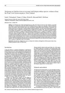
due to increased protein breakdown that is not adequately compensated by an equivalent increment in synthesis. In the whole-body pool, reduced protein balance is accounted for by dialysate amino acid loss and a compensatory reduction in protein synthesis. The discordant protein degradation finding between wholebody turnover and regional kinetics during HD, although counterintuitive, is not surprising because skeletal muscle accounts for only approximately 50% of whole-body protein content. It should be underscored that whereas region- or organspecific kinetics vary, whole-body turnover is a composite results of all taken together. Tessari et al. (17) reported that, in humans, the kidney, the splanchnic compartment, and the leg have different rates of protein degradation and synthesis. Failure to demonstrate increased protein breakdown with the whole-body kinetics can be explained by a compensatory downregulation of proteolysis in other body protein pools. On the basis of the available information, we propose that this dissociation between whole-body and skeletal muscle turnover kinetics is due to the contrasting physiology between the muscle and the liver. In the fasting state, the study condition of all of the experiments listed in Tables 1, 2, and 3, the muscle releases amino acids to replete the plasma pool and the liver extracts these amino acids to synthesizes new proteins. The most direct way to test this hypothesis is to measure simultaneously hepatic vein amino acid concentration and labeled amino acid enrichment as well as hepatic blood flow rate during amino acid turnover study. Although important, these kinds of data are difficult to obtain in humans, but two lines of evidence are already available in the literature to support this concept. First, Felig and Wahren (18) catheterized simultaneously the brachial artery, the femoral vein, and the right main hepatic vein in normal adults and measured amino acid concentrations from these different sites. They found that, in the fasting state, net balance of many amino acids across the leg, i.e., arteriofemoral vein difference, is negative (Figure 1). By contrast, the net balance of many amino acids across the splanchnic bed, that is arteriohepatic vein difference, is positive (Figure 2). These data indicate that, in the fasting state, the muscle releases amino acids, which then are taken up by the liver for de novo protein synthesis. Second, evidence of increased hepatic protein synthesis during HD has already been demonstrated in ESRD patients, and the data are summarized
866
Journal of the American Society of Nephrology
Figure 1. Net balance of individual amino acids across the leg (arteriofemoral venous differences [A-FV]) in 19 subjects in resting, postabsorptive state. The lines at the top or bottom of the bars represent the SEM. The mean A-FV differences were significantly different from zero for all amino acids except taurine and ornithine. Reprinted from reference 18.
Figure 2. Net balance of individual amino acids across the splanchnic bed (arteriohepatic venous differences [A-HV]) in 12 subjects in the resting, postabsorptive state. The lines at the top of the bars represent the SEM. Only those amino acids whose mean A-HV differences were significantly different from zero are shown Reprinted from reference 18.
in Table 4. Technically, amino acid kinetics could also be used to measure fractional synthesis rate of individual proteins, most commonly albumin and fibrinogen (19). In this method, the isotopes are detached from the infused amino acids and subsequently incorporated into new protein molecules. Raj et al. (9) reported that fractional synthesis rates, %/d, of albumin and fibrinogen increase by 49 and 67%, respectively, during HD. Caglar et al. (20) reported that in eight ESRD patients, fractional
J Am Soc Nephrol 16: 862-868, 2005
synthesis rates of albumin and fibrinogen increased by 65 and 35%, respectively, during HD. In another, similar protocol, Pupim et al. (21) reported that HD produced a 54% increase in albumin synthesis. It is not clear whether the two processes—increased muscle protein degradation and increased liver protein synthesis—are intrinsically integrated, or a single HD-related perturbation stimulates both processes. Ikizler and Raj both reported positive correlations between fractional synthesis rates of albumin and fibrinogen and circulating IL-6 levels (9,20,21), suggesting the latter possibility. This kind of reciprocal change between muscle and hepatic protein metabolism is not unique to dialysis patients. Mansoor et al. (22) found that during primed-constant 13C leucine infusion, patients with severe head injuries had a markedly reduced fractional synthesis rate of protein in the muscle, but the incorporation of tracer into albumin and fibrinogen was increased as compared with the control subjects. Similarly, Fearon et al. (23), using an intravenous loading infusion of 2H5 phenylalanine in cachectic cancer patients who had high C-reactive protein concentrations and hypoalbuminemia, found a higher fractional albumin synthesis than seen in normal subjects. Despite the absence of accelerated whole-body protein breakdown, HD is, nonetheless, a catabolic event with regard to the whole-body protein economy as a result of a combination of amino acid loss and a compensatory reduction in protein synthesis. As shown in Table 2, dialysate amino acid losses are considerable, ranging from 12 to 26% of total flux. The reduction in whole-body protein synthesis could be explained by substrate deficiency as circulating essential amino acids are invariably reduced and synthesis could be stimulated with amino acid supplementation (12,14,24,25). Kobayashi et al. (26), in a sophisticated experiment, reported that HD-induced reduction in plasma amino acid concentration in normal swine inhibited leg muscle protein synthesis. This inhibition in synthesis was negated when plasma amino acid reduction during HD was alleviated by amino acid infusion. Muscle protein breakdown, in this study, was not changed by plasma amino acid reduction. Of note, despite unequivocal reduction in plasma amino acids, intracellular amino acid concentration was well maintained during HD. Which organs or tissues downregulate synthesis during HD is not clear. It could be any organ or tissue except the muscle and the central nervous system. Muscle protein synthesis is increased, and central nervous system does not participate much in protein flux because its main source of fuel is glucose. It is possible that the liver, while increasing its synthesis of some urgently needed proteins, might downregulate synthesis of other proteins during a nutrition-deficient situation, such as the fasting state. Discordance between regional muscle and whole-body protein synthesis kinetics have also been documented in non-HD settings. During recombinant human growth hormone infusion in healthy subjects, Fryburg and Barrett (27) found that protein synthesis, although increased in the forearm muscle, remained unchanged in the whole body. Adey et al. (28) found that patients with chronic renal failure, as compared with normal healthy subjects, have lower synthesis rates of mixed muscle
J Am Soc Nephrol 16: 862-868, 2005
Does Hemodialysis Increase Protein Breakdown?
867
Table 4. Fractional synthesis rates of hepatic proteins during HD in ESRD patientsa FSR (%/d) Author (Reference)
Isotope
Raj (9)
13
Caglar (20)
L关1-13C兴leucine
Pupim (21)
L关1-13C兴leucine
a
C6 phenylalanine
Fractional Changes
Protein
Albumin Fibrinogen Albumin Fibrinogen Albumin
Pre-HD
HD
Post-HD
HD/Pre-HD
Post-HD/Pre-HD
6.5 11.8 7.4 11.2 8.3
9.7 19.7 12.2 15.1 16.2
— — 10.9 17.7 11.8
149% 167% 165% 135% 154%
— — 147% 158% 151%
Fractional changes represent increment or decrement comparing HD with pre-HD and post-HD with pre-HD values.
protein and myosin heavy chain but normal whole-body protein synthesis rates. Notwithstanding the catabolic nature of HD, in the absence of concurrent disease, ESRD patients exhibit increased wholebody protein turnover and improved protein synthesis after maintenance dialysis initiation (2). Furthermore, negative protein balance during HD can be reversed with nutritional supplementation. Pupim et al. (14) studied seven ESRD patients at two separate HD sessions, one with and the other without intradialytic parenteral nutrition. Administration of 45 g of protein and 752 kcal resulted in an increase in plasma amino acid concentrations and a change in net protein balance from negative to positive values both in the forearm and in the whole-body pool. In another elegant study, Venneman et al. (12) measured whole-body valine flux in six ESRD patients on two separate dialysis sessions, one performed during fasting and the other during oral feeding. The supplement consisted of 0.62 g/kg protein and 15 kcal/kg energy divided into six oral feedings given every 30 min. They found that feeding during dialysis resulted in reduced proteolysis, increased protein synthesis, and a change in net protein balance from negative to positive values. It is important to note here that insulin secretion after meal ingestion and insulin-induced protein anabolism both are intact in the ESRD patients (29,30).
Conclusion The impetus for writing this article derives from comments made by nephrologists that they have conflicting impressions regarding the effect of HD on protein metabolism. We thus reviewed most of the literature on the studies of protein/amino acid metabolism during HD. We found that the major discordance is in the results obtained between whole-body and regional kinetics. Regional kinetics across the forearm or the leg showed marked increase in protein breakdown, accompanied by an increase in protein synthesis that is inadequate to compensate for the degradation, thus resulting in a reduction in protein balance across the region. Whole-body turnover studies, however, showed no change in proteolysis, but there is dialysate amino acid loss, and protein synthesis is reduced. This combination results in a reduction of net protein balance in the whole-body pool. Reduced synthesis may be related, in part, to diminished substrate availability as plasma essential amino acids are reduced during HD. The organ or tissue that downregulates protein synthesis during HD is not identified.
Failure of the whole-body kinetics study to detect an increase in protein degradation is, we propose, due to contrasting physiology between the muscle and the liver; there seems to be a somatic to visceral recycling of amino acids. During HD, which happens also to be in the postabsorptive state in most of the experiments reported, the muscle releases amino acids and the liver recycles these amino acids for de novo protein synthesis.
Acknowledgments V.S.L.’s work was made possible through the Baxter Extramural Grant, National Institutes of Health (NIH) Grant RR55 (General Clinical Research Center, University of Iowa), and NIH Grant RR00954 (Mass Spectrometry Resource Center, Washington University). T.A.I.’s work is supported, in part, by NIH Grants DK-45604, DK-62849, and RR000951; FDA Orphan Drug Development Grant 000943; and Satellite Health Extramural Grant Program. D.S.C.R. is supported by grants from the Young Investigator Award from the National Kidney Foundation, Research Allocation Committee Grant from the University of New Mexico, and Paul Teschan Research Grant.
References 1. Lim VS, Kopple JD: Protein metabolism in patients with chronic renal failure: Role of uremia and dialysis. Kidney Int 58: 1–10, 2000 2. Lim VS, Yarasheski KE, Flanigan MJ: The effect of uraemia, acidosis, and dialysis treatment on protein metabolism: A longitudinal leucine kinetic study. Nephrol Dial Transplant 13: 1723–1730, 1998 3. Goodship THJ, Mitch WE, Hoerr RA, Wagner DA, Steinman TI, Young VR: Adaptation to low-protein diets in renal failure. Leucine turnover and nitrogen balance. J Am Soc Nephrol 1: 66 –75, 1990 4. Blumenkrantz MJ, Gahl GM, Kopple JD, Kamdar AV, Jones MR, Kessel M, Coburn JW: Protein losses during peritoneal dialysis. Kidney Int 19: 593– 602, 1981 5. Borah MF, Schoenfeld PY, Gotch FA, Sargent JA, Wolfson M, Humphreys MH: Nitrogen balance during intermittent dialysis therapy of uremia. Kidney Int 14: 491–500, 1978 6. Lim VS, Flanigan MJ: The effect of interdialytic interval on protein metabolism: Evidence suggesting dialysis-induced catabolism. Am J Kidney Dis 14: 96 –100, 1989 7. Gutierrez A, Alvestrand A, Wahren J, Bergstrom J: Effect of in vivo contact between blood and dialysis membranes on protein catabolism in humans. Kidney Int 38: 487– 494, 1990 8. Ikizler TA, Pupim LB, Brouillette JR, Levenhagen DK, Farmer K, Hakim RM, Flakoll PJ: Hemodialysis stimulates
868
9.
10.
11.
12.
13.
14.
15.
16.
17.
18. 19.
Journal of the American Society of Nephrology
muscle and whole body protein loss and alters substrate oxidation. Am J Physiol Endocrinol Metab 282: E107–E116, 2002 Raj DSC, Dominic EA, Wolfe R, Shah VO, Bankhurst A, Zager PG, Ferrando A: Coordinated increase in albumin, fibrogen, and muscle protein synthesis during hemodialysis: Role of cytokines. Am J Physiol Endocrinol Metab 286: E658 –E664, 2004 Bier DM, Matthews DE, Young VR: Interpretation of amino acid kinetic studies in the context of whole-body protein metabolism. In: Substrate and Energy Metabolism, edited by Garrow JS, Halliday D, London, John Libbey, 1985, pp 27–36 Lim VS, Bier DM, Flanigan MJ, Sum-Ping ST: The effect of hemodialysis on protein metabolism. J Clin Invest 91: 2429 – 2436, 1993 Veeneman JM, Kingma HA, Boer TS, Stellaard F, DeJong PE, Reijngoud D-J, Huisman RM: Protein intake during hemodialysis maintains a positive whole body protein balance in chronic hemodialysis patients. Am J Physiol Endocrinol Metab 284: E954 –E965, 2003 Gelfand RA, Barrett EJ: Effect of physiology hyperinsulinemia on skeletal muscle protein synthesis and breakdown in man. J Clin Invest 80: 1– 6, 1987 Pupim LB, Flakoll PJ, Brouillette JR, Levenhagen DK, Hakim RM, Ikizler TA: Intradialytic parenteral nutrition improves protein and energy homeostasis in chronic hemodialysis patients. J Clin Invest 110: 483– 492, 2002 Biolo G, Fleming RY, Maggi SP, Wolfe RR: Transmembrane transport and intracellular kinetics of amino acids in human skeletal muscle. Am J Physiol Endocrinol Metab 268: E75–E84, 1995 Raj DS, Zager P, Shah VO, Dominic EA, Adeniyi O, Blandon P, Wolfe R, Ferrando A: Protein turnover and amino acid transport kinetics in end-stage renal disease. Am J Physiol Endocrinol Metab 286: E136 –E143, 2004 Tessari P, Garibotto G, Inchiostro S, Robaudo C, Saffioti S, Vettore M, Zanetti M, Russo R, Deferrari G: Kidney, splanchnic, and leg protein turnover in humans. J Clin Invest 98: 1481–1492, 1996 Felig P, Wahren J: Amino acid metabolism in exercising man. J Clin Invest 50: 2703–2714, 1971 Wolfe RR: Protein synthesis and breakdown. In: Radioactive and Stable Isotope Tracers in Biomedicine, Principles and Prac-
J Am Soc Nephrol 16: 862-868, 2005
20.
21.
22.
23.
24.
25.
26.
27.
28.
29.
30.
tice of Kinetic Analysis, edited by Wolfe RR, New York, Wiley-Liss, 1992, pp 377– 416 Caglar K, Peng Y, Pupim LB, Flakoll PJ, Levenhagen D, Hakim RM, Ikizler TA: Inflammatory signals associated with hemodialysis. Kidney Int 62: 1408 –1416, 2002 Pupim LB, Flakoll PJ, Ikizler TA: Nutritional supplementation acutely increases albumin fractional synthetic rate in chronic hemodialysis patients. J Am Soc Nephrol 15: 1920 – 1926, 2004 Mansoor O, Cayol M, Gachon P, Boirie Y, Schoeffler P, Obled C, Beaufrere B: Albumin and fibrogen syntheses increase while muscle protein synthesis decreases in headinjured patients. Am J Physiol Endocrinol Metab 273: E898 – E902, 1997 Fearon KCH, Falconer JS, Slater C, McMillan DC, Ross JA, Preston T: Albumin synthesis rates are not decreased in hypoalbuminemic cachectic cancer patients with an ongoing acute-phase protein response. Ann Surg 227: 249 –254, 1998 Ikizler TA, Flakoll PJ, Parker RA, Hakim RM: Amino acid and albumin losses during hemodialysis. Kidney Int 46: 830 – 837, 1994 Veeneman JM, Kingma HA, Boer TS, Stellaard F, deJong PE, Reijngoud D-J, Huisman RM: The metabolic response to ingested protein is normal in long-term hemodialysis patients. Am J Kidney Dis 43: 330 –341, 2004 Kobayashi H, Borsheim E, Anthony TG, Traber DL, Badalamenti J, Kimball SR, Jefferson LS, Wolfe RR: Reduced amino acid availability inhibits muscle protein synthesis and decreases activity of initiation factor eIF2B. Am J Physiol Endocrinol Metab 284: E488 –E498, 2003 Fryburg DA, Barrett EJ: Growth hormone acutely stimulates skeletal muscle but not whole-body protein synthesis in humans. Metabolism 42: 1223–1227, 1993 Adey D, Kumar R, McCarthy JT, Nair KS: Reduced synthesis of muscle proteins in chronic renal failure. Am J Physiol Endocrinol Metab 278: E219 –E225, 2000 Deshmukh S, Phillips BG, O’Dorisio T, Flanigan MJ, Lim VS: Hormonal responses to fasting and refeeding in chronic renal failure patients. Am J Physiol Endocrinol Metab 288: E47–E55, 2005 Lim VS, Yarasheki KE, Crowley JR, Fangman J, Flanigan M: Insulin is protein-anabolic in chronic renal failure patients. J Am Soc Nephrol 14: 2297–2304, 2003