Missing:
Proc. Natl. Acad. Sci. USA
Vol. 88, pp. 5944-5948, July 1991 Biochemistry
Domain of a yeast site-specific recombinase (Flp) that recognizes its target site JING-WEN CHEN*, BARBARA R. EVANS*, SANG-HWA YANG*, DAVID B. TEPLOWt,
AND MAKKUNI JAYARAM*
*Department of Microbiology, University of Texas, Austin, TX 78712; and tDivision of Biology, California Institute of Technology, Pasadena, CA 91125
Communicated by Esmond E. Snell, April 1, 1991
domain could be operationally divided into three subdomains-IA, IB, and IC (see Fig. 1 for details). In this paper, we demonstrate that the substrate binding potential of Flp is extraneous to IA and is likely contained within IlB-IC. In addition, a fusion protein that contains the amino-terminal one-third of the Zygosaccharomyces R recombinase (equivalent to domain IA) and the carboxylterminal two-thirds of Flp (domains IB-IC and II) shows specific binding to the Flp target site. In vivo results indicate that the chimeric protein is not catalytically active.
Binding of a partial proteolytic digest by V8 ABSTRACT enzyme of the yeast site-specific recombinase Flp to its target site gives rise to DNA-protein complexes that are smaller than those produced by the full-sized protein. The smallest of these complexes (occupancy of one peptide monomer per site) contains either one of two polypeptides (32 and 28 kDa) of the V8 digestion mixture. The amino termini of both polypeptides map to Ser-129 of Flp, corresponding to V8 cleavage at Glu-128. The relative mobilities of the complexes formed by the V8 peptides indicate that they lack the sharp substrate bend that is characteristic of Flp-derived complexes. A hybrid protein consisting of the amino-terminal one-third of the R recombinase (from Zygosaccharomyces rouxii) and the carboxyl-terminal twothirds of Flp recognizes the Flp target site.
MATERIALS AND METHODS Plasmids and Strains. The plasmid expressing the R recombinase from the tac promoter was a gift from H. Araki and Y. Oshima (University of Osaka). To construct the R-Flp hybrid gene, the plasmid was linearized by Nde I partial digestion. After gel isolation, the linear plasmid was cut with Sal I. The desired backbone fragment was gel isolated and ligated to the oligodeoxynucleotide adaptor:
The Flp protein encoded by the 2-,um circle plasmid of Saccharomyces cerevisiae is a site-specific recombinase. The physiological significance of the recombination reaction appears to be in maintaining plasmid copy number via replicative amplification (1-3). Sequentially, the steps of recombination are as follows: binding of a Flp monomer to each of the two attachment sites within the minimal target site, cleavage of DNA and transient covalent attachment of Flp to the 3'-phosphate through the active site tyrosine (Tyr-343), and transfer of DNA strands to complete the reaction (reviewed in refs. 4 and 5). Strand exchange takes place in two steps of pairwise swaps with a Holliday junction as an obligatory reaction intermediate (6, 7). Several features of the DNA substrate that are essential for normal interactions with Flp (critical bases that are contacted by Flp or protected by it from attack by chemical probes, phosphates whose ethylation interferes with recombination, and the face of the helix from which the protein monomers approach their target site) are known (reviewed in ref. 4; A. Kimball and T. D. Tullius, personal communication). However, there is little information on what portions of the Flp protein or what amino acid residues might be responsible for substrate specificity and/or substrate binding. The primary structure of Flp does not display any of the three most common motifs that identify DNA binding proteins-helixturn-helix, zinc finger, or leucine zipper (reviewed in ref. 8; see also ref. 9). A recent search for the DNA binding domain of Flp by using an in vitro transcription/translation system in conjunction with a gel-retardation binding assay was not successful (10). Polypeptides missing only a few amino acids from the amino or carboxyl terminus of Flp were inactive in substrate binding. Partial proteolysis of Flp with trypsin, chymotrypsin, or V8 protease suggested that the Flp protein was organized into rather well-defined structural domains (5). A short flexible peptide segment that contained the active-site tyrosine separated a large amino-terminal domain from the small carboxyl-terminal domain. Furthermore, the amino-terminal
5'-TATGGATATCTCTAGAG-3' 3'-ACCTATAGAGATCTCAGCT-5' The resultant plasmid was cut with EcoRV and Xba I and ligated to the EcoRV/Xba I fragment from the 2-,um circle that includes the carboxyl-terminal two-thirds of the FLP gene. Genetic manipulations were carried out in Escherichia coli strains HB101 [F-, hsdS20 (ri, m-), recA13, ara-14, proA2, galK2, supE44], JM103 [(Alac pro), thi, strA, supE, endA, sbcB, hsdR-, lacIq], or DH1 [F-, recAl, endAl, gyrA96, thi-l, hsdRJ7 (r17, mk), supE44]. Partial Proteolysis. Digestions with V8 protease or trypsin were done by slight modifications ofdescribed procedures (5). A large-scale V8 digestion contained in a total vol of 180 pd, 30-40 ,g (600-800 pmol) of Flp(Y343F), 50 mM Tris-HCl (pH 7.5), 200 mM NaCl, 18% (vol/vol) glycerol, 40-50 ,ug of salmon sperm DNA, and 4 ,tg of V8 (Boehringer Mannheim, catalog number 791156). The digestion mixture was incubated for 1 hr at 30°C. After addition of 2 ,ul of L-1-tosylamido-2phenylethyl chloromethyl ketone (4 mg/ml), an aliquot was prepared for SDS/polyacrylamide gel electrophoresis, and the remainder was immediately used for a preparative binding reaction. Binding Reactions. Conditions for binding were roughly the same as those used by Parsons et al. (11, 12). The final NaCl concentration in the binding mixture was 75-100 mM. Incubations were done on ice for 20 min prior to electrophoresis of samples. Two substrates were used in the binding assay. One was a DNA fragment of approximately 100 base pairs (bp) that contained a copy of the Flp site with three copies of the binding element (5, 11). The other was roughly 400 bp long and contained the minimal Flp site (two copies of the binding element) placed almost in the middle. Preparation of Crude Flp or R-Flp Hybrid Extracts. The E. coli host containing the expression plasmid was grown to an
The publication costs of this article were defrayed in part by page charge payment. This article must therefore be hereby marked "advertisement" in accordance with 18 U.S.C. §1734 solely to indicate this fact.
Abbreviation: IPTG, isopropyl 8-D-thiogalactopyranoside.
5944
Biochemistry: Chen et al. E-128
Proc. Natl. Acad. Sci. USA 88 (1991)
E-271 E-370
H2N -FIQ-CjCOOH R-340' Z IA
~I 1B
"Y-343
*II-. IC
V32 V28 128
Flp:
ESSEEADKGNSHSKKML ---
V32:
S?S?E E A D K G N S X S?K K M L---
V28:
S?S?E E A D K G N S?X S K K M L---
sociation with the substrate. Patterns of partial digestions of Flp and Flp(Y343F) by trypsin, chymotrypsin, and V8 are also identical (R. L. Parsons, personal communication). Hence, throughout the rest of this paper, Flp(Y343F) is referred to as Flp. Two types of partial proteolysis/binding experiments were done with trypsin and V8. First, 32P-labeled Flp substrate was incubated with Flp under standard binding conditions and treated with protease, and the DNA-protein complexes were revealed by electrophoresis in polyacrylamide gels. Second, Flp was pretreated with the protease, aliquots of the partial digest were added to the labeled substrate, and the resultant complexes were fractionated as in the first experiment. The results of these experiments were strikingly similar (Fig. 2). In both types of digestions with each of the two proteases,
la A
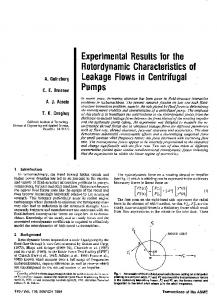
FIG. 1. Domain organization of Flp. (Upper) On a schematic representation of Flp, the positions of V8 cleavage are indicated by solid arrows (this study and ref. 5). Previous studies (5) showed primary tryptic and chymotryptic cleavage sites at Arg-340 and Tyr-343 (broken arrows). These results permit the Flp protein to be divided operationally into two domains, I and 11. Based on the V8 cleavage sites, domain I is subdivided into IA, 1B, and IC. Of particular interest to the present investigation are the V8 peptides, V32 and V28. The amino-terminal sequence of V32 has been determined (5). (Lower) In this study, the amino-terminal residues of V28 were sequenced, and those of V32 were reconfirmed. Question marks denote weak signals. X signifies an undetermined residue.
OD650 of -1.0. Isopropyl /8-D-thiogalactopyranoside (IPTG) was added to a final concentration of 1 mM and the culture was incubated for 1 hr. Cells were harvested, washed, and resuspended in 5 vol of 50 mM TrisHCI, pH 7.4/50 mM ammonium sulfate/1 mM EDTA/0.5 mM dithiothreitol/1 mM each phenylmethylsulfonyl fluoride, leupeptin, and pepstatin (S buffer). After treatment with lysozyme (20 mg/ml) for 30 min on ice, the suspension was subjected to three quick freeze-thaw cycles. After spinning in the cold for 15 min at 15,000 X g, the supernatant was collected, diluted with an equal volume of S buffer containing 40% (vol/vol) glycerol, and kept frozen in aliquots at -70°C. Electroelution of Peptides. A gel slice containing a DNAprotein complex of interest was cut into fragments (approximately 3 x 1 x 1 mm), placed on a nylon net, and electroeluted in an ISCO apparatus (model 1750 concentrator). The electrolyte for elution was 10 mM ammonium carbonate adjusted to pH 10.5 with ammonium hydroxide and containing 0.01% SDS. Elution was carried out at 3 W for 3.5 hr with one change of electrolyte at 2 hr. At the end of the procedure, the current was reversed for 20 sec, the eluate was collected with a Pasteur pipette, concentrated in a Savant Speed-Vac system, and prepared for Laemmli electrophoresis. Electroblotting of Peptides and Amino Acid Sequencing. After SDS/polyacrylamide gel electrophoresis, polypeptides were transferred to poly(vinylidene difluoride) membranes (Immobilon, Millipore) as described by Matsudaira (13) and stained with Coomassie blue. Strips of the membrane containing peptides of interest were directly used for amino acid sequencing in an ABI model 477A/120A protein sequenator/ analyzer. General Methods. Bacterial transformations, isolation of plasmid DNA, restriction enzyme digestions, and other miscellaneous methods followed published procedures (14).
RESULTS Proteolytic Fragments of Flp Can Bind Substrate. All experiments were carried out with a Flp variant [Flp(Y343F)] whose substrate binding characteristics are virtually indistinguishable from those of wild-type Flp (11). However, this protein cannot cleave DNA and thereby form covalent as-
5945
Va
Il'a
V8. ig
0
W
0.1 0.5 1.0 2.0
III u I* II 64 "', bd_.
"II
*,
1_4
B
Flp
V8 DIGEST, jil 0 6 12 20
III,
1lb
I
I
Ill' 1
4
O'. I4t:.-
ll'b
I
64ww I,1 6%. 4 -*
C
D
III
iiil
II
II
Iifb I1'a
I
ad
It
III, IIb II'a
I
-
aI
IP
0 0.02 0.2 0.4 1.0
0 0.02 0.2 0.4 1.0
TRYPSIN, Vg
TRYPSIN, pg
FIG. 2. Complexes formed between a Flp substrate and V8 or tryptic peptides of FIp. The substrate for the binding assay is schematically shown at the top. It contains three Flp binding elements labeled la, 1'a, and 1'b. The spacer region of the substrate is shown by the broken line. (A) Approximately 60 pmol of Flp was digested with V8 protease (0.1-0.2 ,g) in 50 Al in the presence of salmon sperm DNA (1 ,ug) for 1 hr at 30°C. Aliquots of the digestion mixture (6, 12, and 20 Al in lanes 2, 3, and 4, respectively) were assayed for DNA binding with 0.3-0.6 pmol of labeled substrate per assay. Reaction mixtures were analyzed on a 5% polyacrylamide bindinggel. Lanes: 1, free substrate; 5, intact Flp. Complexes formed by Flp are labeled I, 11, and III and those formed by V8 peptides are labeled I', ll'a, I1'b, and III'. (B) FHp (5-10 pmol) was mixed with 0.3-0.6 pmol of labeled substrate and incubated on ice for 30 min. The indicated amounts of V8 were added (lanes 1-5), incubated at 30°C for 30 min, and samples were run on a 5% polyacrylamide binding gel. Analogous experiments with trypsin are shown in C (proteolysis prior to binding) and D (postbinding proteolysis). Each reaction was done with 10-20 pmol of Flp and the indicated amounts of trypsin.
Proc. Natl. Acad. Sci. USA 88 (1991)
Biochemistry: Chen et al.
5946
DNA-protein complexes with faster mobilities (I'-III') than the normal Flp complexes (I-III) were produced. Furthermore, the mobility patterns of the new complexes were virtually the same, irrespective of whether substrate-bound Flp was proteolyzed or preproteolyzed Flp was used for binding substrate. These results strongly indicated that Flp contained a DNA-binding/specificity domain. Association of Specific FIp Peptides with Substrate. To identify the putative Flp domain(s) responsible for substrate binding, we bound V8-treated Flp to its substrate, isolated the smallest of the complexes (presumably, association of one peptide molecule per site), and identified the peptide(s) present in the complex. Flp was treated with V8 under standardized conditions in the presence of salmon sperm DNA for 1 hr at 30TC. The digestion resulted in a mixture composed of -25% intact Flp (as judged by its comigration with untreated Flp; 45 kDa) and peptides of 39, 32, 30, 28, and 15 kDa (Fig. 3 B and C, lanes 4 and 5). The gel system would not have identified peptides smaller than 10 kDa. Previous experiments had determined, by direct sequencing of the amino terminus or by inference from the known sequence of Flp, the origins of all of these peptides except the 28-kDa peptide (5). Our digestion pattern differed from that of Evans et al. (5) by the presence of an increased amount ofthe 28-kDa peptide with a corresponding decrease in the yield of the 30-kDa peptide. We suspect that the presence of salmon sperm DNA in our digestion was likely responsible for the observed difference. Aminoterminal sequencing showed that the 32- and 28-kDa peptides had identical amino termini, beginning at Ser-129. This was consistent with V8 cleavage at Glu-128 of Flp. The V8 digest of 600-800 pmol of Flp (30-40 I&g) was incubated with 80-100 pmol of substrate DNA in a 500-plI binding reaction mixture. The binding mixture was electrophoresed in two lanes of a preparative polyacrylamide gel (Fig. 3A, lanes 1 and 2). For reference, a standard binding mixture with whole Flp was run alongside (lane 3). The portion of the gel corresponding to I' was excised, and the bound peptide(s) was electroeluted and then run on a SDS/ 12% polyacrylamide gel. The 32- and 28-kDa peptides were the only ones associated with the complex as inferred from the Coomassie blue (Fig. 3B, lanes 2 and 3) or the silver (Fig. 3C, lanes 2 and 3) stain profiles. When a mock binding A V8 DIGEST 2 1
1
2
3
4
5
6
7
8
10
9
C
B
Flp 3
reaction containing the Flp digest, but no DNA substrate, was treated as described above and the gel slice corresponding to the position of F' was electroeluted and gel fractionated, no peptides were detectable (data not shown). We conclude that the recovery of the 32- and 28-kDa peptides reflects the ability of each of these peptides to form a complex with the Flp substrate. The possibility that one of the two peptides is part of the complex because of its fortuitous association with the other can be ruled out. The size of such a complex (32 + 28 = 60 kDa of protein) would be inconsistent with the observed relative migration of I'. Absence of Pronounced DNA Bending in Complexes Formed by Flp Peptides. One striking feature of a Flp-DNA complex, in which the two binding elements of the substrate bordering the strand exchange region (or the spacer) are filled by protein (complex labeled II in Figs. 2 and 4), is the sharp bend of >1440 introduced within the spacer (15). The bend is thought to be the result of protein-protein interactions between Flp monomers bound across the spacer. To test whether complexes formed by the V8 peptides contained DNA in a bent configuration, a 400-bp DNA fragment, in which the Flp site was placed almost in the middle, was used as the substrate for binding. The proteininduced bend significantly diminishes the end to end distance in such a fragment and magnifies its gel retardation relative to the unbent fragment, or a similar-sized fragment bent near its end (16). The gel-retardation pattern of this fragment upon binding to aliquots of a V8 digest of Flp is shown in Fig. 4 (lanes 2-4). The digestion conditions were the same as those for the peptide recovery experiment shown in Fig. 3. In a control experiment, aliquots identical to those in lanes 2-4 were bound to a variant substrate containing a 4-bp insertion in the spacer that offset the phasing of the bound protein monomers (lanes 6-8). We assume that Flp-induced DNA bending is eliminated in this substrate. The binding of intact Flp to the bending-competent, or the nonbending substrate is shown in lanes 9 and 10, respectively. Since there were only two binding sites in the substrate (as opposed to three in the one used for experiments of Figs. 2 and 3), only two complexes, I and II, were formed with Flp. The corresponding complexes formed by the V8 peptides were I' and II'. The relative retardation of a complex containing the bending-
II
kD 1 45
23 4 5
1
2 3
45
-. .-Flp -*V39 V-32
.
J30a.
-
30
=v28
II --A
AL...
-40.-
_
6
so
'JmI
11 18
14;
_
4-V.5
FIG. 3. Electroelution of substrate-bound peptides. (A) Approximately 80-100 pmol of the Fip substrate containing three Flp binding elements (10W cpm of 32p) was incubated with a V8 digest of 600-800 pmol of Flp (30-40 jg), and the reaction mixture was run on a 5% polyacrylamide binding gel. A 45-min exposure of the gel is shown. Lanes: 1 and 2, V8 digests; 3, Flp control. (B) Complex I' was excised, the bound peptide(s) was electroeluted, separated on a 12% Laemmli gel, and stained with Coomassie blue. Lanes: 1, molecular size markers; 2 and 3, electroeluted peptides; 4 and 5, aliquots of the V8 digest of Flp used for binding. (C) Samples in B stained with silver. The names of the V8 peptides correspond to those used by Evans et al. (5). We estimate that -30% of the substrate (30-35 pmol) was converted to complex 1'. The amount of recovered peptides (200-250 ng; 5-7 pmol) corresponds to 15-25% efficiency of electroelution.
I_I _i
FIG. 4. The V8 peptide-DNA complexes are bending incompetent. The complexes formed between the V8 digest of Flp and a 400-bp 32P-labeled substrate containing the Flp site in the middle were run on a 5% polyacrylamide binding gel (lanes 1-4). The site contained only two binding elements, la and 1a (cf. Fig. 2). The binding pattern obtained when the substrate included a 4-bp insertion in the spacer is shown in lanes 5-8. Binding of intact Flp to the normal and the insertion substrate are shown in lanes 9 and 10, respectively. Lanes: 1, free substrate; 2-4, increasing aliquots of the V8 digest; 5, the insertion substrate; 6-8, aliquots of V8 digest corresponding to lanes 2-4 (lane 2 corresponds to lane 6, etc.).
Biochemistry: Chen et A
Proc. Natl. Acad. Sci. USA 88 (1991)
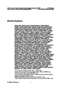
competent substrate was calculated as the ratio of its mobility to that of the corresponding complex containing the nonbending substrate. Bend angles could be derived from the values for relative retardation by using a calibration curve prepared with a set of standard bent DNA fragments (26). The DNA bend angles in the Flp complexes I and II were estimated to be approximately 400 and 1400, respectively. The corresponding values for the peptide-DNA complexes I' and II' were roughly 200 and 400, respectively. A faint complex above II' was observed in the binding reactions with the V8 digests (see lanes 4 and 8). Assuming the complex in lane 4 to be the bent equivalent of the complex in lane 8, the DNA bend in this complex was derived as 50°-60°. The overall conclusion from these results is that the DNA within the complexes formed by the V8 peptides is devoid of the large bend that is characteristic of the normal Flp complexes. A Chimeric Protein Derived from R Recombinase and Flp with FHp-Substrate Specificity. The hypothesis that the aminoterminal portion of Flp is not required for substrate specificity was further tested by constructing a fusion between the R recombinase of Zygosaccharomyces (17, 18) and Flp. The R-Flp hybrid contained the first 127 or 142 amino acids of R (the translation start in R has not been precisely determined) linked in-frame to the last 309 amino acids of Flp (Fig. 5). The expression of the chimeric protein, driven by the tac promoter, was induced in E. coli by addition of IPTG. Crude extracts prepared from induced cells were used to monitor substrate binding in gel retardation assays. The results of such an assay are shown in Fig. 5. The extracts expressing wild-type Flp from the lacuvS promoter (ref. 19; lanes 2 and 3) or from the A Pr promoter (ref. 20; lane 6) bound the Flp substrate; so did the R-Flp chimera (lanes 4 and 5). The binding to purified Flp is shown in lane 7. Under the assay conditions with crude extracts from IPTG-induced cells, complexes I and II were clearly formed, but complex III was barely detectable. Increasing the amounts of extract resulted in nonspecific binding and smearing of the label. Neither the H2N
R-F= p
COOH
SAVHM-DITDIV R-Flp Fip 4 5 2 3
4
I
Flp
Flp
6
7
jX11~~-II
; ~~~ ~ ~ ~ ~ ~ ~ ~ ~ ~ z~~~~~~~~~i L-*
I I
FIG. 5. Substrate binding by a hybrid between R and Flp. (Upper) Amino acids in Flp and R recombinases spanning the sites of fusion are shown. The Flp amino acids are italicized. The methionine residue at position 127 (or 142; refs. 17 and 18) of R is joined to the aspartic acid residue of Flp at position 115. (Lower) Crude extracts were used in a binding assay with a Flp substrate containing three binding elements. Lanes: 1, free substrate; 2 and 3, aliquots of Flp extract; 4 and 5, aliquots of hybrid extract; 6, extract of Ftp expressed from the A Pr promoter; 7, purified Flp. In the binding assays with IPTG-induced crude extracts, complex III was not readily detectable. The Flp used in lane 7 was an old preparation. The band below I in lane 7 likely results from substrate binding by a breakdown product of Flp. This interpretation would be consistent with the data shown in Figs. 2 and 3.
5947
host extract nor the extract from cells expressing R showed binding to the Flp substrate (data not shown). Previous experiments with partially purified R had shown that it does not recognize the Flp site (B.R.E., unpublished result). We wished to test whether the R-Flp hybrid, with its Flp target specificity, could catalyze recombination between two Flp sites. This is not an unreasonable expectation, since the Flp portion of the hybrid protein is expected to include the catalytic domain of the recombinase (5, 11, 12, 21). Recombination was assayed in a RecA- E. coli strain expressing the R-Flp fusion protein by monitoring the loss of a kanamycinresistance marker sandwiched between two directly oriented Flp sites on a substrate plasmid (19, 20). Our failure to observe kanamycin sensitivity suggests that, at least under conditions of the in vivo assay, the protein chimera was not active as a recombinase (data not shown).
DISCUSSION Two peptides obtained by limited proteolysis of Flp by V8 protease retain the ability to bind to the Flp target site. The amino-terminal residue in both of these peptides is Ser-129 of Flp. The carboxyl termini of the peptides (although not sequenced) may be inferred from their sizes. The 32-kDa peptide likely extends to the carboxyl terminus of Flp, while the 28-kDa peptide likely ends at Glu-370 (see Fig. 1). Previous studies had identified this residue to be a moderately strong target for cleavage by V8 (5). Earlier results also showed that substrate recognition by Flp was not affected by short in-frame peptide insertions at Arg-340 (5). Taken together, our data identify the Flp segment spanning domains IB-IC as the substrate binding/specificity domain (Fig. 1). This conclusion agrees with the recent findings of Pan et al. (27). They purified peptides obtained by proteinase K treatment of Flp and showed that a 21-kDa peptide specifically binds the Flp target site. The amino terminus of this peptide maps to Leu-148 of Flp. The IB-IC region of Flp, in addition to harboring substrate specificity, is interesting for a number of reasons. The "yeast family" site-specific recombinases, to which Flp and R proteins belong, show a remarkably high degree of homology within this segment (18). It also harbors the 40-amino acid motif that is characteristic of the "integrase family" of recombinases and the invariant family tetrad (Arg-191-His305-Arg-308-Tyr-343 of Flp; refs. 17 and 18; K. Abremski and R. H. Hoess, personal communication). Three of four mutations that improve binding of Flp to its target DNA sequence (revealed by the P22 challenge phage assay in Salmonella; ref. 22) also form part of the IB-IC region (Arg-258 to Gln, Ile-298 to Met, and Tyr-343 to Phe). Note that Tyr-343 is the active site tyrosine (5, 23) and its potential role in stabilizing Flp-DNA contacts is not surprising. The DNA within the complexes formed by the V8 peptides of Flp is bent poorly. The amino-terminal subdomain IA may be required for at least part of the protein-protein interactions that cause substrate bending. Consistent with this notion is the observation that one of the key residues required for substrate bending is Tyr-60, an invariant residue within the yeast family recombinases (19, 24). Some of the residues within the IB-IC domain also contribute to substrate bending interactions. Mutations of Gly-328 or of Tyr-343 have been shown to lead to bending incompetence (25). Although the R-Flp hybrid that we constructed shows Flp site specificity in binding, it shows no recombinase activity in an in vivo assay. It is possible that interdomainal interactions within or between recombinase protomer that contribute to catalysis are at least partially lost in the hybrid protein. Nevertheless, exchange of more limited regions between the two proteins may define the substrate specificity domain more precisely. A smaller DNA binding peptide would pro-
5948
Biochemistry: Chen et al.
Proc. Natl. Acad. Sci. USA 88 (1991)
vide a better target for site-directed manipulations or for physical studies. The structural and functional relatedness between the two recombinases (18) is likely to minimize potential problems of protein folding that might be encountered in fusions between segments of unrelated proteins.
12. Parsons, R. L., Evans, B. R., Zheng, L. & Jayaram, M. (1990) J. Biol. Chem. 265, 4527-4533. 13. Matsudaira, P. (1987) J. Biol. Chem. 262, 10035-10038. 14. Maniatis, T., Fritsch, E. F. & Sambrook, J. (1982) Molecular Cloning: A Laboratory Manual (Cold Spring Harbor Lab., Cold
We acknowledge the gift of the R recombinase expression plasmid from H. Araki and Y. Oshima. We thank Lei Zheng, Christine Acklin, and Tammy Bauer for excellent technical assistance. This work was supported by National Institutes of Health funds to M.J. and by National Science Foundation Grant DIR 86-18937 to D.B.T.
15.
1. Futcher, A. B. (1986) J. Theor. Biol. 119, 197-204. 2. Reynolds, A. E., Murray, A. W. & Szostak, J. W. (1987) Mol. Cell. Biol. 7, 3566-3573. 3. Volkert, F. C. & Broach, J. R. (1986) Cell 46, 541-550. 4. Cox, M. M. (1989) in Mobile DNA, eds. Berg, D. E. & Howe, M. M. (Am. Soc. Microbiol., Washington), pp. 661-670. 5. Evans, B. R., Chen, J. W., Parsons, R. L., Bauer, T. K., Teplow, D. B. & Jayaram, M. (1990) J. Biol. Chem. 265, 18504-18510. 6. Jayaram, M., Crain, K., Parsons, R. L. & Harshey, R. M. (1988) Proc. Natl. Acad. Sci. USA 85, 7902-7906. 7. Meyer-Leon, L., Huang, L. C., Umlauf, S. W., Cox, M. M. & Inman, R. B. (1988) Mol. Cell. Biol. 8, 3784-37%. 8. Schleif, R. (1988) Science 241, 1182-1187. 9. Landschulz, W. H., Johnson, P. F. & McKnight, S. L. (1988) Science 240, 1759-1764. 10. Amin, A. A. & Sadowski, P. D. (1989) Mol. Cell. Biol. 9, 1987-1995. 11. Parsons, R. L., Prasad, P. V., Harshey, R. M. & Jayaram, M. (1988) Mol. Cell. Biol. 8, 3303-3310.
Spring Harbor, NY). Schwartz, C. J. E. & Sadowski, P. D. (1990) J. Mol. Biol. 216,
289-298.
16. Wu, H. & Crothers, D. (1984) Nature (London) 308, 509-513. 17. Araki, H., Jearnpipatkul, A., Tatsumi, H., Sakurai, T., Ushio, K., Muta, T. & Oshima, Y. (1985) J. Mol. Biol. 182, 191-203. 18. Utatsu, J., Sakamoto, S., Imura, T. & Toh-E, A. (1987) J.
Bacteriol. 169, 5537-5545. 19. Govind, N. S. & Jayaram, M. (1987) Gene 51, 31-41. 20. Jayaram, M. (1985) Proc. Natl. Acad. Sci. USA 82, 5875-5879. 21. Argos, P., Landy, A., Abremski, K., Egan, J. B., HaggardLjungquist, E., Hoess, R. H., Kahn, M. L., Kalionis, B., Narayana, S. V. L., Pierson, L. S., III, Sternberg, N. & Leong, J. M. (1986) EMBO J. 5, 433-440. 22. Lebreton, B., Prasad, P. V., Jayaram, M. & Youderian, P. (1988) Genetics 118, 393-440. 23. Prasad, P. V., Young, L. J. & Jayaram, M. (1987) Proc. Natl. Acad. Sci. USA 84, 2189-2193. 24. Chen, J. W., Evans, B. R., Zheng, L. & Jayaram, M. (1991) J. Mol. Biol. 218, 107-118. 25. Schwartz, C. J. E. & Sadowski, P. D. (1989) J. Mol. Biol. 205, 647-658. 26. Thompson, J. F. & Landy, A. (1988) Nucleic Acids Res. 16,
9687-9705. 27. Pan, H., Clary, D. & Sadowski, P. D. (1991) J. Biol. Chem., in press.