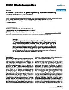
Feb 19, 2014 - From the âModel. Navigatorâ window, select a 3D model and, in the proposed ...... Deniau, J. M., Degos, B., Bosch, C., and Maurice, N. (2010).
May 12, 2015 - Reading Instruction Supported by LDA. Given appropriate instruction, most children learn to read quickly
NEW ZEALAND: Regulation and Management of Cyanobacteria . ... Denmark, Germany, Hungary and South Africa as well as to Tables 1 and 2 below for updates of detail. The 2005 ... ters are used for assessing situations and which values guide responses to
Feb 8, 2013 - Acquired von Willebrand syndrome (AVWS) is an acquired bleeding disorder, first .... Malformation of vessels (M. Osler, Kasabach–Merritt.
âWhere was Elvis born?â is Tupelo, MS. 'Elvis' is automatically interpreted as 'Elvis Presley' according to the highest-ranked results; no other Elvis is mentioned.
At best, we can extract templates that might work in sync with such ..... 1996a. http://www.cogsci.ed.ac.uk/hcrc/publications/wp-2.html ... ICAME Newsletter,.
The success of programmes that relate to disarmament, demobilisation and reintegration (DDR) of former combatants into civilian life is dependent on four.
However, dur- ing the a-fluorination of 1,3-oxazolidines, the yield of fluoro compound is higher in CH3CN than in DME [43]. Other solvents such as sulfolane and ...
Sep 27, 2007 - The binding sites in the FLSM are comparable to. DNA binding sites for transcription factors in the pro- moter regions of genes. A combination ...
Dec 9, 2016 - 1700- 1800 Closing Public Keynote Address by Mr. Henry Marsh. Booking details and biographies of speakers are available on the website.
ting chemotherapy is limited by MGMT in cancer cells and adverse toxic side effects in .... *The platinum analogues, as well as the tetrazines (dacarbazine, ...
1953; Friedman, 1979; Hochberg, 1986; Kuleshov, 1987; McConkie & Zola, ..... most often assumed to be true in the literatureâis that the initial representation.
endothelium is only 5 ml in the entire human brain and 1 μl in the rat brain. ... cellular barrier has some of the most restrictive permeability properties of .... layers of liposomes are similar in structure to those found in living cell ... body (
regulations in different countries compiled and edited by. Dr. Ingrid Chorus. Federal Environment Agency, Germany. UMWELTBUNDESAMT. | TEXTE | 63/2012 ...
(Dardes et al., 2002), and lasofoxifene (Cohen et al., 2001). Steroid aromatase inhibitors block estrogen biosynthesis and thus modulate the estrogen levels in ...
ecules across a cell membrane against its concentration gradient with cellular .... Cell-penetrating peptide (CPP)-mediated drug delivery ... (human immunodeficiency virus [HIV] type 1) trans-activat- ... great promise as intracellular drug delivery
laneous techniques â have been developed to enhance drug delivery to the CNS (Figure ...... reduced.127 This technique makes the procedure more practi- cal for application ..... 2007;14:939â949. 41. Strayer DS, Agrawal L, Cordelier P, et al.
XML management systems vary widely in their expressive ... ment-centric uses of XML management .... Timeline of database management system evolution.
May 24, 2001 - for cervical neoplasia. THE CLINICAL PROBLEM. Although screening for cervical cancer with the. Pap smear is one of the most effective ...
Nov 23, 2011 - CD4+T cell population.2 They showed that generalized auto- immunity ... *Correspondence to: Eric Tartour; Email: [email protected]. Submitted: .... induction of CD8+T cells response for priming of anti-tumor. T cells.
Bennett, K.D. (1989). A provisional map of forest types for the British Isles 5000 ..... Argyll and Bute Countryside Trust Charity. Stornoway Trust Millenium Forest.
Mar 19, 2013 - Anti-angiogenesis strategies to block. CSC expansion have been utilized. However, the benefit of anti- angiogenesis therapy has been ...
Depot pipotiazine palmitate and undecylenate for schizophrenia. Cochrane Database Syst Rev. 2004;(3):CD001720. 96. David A, Quraishi SN, Rathbone J.
Saratoga Springs, New York, May 3-7, 2009, on CD-ROM, American Nuclear Society, LaGrange ... a vector displacement based on a combination of tissue (adjoint) and seed (forward) currents acting ... further work in 3-D should be performed. ..... x (i)|
May 15, 2018 - androgenic alopecia, dermal papilla, hair cycling, hair follicle, male .... preventative therapies, and neither finasteride, minoxidil nor current.
Accepted: 15 May 2018 DOI: 10.1111/exd.13690
VIEWPOINT
A bald statement — Current approaches to manipulate miniaturisation focus only on promoting hair growth Nikolaos Pantelireis
| Claire A. Higgins
Department of Bioengineering, Imperial College London, London, UK Correspondence Claire A. Higgins, Royal School of Mines, London, UK. Email: [email protected] Funding information British Skin Foundation
Abstract Hair plays a large part in communication and society with its role changing through time and across cultures. Most people do not leave the house before combing their hair or shaving their beard and for many hair loss or irregular hair growth can have a significant impact on their psychological health. Somewhat unsurprisingly, according to GMR Data, today’s global hair care industry is worth an estimated $87 Billion, with hair loss estimated at $2.8 Billion. Considering that no current hair loss-related products can completely reverse hair loss, it is reasonable to believe this market could expand significantly with the discovery of a comprehensive therapy. As such, a great deal of research focuses on overcoming hair loss, and in particular, a common form of hair loss known as androgenetic alopecia (AGA) or male pattern baldness. In AGA, hair follicles miniaturise in a large step change from a terminal to a vellus state. Within this viewpoint article, we discuss how influx and efflux of cells into and out from the dermal papilla (DP) can modulate DP size during the hair cycle. As DP size is positively correlated with the size of the hair fibre produced by a follicle, we argue here that therapies for treating AGA should be developed which can alter DP size, rather than just promote hair growth. We also discuss current therapeutics for AGA and emphasise the importance of using the right model systems to analyse miniaturisation. KEYWORDS
TA B L E 1 Brief overview of phases of the hair cycle in human scalp follicles Phase
Description
Anagen (growth phase)
Characterized by the proliferation and differentiation of epithelial cells, originating directly or indirectly from the bulge, that form the follicle and eventually hair itself. The duration of anagen varies across follicles and determines the length of the hair fibre produced. It can last from a few months in eyelashes to a several years in scalp hairs
Catagen (regression phase)
Often referred to as the first stage of the hair cycle, catagen is a 2-week period of regression and preprogrammed apoptosis. The hair shaft itself becomes a club hair, which is no longer actively growing. During catagen, the dermal papilla (DP) reduces in size, and there is apoptosis within the dermal sheath and epithelium,[20] resulting in movement of the DP so it rests beneath the regressing epidermal compartment
Telogen (resting phase)
After catagen, the follicle transitions through to telogen, a resting stage of low metabolic activity that lasts for around 3 months in scalp follicles. The club hair resides in a trichilemmal sac, or old bulge.[72] It is thought that the DP regulates the return of the follicle back to anagen by signalling the overlying epithelial cells, known as the hair germ, to proliferate and differentiate in a process reminiscent of development[73]
Exogen (shedding phase)
Exogen is a phase independent of the main hair follicle cycle where the club hair is actively shed from the old bulge.[72,74,75] Exogen refers to the state of the club hair rather than the hair follicle
Kenogen (latent lag phase)
Kenogen is not observed in all hair follicles; however, frequency and duration increases in scalp follicles of individuals with AGA.[76,77] It refers to a lag phase, or a follicle in telogen which has lost its club hair (exogen) prior to re-entry into anagen
Hair follicles are extremely heterogeneous across body sites in
humans, apoptosis has been observed in the DS, reinforcing this sug-
terms of morphology, cycling and gene expression profile.[13] Almost
gestion that DP cells apoptose once outside the DP.[20] Transitioning
all of the human body bears hair follicles; however most of these are
from telogen-anagen, there is a small amount of cell division within
tiny, unpigmented and almost invisible hairs known as vellus hairs.
the DP itself,[20] however, the majority of the increase in cell number
In pattern baldness, we see terminal hairs (thick and pigmented hair
is due to an influx of cells from the DS, populated by proliferation of
shafts) on the scalp transition to a vellus state through a process
hfDSCs in late telogen and early anagen.[19,21] Thus, if miniaturisation
known as miniaturisation. This gives the appearance of hair loss
occurs during one cyclic transition, either the efflux from the DP is
when in most people (90%), the quantity of hair follicles remains un-
too great or the influx from the DS is too little (Figure 1).
changed.
[14,15]
During miniaturisation we see heterogeneity in fibre
Increased cell efflux in the anagen-catagen transition may be due
size, but this is between follicles and not within a single fibre.[16]
to loss of cell adhesion molecules such as N-C AM, tenascin or inte-
Thus, it seems clear that miniaturisation of the fibre occurs during
grin within the DP,[22] increased mechanical forces acting on the DP,
transitions through the cycle phases, rather than part way though
or a greater migratory response of DP cells attempting to replenish
anagen (Figure 1). Original theories proposed that miniaturisation
the DS which undergoes apoptosis during catagen.[20] Reduced in-
occurred gradually over several hair cycles, with terminal hair tran-
flux during the telogen-anagen transition may be due to an inability
sitioning to a vellus state through a series of shorter and shorter an-
of hfDSCs to proliferate and thus replenish the DP. Alternatively,
agen phases. However, in 2001, Whiting argued that a reduction in
there may be a reduction in adhesive or aggregative cues from the
anagen phase duration over a number of cycles could not be the sole
DP which would signal to promote its enlargement.
mechanism responsible for miniaturisation as the process would take
Hair loss is not entirely dependent on a loss of cells from the
years to occur, which is significantly longer than clinical observations
DP, and cells within the epithelial portion of the follicle have also
of miniaturisation (6-12 months).[17] It is now generally accepted that
been implicated with male pattern baldness. In human scalp folli-
miniaturisation occurs as an abrupt large step process during one
cles, highly expressed cytokeratin 15 (KRT15hi) is a well described
cycle transition instead of over several cycles. The question is now
positive marker of bulge stem cells[23–25] while low levels are found
whether miniaturisation is perpetuated as a result of disruption in
within the proximal ORS of anagen follicles and secondary germ in
the anagen-catagen transition or the telogen-anagen transition, or
telogen.[24] Analysis of epithelial cells (bulge and progenitor) from
both.
balding and non-balding follicles using flow cytometry revealed that
During the hair follicle cycle, there are also cyclic changes in the
while KRT15hi bulge cells were present in the bald scalp at similar
number of cells within the DP. At the end of anagen, there is a de-
numbers to non-balding scalp, CD200 hiItga6hi and CD34hi, which are
crease in the number of cells in the DP, however, with anti-apoptotic
believed to represent progenitor cells, were markedly diminished.[25]
factors expressed and little evidence of cell death,
[18]
cell migration
There are 2 possible explanations for the loss of progenitor cells in
from the DP is thought to drive this reduction. Recently, serial imag-
balding scalp which depend on whether the shortcoming is epithelial
ing of labelled hair follicles in mice during the transition from late an-
or mesenchymal. Potentially, the ability of bulge stem cells to con-
agen to telogen shows that DP cells efflux from the DP into the DS.
vert into progenitor cells is perturbed, either due to an autonomous
Here, it is thought they either undergo apoptosis or incorporate into
defect or an inability to respond to signalling initiating the conver-
the hair follicle dermal stem cell (hfDSC) niche,[19] while similarly in
sion.[25] Alternatively, the DP signal initiating the conversion of bulge
|
3
PANTELIREIS and HIGGINS
determining it.[28] Lending weight to the idea that there is communication between the follicle bulge and the APM are mouse studies where loss of matrix proteins produced by the bulge resulted in a shift in the site of attachment of the APM.[29]
2 | G E T TI N G TE S T Y—A N D RO G E N S A N D CO M PA N Y The cellular mechanism of how miniaturisation occurs is complicated; however, a clue that androgen action is fundamental to this process lies in the clinical name for male pattern baldness; androgenic alopecia (AGA). Somewhat paradoxically androgens are also required for terminal hair maturation during puberty (pubarche). In individuals with testicular feminisation, functional androgen receptors (ARs) fail to develop which results in no pubarche or AGA.[30] The AR is a ligand-activated transcription factor,[31,32] found in both DP and DS.[33–35] The focus on androgens in male pattern baldness has been intense over the years as genetic variability in this receptor was shown to be a prerequisite for early onset of AGA.[36] Since identifying variants in the AR as predisposing for AGA, several other potential loci have been identified in further genetic studies. Most recently, a GWAS assessing AGA was performed which identified 71 possibly causative loci, most notably associated with 3 pathways; the Wnt signalling pathway, apoptosis and the AR signalling pathway.[37,38] Aside from GWAS, transcriptomic studies, or those F I G U R E 1 Simplified dermal mechanisms underlying terminal follicle cycling and miniaturisation. In catagen, dermal papilla (DP) cells migrate out of the DP into the dermal sheath (DS) which is largely degraded. In telogen, the dermal compartment of the hair follicle is reduced with a small number of hfDSC surrounding the DP. In homoeostasis, the DP is fully restored via proliferation and migration of these hfDSC into the DP during anagen resulting in the same number of cells in the DP as in the previous anagen. In miniaturisation, increased efflux of DP cells may occur in the anagen to catagen transition. Alternatively, or conjointly, hfDSCs might not replenish the DP fully in the telogen to anagen transition. Loss of hair follicle homoeostasis may be due to external factors such as DHT and/or perturbations to self-regulating modulators
analysing serum content in men with AGA, have identified a myriad of other factors associated with hair loss.[39,40] A number of androgens can bind to the AR leading to differential binding of the AR to genomic locations regulating gene expression. In balding, the necessary androgen is 5a-Dihydrotestosterone (DHT). DHT is a metabolite of testosterone that is converted in the hair follicle mesenchyme by the action of 5α-reductase.[35] The capacity of a hair follicle to convert circulating testosterone determines whether or not it will miniaturise, and DP from balding scalp are known to express higher levels of 5a-reductase than non-balding scalp.[41,42] This heterogeneity in 5a-reductase expression in hair follicles explains the classic pattern of balding on the frontal scalp, but also explains how a hair follicle from occipital non-balding scalp can be utilised in
cells into progenitors may be altered or weakened. Evidence in sup-
hair transplant surgeries and why a balding hair follicle will still min-
port of this second proposal comes from work performed in vitro
iaturise at the same rate even if transplanted away from the scalp.[43]
where balding “induced” DP cells, unlike non-balding DP cells, were
However, as alluded to earlier, androgens have a paradoxical effect
unable to initiate expression of the differentiated follicular keratin,
on hair follicles. DHT is required for hair follicles on the scalp to min-
K6hf, in cultured bulge cells,[26] suggesting differences in the ability
iaturise, but it is also required for beard and chest hair follicles to
of the 2 DP types to signal to epithelial cells.
mature.[34,44] So DHT is able to elicit the exact opposite response
In recent years, other components of the follicle have been sug-
in different hair follicles. Some clues as to why this might be can be
gested to be involved in the onset of androgenetic alopecia (AGA).
found in expression studies comparing beard DP and occipital scalp
In particular, observational studies on the arrector pili muscle (APM)
DP[45] and while the definitive reason for this inverse response is yet
attachment and AGA remarked that miniaturised follicles within a
to be elucidated, it may well hold the key to reversing miniaturised
follicular unit have had their APM replaced by fat.
[27]
This phenom-
hair follicles.
enon is not observed in alopecia areata nor in standard vellus hair
While androgens seem to initiate miniaturisation, the molecular
follicles, so it poses an interesting question as to whether APM
mechanism by which miniaturisation occurs is not well understood.
loss is simply a consequence of AGA miniaturisation or a factor in
Recently, elevated expression levels of AR associated Prostaglandin
|
PANTELIREIS and HIGGINS
4
D2 Synthase (PTGDS) and its product prostaglandin D2 (PGD2)
female C57BL/6 lab mice at 7 weeks of age.[46,56,59] At this stage, all
were identified in balding scalp skin compared to haired occipital
hair follicles are in telogen and skin appears pink rather than black;
scalp skin.[46] PGD2 is thought to mediate apoptosis of the non-
depilation induces a homogenous re-entry into anagen which can
permanent hair follicle keratinocytes through Prostaglandin D2 re-
then be observed and potentially perturbed.[60] Alternatively, if mice
ceptor 2 (DP2).
are shaven mice one can assess whether anagen re-entry, which would not normally occur for another 4-5 weeks, can be brought for-
3 | A R E W E M A N I PU L ATI N G M I N I AT U R I S ATI O N O R S I M PLY ACC E LE R ATI N G TH E H A I R C YC LE ?
ward[61] by application of candidate therapeutics.[56,59] While these mouse models are incredibly useful to observe signalling during hair follicle cycling in mice, a drug’s ability to reverse miniaturisation cannot be truly observed. Probably the most relevant model for AGA is the stump tailed
While much research has been carried out into the causes of hair
macaque,[62,63] due to the similar physiology of androgen-induced
loss, currently, there are only 2 drugs that have been FDA approved
miniaturisation, however, its use is severely limited due to availabil-
for AGA; finasteride and minoxidil. However, these are viewed as
ity, size, cost and ethical implications. As an alternative, transgenic
preventative therapies, and neither finasteride, minoxidil nor current
K5-hAR mice, which express hAR in the ORS and basal epidermis,
drugs under clinical trials have shown the ability to reverse termi-
have also been developed, where cycling is inhibited temporarily by
nally miniaturised hair follicles[47,48] underscoring that we are yet to
introducing DHT.[64] While this model is androgen dependant, the
see a comprehensive therapy for reversing AGA.
primary measure of efficacy would be re-establishing normal cycling.
Finasteride inhibits the action of 5α-reductase and therefore re-
A perhaps more relevant mouse model for studying miniaturisation
duces the upstream action of DHT effectively halting the progression
is the recently utilised K14-P tgs2 mice, which over expresses PTGS2
of miniaturisation.[49] Finasteride’s effect on the cellular mechanism
resulting in higher PGD2 levels. These mice develop alopecia due to
of miniaturisation is not clear; however, an increase in anagen to tel-
miniaturisation of hair follicles, which have a marked resemblance to
ogen ratio is observed in patients receiving finasteride[50] indicating
human miniaturised follicles.[46]
that hair follicles are promoted to either stay in anagen or transition into anagen from telogen or kenogen.
The focus on manipulating the hair cycle is also the case with clinical trials where total anagen hair count is the primary measured
Minoxidil, in its approved topical form, is believed to act by
outcome. While a prospective drug may be able to accelerate the
prompting hair follicles to transition from telogen to anagen.
induction of anagen in hair follicles, this does not with certainty indi-
However, lengthier body hairs in individuals using minoxidil would
cate that a miniaturised follicle will be resurrected. We see this with
indicate that anagen duration may also be prolonged.[51] There is
current therapies where none are able to restore terminally minia-
confusion as to the exact mechanism by which minoxidil works;
turised hair follicles.
opening potassium ion channels promoting vasodilation, upregulation of VEGF and upregulation of prostaglandin E2 have all been proposed.[52–55] Regardless, if minoxidil simply accelerates hair cycle progression and does not impact efflux or influx, it would only slow
4 | C E LLU L A R TH E R A PI E S A N D M O D E L S TH AT LO O K B E YO N D TH E H A I R C YC LE
the progression of miniaturisation and/or reduce the appearance of baldness by increasing the number of hair follicles in anagen at any
As we have introduced during this viewpoint, there are 3 main cel-
given point in time.
lular mechanisms which can be possible causes for follicular min-
Prostaglandin/Prostamide F2a analogues such as latanoprost
iaturisation. Firstly, there may be too much efflux of cells from the
and bimatoprost have also shown an ability to increase hair growth
DP in the anagen to catagen transition. Secondly, and in combina-
by stimulating hair follicles to transition from telogen to anagen in
tion with the above point, there may be too little influx of cells
mice.[56] As of yet, subsequent research into these analogues for
into the DP in the telogen to anagen transition. Thirdly, there may
AGA has not led to an approved therapy for AGA but this is perhaps
be an impairment in the conversion of bulge cells into their pro-
expected as the progression from bench to bedside can take sev-
genitors. Thus, while research focus is often on manipulating the
eral years.[57,58] Setipiprant is a more recently developed orally ad-
signalling pathways associated with male pattern baldness, such as
ministered antagonist of DP2 which is currently undergoing clinical
AR signalling, we believe that modulating the cellular mechanisms
trials (NCT02781311) due to finish mid-2018. DP2 antagonists are
behind miniaturisation will help identify treatments which can
proposed to reduce PGD2-mediated apoptosis, effectively delaying
reverse AGA in addition to preventing its progression. However,
catagen.
while targeting all 3 of these mechanisms may help prevent minia-
For the most part manipulating the hair cycle seems to be the
turisation, they will not necessarily reverse it. For example, reduc-
mode by which newer therapies look to treat alopecia. The main rea-
ing efflux from an already miniaturised DP in the anagen-c atagen
son for this could be down to the animal models used to evaluate ef-
transition may result in a gradual increase in DP size over time so
ficacy of drugs to take forward for trials. The main animal model for
long as hfDSCs migrate into the DP, but not a step change such as
research in hair growth revolves around either shaving or depilating
the one observed during miniaturisation (Figure 2). We believe the
|
5
PANTELIREIS and HIGGINS
intriguing targets through which we can manipulate the size of the fibre produced by a hair follicle.
5 | CO N C LU S I O N In summary, to identify therapeutics to reverse AGA we believe focus should be on modulating the cellular processes perturbed during transitions through the phases of the cycle, rather than the specific phases. Inter-p hase migration between the mesenchymal compartments and the capacity of hfDSCs to replenish the DP, either through epithelial signalling from bulge (similar F I G U R E 2 Schematic with possible dermal cellular mechanisms modulating a vellus-terminal transition. Increased influx into the dermal papilla (DP) during the telogen-anagen transition would trigger a large step change that would take a vellus hair to terminal state in one cycle. Comparatively, reduced DP cell efflux during catagen would over time result in a gradual increase in DP size assuming that influx of hfDSCs remains unchanged. Alternatively, both of these mechanisms may be required to act in tandem with one another to achieve a reversal of male pattern baldness
to placodal signalling seen in development) or through self-
key to reversing miniaturisation lies in the telogen-anagen transi-
The authors are grateful for funding from the British Skin Foundation
tion, and influx of cells into the DP. Thus, to increase DP size over
to CAH.
modulating mechanisms, can determine whether or not a follicle will miniaturise. Our viewpoint is that an ideal therapy for AGA is one which can promote influx into the DP from the hfDSCs, resulting in DP enlargement and a step change reversal of miniaturisation.
AC K N OW L E D G E M E N T S
and above the increase normally observed during a cycle transition, there has to (i) be an active recruitment by the DP or (ii) be an increased number of cells migrating towards the DP. We know
C O N FL I C T O F I N T E R E S T
DP enlargement is possible as hirsutism is seen in individuals after
The authors have declared no conflicting interests.
taking of certain therapies including oral minoxidil.[55,65] In addition, as mentioned previously, beard follicles undergo a reverse miniaturisation process during puberty, transitioning from a vellus
AU T H O R C O N T R I B U T I O N
state with a small DP to a terminal state with a larger DP (Figure 2).
NP and CAH wrote, read and approved the final manuscript.
Assuming DP cells do not proliferate significantly during this transition, net migration of cells into the DP must be positive. Possible avenues to promote net migration into the DP may be inspired by our understanding of hair development and hair follicle cycling where WNT, FGF and BMP signalling all play roles in promoting dermal condensation and maintaining the inductive properties of the DP.
Whether or not increasing DP influx will act as a permanent therapy for AGA may depend on whether a miniaturised state is the new normal. DP in mice have been shown to self-regulate their size during transitions through the follicular cycle, returning to their original size in anagen subsequent to partial ablation in a previous anagen.[68] So while promoting influx may increase DP size, intrinsic properties may revert the DP to miniaturised state in a subsequent cycle. Recently, the Hippo pathway has been implicated and proposed as a potential modulator of the hair follicle due to its ability to guide cell proliferation, differentiation and stemness.[69,70] Moreover, the Hippo pathway is well established in organ size control and tissue homeostasis.[71] While it has not been comprehensively shown to play a role in AGA if indeed Hippo or another signalling pathway enables DP to self-regulate their size or the bulge cells to maintain their stemness, then these may provide
REFERENCES [1] C. A. B. Jahoda, A. M. Christiano, Cell 2011, 146, 678. [2] M. Ito, G. Cotsarelis, K. Kizawa, K. Hamada, Differentiation 2004, 72, 548. [3] T. S. Purba, I. S. Haslam, E. Poblet, F. Jiménez, A. Gandarillas, A. Izeta, R. Paus, BioEssays 2014, 36, 513. [4] H. Yang, R. C. Adam, Y. Ge, Z. L. Hua, E. Fuchs, Cell 2017, 169, 483. [5] E. Legue, J.-F. Nicolas, Development 2005, 132, 4143. [6] E. J. Van Scott, T. M. Ekel, J. Invest. Dermatol. 1958, 31, 281. [7] K. Elliott, T. J. Stephenson, A. G. Messenger, J. Invest. Dermatol. 1999, 113, 873. [8] C. A. B. Jahoda, Exp. Dermatol. 1998, 7, 235. [9] M. Ito, Z. Yang, T. Andl, C. Cui, N. Kim, S. E. Millar, G. Cotsarelis, Nature 2007, 447, 316. [10] R. Sennett, M. Rendl, Semin. Cell Dev. Biol. 2012, 23, 917. [11] S. E. Millar, J. Invest. Dermatol. 2002, 118, 216. [12] R. Schmidt-Ullrich, R. Paus, BioEssays 2005, 27, 247.
|
6
[13] M. H. Kwack, J. S. Ahn, J. H. Jang, J. C. Kim, Y. K. Sung, M. K. Kim, Exp. Dermatol. 2016, 25, 813. [14] D. A. Whiting, Int. J. Dermatol. 1998, 37, 561. [15] A. M. Kligman, Clin. Dermatol. 1988, 6, 108. [16] M. P. Birch, J. F. Messenger, A. G. Messenger, Br. J. Dermatol. 2001, 144, 297. [17] D. A. Whiting, J. Am. Acad. Dermatol. 2001, 45, 81. [18] K. S. Stenn, L. Lawrence, D. Veis, S. Korsmeyer, M. Seiberg, J. Invest. Dermatol. 1994, 103, 107. [19] W. Rahmani, S. Abbasi, A. Hagner, E. Raharjo, R. Kumar, A. Hotta, S. Magness, D. Metzger, J. Biernaskie, Dev. Cell 2014, 31, 543. [20] D. J. Tobin, M. Magerl, B. Handijski, R. Paus, J. Invest. Dermatol. 2003, 120, 895. [21] N. A. Agabalyan, N. L. Rosin, W. Rahmani, J. Biernaskie, Exp. Dermatol. 2017, 26, 505. [22] C. Chuong, H. Chen, T. Jiang, J. Chia, Ann. N. Y. Acad. Sci. 1991, 642, 263. [23] R. J. Morris, Y. Liu, L. Marles, Z. Yang, C. Trempus, S. Li, J. S. Lin, J. A. Sawicki, G. Cotsarelis, Nat. Biotechnol. 2004, 22, 411. [24] J. E. Kloepper, S. Tiede, J. Brinckmann, D. P. Reinhardt, W. Meyer, R. Faessler, R. Paus, Exp. Dermatol. 2008, 17, 592. [25] L. A. Garza, C. Yang, T. Zhao, H. B. Blatt, M. Lee, H. He, D. C. Stanton, L. Carrasco, J. H. Spiegel, J. W. Tobias, G. Cotsarelis, J. Clin. Invest. 2011, 121, 613. [26] G. J. Leirõs, A. I. Attorresi, M. E. Balañá, Br. J. Dermatol. 2012, 166, 1035. [27] N. Torkamani, N. W. Rufaut, L. Jones, R. Sinclair, Br. J. Dermatol. 2014, 170, 1291. [28] S. Tiede, J. E. Kloepper, D. A. Whiting, R. Paus, Br. J. Dermatol. 2007, 157, 1013. [29] H. Fujiwara, M. Ferreira, G. Donati, D. K. Marciano, J. M. Linton, Y. Sato, A. Hartner, K. Sekiguchi, L. F. Reichardt, F. M. Watt, Cell 2011, 144, 577. [30] V. A. Randall, M. Thornton, K. Hamada, C. Redfern, M. Nutbrouwn, J. Ebling, A. Messenger, Ann. N.Y. Acad. Sci. 1991, 642, 355. [31] P. L. Shaffer, A. Jivan, D. E. Dollins, F. Claessens, D. T. Gewirth, Proc. Natl Acad. Sci. USA 2004, 101, 4758. [32] T. Gao, K. Brantley, E. Bolu, M. J. McPhaul, Mol. Endocrinol. 1999, 13, 1645. [33] R. Choudhry, M. B. Hodgkins, T. H. Van Der Kwast, A. O. Brinkmann, W. J. A. Boersma, J. Endocrinol. 1992, 133, 467. [34] V. A. Randall, Dermatol. Ther. 2008, 21, 314. [35] Y. Asada, T. Sonoda, M. Ojiro, S. Kurata, T. Sato, T. Ezaki, S. Takayasu, J. Clin. Endocrinol. Metab. 2001, 86, 2875. [36] A. M. Hillmer, S. Hanneken, S. Ritzmann, T. Becker, J. Freudenberg, F. F. Brockschmidt, A. Flaquer, Y. Freudenberg-Hua, R. A. Jamra, C. Metzen, U. Heyn, N. Schweiger, R. C. Betz, B. Blaumeiser, J. Hampe, S. Schreiber, T. G. Schulze, H. C. Hennies, J. Schumacher, P. Propping, T. Ruzicka, S. Cichon, T. F. Wienker, R. Kruse, M. M. Nöthen, Am. J. Hum. Genet. 2005, 77, 140. [37] S. Heilmann, A. K. Kiefer, N. Fricker, D. Drichel, A. M. Hillmer, C. Herold, J. Y. Tung, N. Eriksson, S. Redler, R. C. Betz, R. Li, A. Kárason, D. R. Nyholt, K. Song, S. H. Vermeulen, S. Kanoni, G. Dedoussis, N. G. Martin, L. A. Kiemeney, V. Mooser, K. Stefansson, J. Brent Richards, T. Becker, F. F. Brockschmidt, D. A. Hinds, M. M. Nöthen, J. Invest. Dermatol. 2013, 133, 1489. [38] A. M. Hillmer, F. F. Brockschmidt, S. Hanneken, S. Eigelshoven, M. Steffens, A. Flaquer, S. Herms, T. Becker, A.-K. Kortüm, D. R. Nyholt, Z. Z. Zhao, G. W. Montgomery, N. G. Martin, T. W. Mühleisen, M. A. Alblas, S. Moebus, K.-H. Jöckel, M. BröckerPreuss, R. Erbel, R. Reinartz, R. C. Betz, S. Cichon, P. Propping, M. P. Baur, T. F. Wienker, R. Kruse, M. M. Nöthen, Nat. Genet. 2008, 40, 1279. [39] A. Vogt, E. K. B. Pfannes, S. Fimmel, S. Hadam, A. Andruck, J. Kottner, U. Blume-Peytavi, Exp. Dermatol. 2017, 26, 518.
PANTELIREIS and HIGGINS
[40] C. C. Yang, P. L. Chung, L. Y. Lin, M. W. Hughes, Y. S. Tsai, Exp. Dermatol. 2017, 26, 524. [41] M. E. Sawaya, V. H. Price, J. Invest. Dermatol. 1997, 109, 296. [42] N. A. Hibberts, A. E. Howell, V. A. Randall, J. Endocrinol. 1998, 156, 59. [43] R. E. A. Nordstrom, Acta Derm. Venereol. 1979, 59, 266. [44] J. D. Wilson, J. E. Griffin, D. W. Russell, Endocr. Rev. 1993, 14, 577. [45] S. E. Rutberg, M. L. Kolpak, J. A. Gourley, G. Tan, J. P. Henry, D. Shander, J. Invest. Dermatol. 2006, 126, 2583. [46] L. A. Garza, Y. Liu, Z. Yang, B. Alagesan, J. A. Lawson, S. M. Norberg, D. E. Loy, T. Zhao, H. B. Blatt, D. C. Stanton, L. Carrasco, G. Ahluwalia, S. M. Fischer, G. A. FitzGerald, G. Cotsarelis. Sci. Transl. Med. 2012: 4: 126ra34. [47] V. H. Price, N. Engl. J. Med. 1999, 341, 964. [48] K. J. McElwee, J. S. Shapiro, Ski. Ther. Lett. 2012, 17, 1. [49] K. D. Kaufman, E. A. Olsen, D. Whiting, R. Savin, R. Devillez, W. Bergfeld, V. H. Price, D. Van Neste, J. L. Roberts, M. Hordinsky, J. Shapiro, B. Binkowitz, G. J. Gormley, J. Am. Acad. Dermatol. 1998, 39, 578. [50] D. Van Neste, V. Fuh, P. Sanchez-Pedreno, E. Lopez-Bran, H. Wolff, D. Whiting, J. Roberts, D. Kopera, J. J. Stene, S. Calvieri, A. Tosti, E. Prens, M. Guarrera, P. Kanojia, W. He, K. D. Kaufman, Br. J. Dermatol. 2000, 143, 804. [51] J. L. Burton, A. Marshall, Br. J. Dermatol. 1979, 101, 593. [52] K. Shorter, N. P. Farjo, S. M. Picksley, V. A. Randall, FASEB J. 2008, 22, 1725. [53] S. Lachgar, M. Charveron, Y. Gall, J. L. Bonafe, Br. J. Dermatol. 1998, 138, 407. [54] S. Lachgar, M. Charvéron, N. Bouhaddioui, Y. Neveux, Y. Gall, J. L. Bonafé, Arch. Dermatol. Res. 1996, 288, 469. [55] A. G. Messenger, J. Rundegren. Br. J. Dermatol. 2004: 150: 186. [56] S. Sasaki, Y. Hozumi, S. Kondo, Exp. Dermatol. 2005, 14, 323. [57] H. Uno, M. L. Zimbric, D. M. Albert, J. Stjernschantz, Acta Derm. Venereol. 2002, 82, 7. [58] U. Blume-Peytavi, S. Lönnfors, K. Hillmann, N. Garcia Bartels, J. Am. Acad. Dermatol. 2012, 66, 794. [59] K. G. Khidhir, D. F. Woodward, N. P. Farjo, B. K. Farjo, E. S. Tang, J. W. Wang, S. M. Picksley, V. A. Randall, FASEB J. 2013, 27, 557. [60] S. Müller-Röver, B. Handjiski, C. Van Der Veen, S. Eichmüller, K. Foitzik, I. A. McKay, K. S. Stenn, R. Paus, J. Invest. Dermatol. 2001, 117, 3. [61] S. Harel, C. A. Higgins, J. E. Cerise, Z. Dai, J. C. Chen, R. Clynes, A. M. Christiano, Sci. Adv. 2015, 1, e1500973. [62] W. Montagna, H. Uno, J. Soc. Cosmet. Chem. 1968, 19, 173. [63] J. P. Sundberg, W. G. Beamer, H. Uno, D. Van Neste, L. E. King, Exp. Mol. Pathol. 1999, 67, 118. [64] J. S. Crabtree, E. J. Kilbourne, B. J. Peano, S. Chippari, T. Kenney, C. McNally, W. Wang, H. A. Harris, R. C. Winneker, S. Nagpal, C. C. Thompson, Endocrinology 2010, 151, 2373. [65] A. Tosti, C. Misciali, B. M. Piraccini, A. M. Peluso, F. Bardazzi, Drug Saf. 1994, 10, 310. [66] G. Cotsarelis, S. E. Millar, Trends Mol. Med. 2001, 7, 293. [67] S. H. Huh, K. Närhi, P. H. Lindfors, O. Häärä, L. Yang, D. M. Ornitz, M. L. Mikkola, Genes Dev. 2013, 27, 450. [68] W. Chi, E. Wu, B. A. Morgan, Development 2013, 140, 1676. [69] H. Zhang, H. A. Pasolli, E. Fuchs, Proc. Natl Acad. Sci. 2011, 108, 2270. [70] R. K. Severin, X. Li, K. Qian, A. C. Mueller, L. Petukhova, Sci. Rep. 2017, 7, 1. [71] F. X. Yu, B. Zhao, K. L. Guan, Cell 2015, 163, 811. [72] Y. C. Hsu, H. A. Pasolli, E. Fuchs, Cell 2011, 144, 92. [73] V. Greco, T. Chen, M. Rendl, M. Schober, H. A. Pasolli, N. Stokes, J. dela Cruz-Racelis, E. Fuchs. Cell Stem Cell 2009: 4: 155.
|
7
PANTELIREIS and HIGGINS
[74] K. S. Stenn, S. Parimoo, in Molecular Basis of Epithelial Appendage Morphogenesis (Ed: C. M. Chuong). RG Landes Co, Austin, TX 1998, pp. 111–130. [75] C. A. Higgins, G. E. Westgate, C. A. B. Jahoda, J. Invest. Dermatol. 2009, 129, 2100. [76] A. Rebora, M. Guarrera, Arch. Dermatol. 2004, 140, 619. [77] M. Guarrera, A. Rebora, Dermatology 2005, 210, 18.
How to cite this article: Pantelireis N, Higgins CA. A bald statement — Current approaches to manipulate miniaturisation focus only on promoting hair growth. Exp Dermatol. 2018;00:1–7. https://doi.org/10.1111/exd.13690