Annals of Clinical & Laboratory Science, vol. 32, no. 1, 2002
3
A Testing Algorithm for Determination of HER2 Status in Patients with Breast Cancer Dawn W. Nichols,1* Daynna J. Wolff,1 Sally Self,1 John S. Metcalf,1 Donna Jacobs,1 Rayna Kneuper-Hall,2 and John C. Cate, IV 1 1 Department of Pathology and Laboratory Medicine, 2 Department of Medicine, Medical University of South Carolina, and Hollings Cancer Center, Charleston, South Carolina *Current address: Anderson Memorial Hospital, Anderson, South Carolina Abstract. The HER2 gene is amplified and overexpressed in 25–30% of breast carcinomas. Assessment of HER2 status for prognosis and treatment of breast cancer patients can be performed by immunohistochemistry and/or fluorescence in situ hybridization (FISH). To develop a testing algorithm for HER2 in breast cancers, we used FISH analysis to determine the HER2 gene copy number and immunostaining to detect the p185 protein. Interlaboratory, interobserver, and intermethod variabilities of immunohistochemistry were assessed. In 24 invasive breast carcinomas, the indices of HER2 status obtained by FISH and by a reference laboratory’s DAKO HercepTest® (immunostain) gave an overall concordance of 96%. The reference laboratory’s stained slides were re-interpreted by an in-house panel of pathologists; the interpretation differed in one case. The panel’s interpretations were concordant with the FISH results in all 24 cases. Interobserver variability for the panel’s immunohistochemistry interpretations was assessed using three different immunostaining methods on 70 slides. The numerical (0-1+, 2+, 3+) scores showed greater variability among observers than did the overall positive/negative results. One pathologist reported inconsistent results in >30% of the slides evaluated. Borderline scoring of 1-2+ was reported in 18 slides (23%) by at least one observer. Incongruent interobserver immunohistochemistry scores, leading to different positive and negative interpretations, were obtained with 5 slides (7%). The majority of consensus positive cases exhibited strong membrane staining (3+). The majority of consensus negative cases scored as 0. Based on these observations, we developed a testing algorithm that maximizes the benefits of FISH and immunohistochemistry, providing physicians with accurate results for appropriate clinical care. (received 24 September 2001, accepted 22 October 2001)
Keywords: fluorescence in situ hybridization (FISH), immunohistochemistry, breast cancer, HER2 gene Introduction The human epidermal growth factor receptor 2 (HER2) gene encodes a 185 kd protein that acts as a cell surface receptor with tyrosine kinase activity [1]. Amplification of the HER2 gene and/or overexpression of its protein product have been demonstrated in 25–30% of breast cancers [2-3]. This up-regulation is associated with poor prognosis, increased risk for recurrence, and shortened survival Address correspondence to Daynna J. Wolff, Ph.D., Department of Pathology and Laboratory Medicine, Medical University of South Carolina, P.O. Box 250908, Charleston, SC 29425, USA; tel 843 792 3574; fax 843 792 1248; e-mail
[email protected].
in breast cancer patients [3-5]. Several studies have shown that HER2 status correlates with sensitivity or resistance to certain chemotherapy regimens [69]. Furthermore, an effective therapy that is targeted specifically to cells that overexpress HER2 protein has been approved for use in HER2-positive patients with metastatic breast cancer. Numerous methods can be employed to detect the HER2 gene and its protein product. Gene-based assays include Southern blotting, slot blotting, polymerase chain reaction methods, and in situ hybridization techniques. The HER2 protein product is detected using immunohistochemistry, Western blotting, and enzyme immunoassays. The FDA has
0091-7370/02/0100/0003 $2.25. © 2002 by the Association of Clinical Scientists, Inc.
4 Annals of Clinical & Laboratory Science
approved methods for FISH (INFORM®, Ventana, Inc.; PathVision®, Vysis, Inc.) and immunohistochemistry (HercepTest®, DAKO, Inc.). FISH has the benefit of being highly sensitive (96%) and specific (100%) [10], but is expensive, laborintensive, and not universally available. Immunostaining, on the other hand, is available in most pathology laboratories. The major problems with quantitative immunohistochemistry of HER2 stem from lack of standardization in methodology and interpretation. Even when most technical factors are kept constant, eg, use of the HercepTest® on internal samples, subjective interpretation may lead to high false positive and/or negative rates. This is particularly troublesome for samples that stain in the mid-range (1+, 2+) using the HercepTest® kit. Clinical trials are underway to determine the predictive values and appropriate uses of the FDAapproved assays. Preliminary data suggest that results of FISH testing correlate better with clinical response to the targeted therapy, compared to results of the HercepTest® [11-15]. Our strategy was to develop a testing algorithm that maximizes the benefits of FISH and immunohistochemistry testing. We believe that this integrated approach can provide the most accurate results, while minimizing costs and optimizing therapeutic options. Materials and Methods Patients. Cases were selected from patients with invasive breast carcinoma who were being evaluated for HER2 status. Tissue samples were fixed in formalin and embedded in paraffin using standard laboratory protocols. Fixation time ranged from 2 to 24 hr. Fluorescence in situ hybridization. FISH was performed according to manufacturer’s instructions (Ventana, Inc., formerly Oncor). In brief, slides were baked overnight at 65°C and deparaffinized in xylene. Slides were subjected to proteinase treatment for 30-40 min, followed by DNA denaturation at 75°C for 8 min. After dehydration, the biotinylated HER2 cosmid probe was applied to the slide, coverslipped, and hybridized overnight at 37°C. Hybridized slides were stringently washed to remove
unbound probe. Bound probe was detected using fluorescein isothiocyanate (FITC)-strepavidin. Nuclei were counterstained with 4’,6-diamidine-2phenylindole (DAPI). The preparations were visualized using an epifluorescence microscope (Axioskop®, Zeiss, Inc., Thornwood, NY) with a triple dichroic filter (DAPI; fluoroscein isothiocyanate; Texas red). For scoring FISH slides, two areas of invasive tumor were identified using corresponding hematoxylin-eosin stained slides. The number of green fluorescent signals was determined for 20 intact cells in both areas. The average number of signals per nucleus was calculated and the specimen was labeled “amplified” if the average was ≥4. Immunohistochemistry. Paraffin blocks from 24 breast carcinomas with known FISH results (12 amplified and 12 unamplified cases) were sent to a reference laboratory experienced in the HercepTest®. For 22 cases with tissue remaining in the blocks, re-cuts were made in our laboratory and stained for HER2 using two internal procedures: a NexES® automated staining system and the FDAapproved HercepTest®. Tissue was cut at 4 µm and mounted on positively charged glass slides. Slides were dried overnight at 60°C, deparaffinized, and then hydrated [two changes of xylene (10 min each); two changes of 100% ethanol (3 min each); two changes of 95% ethanol (3 min each); and 2 changes of distilled water (2 min each)]. Testing was performed with the NexES® automated immunostainer, standard Ventana diaminobenzadine detection system, and Ventana prediluted HER2 antibody. The staining protocol was: primary antibody (32 min) with amplification; hematoxylin (2 min); and bluing (2 min). Before staining on the NexES® immunostainer, the slides were immersed in Ventana’s epitope retrieval solution, placed in a steamer (20 min), and cooled at room temperature (10 min). The DAKO HercepTest® package insert was followed without exception in the second method. Nuclei were counter-stained (1 min) with Harris hematoxylin, rinsed in distilled water, and blued with 0.1% NH4OH solution (30 sec). All slides were rinsed in distilled water, dehydrated, cleared, and mounted with synthetic media.
HER2 testing algorithm for breast cancer
Table 1. HER2 results obtained by fluroescence in situ hybridization (FISH) in 24 cases of breast cancer (12 FISH-amplified; 12 FISH-unamplified), versus immunohistochemistry (Hercept-Test®) scores reported by a reference laboratory. FISH results
HerceptTest® results 0 1+ 2+ 3+
unamplified
12
0
0
0
amplified
0
1
7
4
Table 3. Interobserver variation of scoring of 70 immunohistochemisty (IHC) slides by a 5-member panel of pathologists. Discordant results
“Gray” zone results
0,0,0,2,2
1,1,1-2,1,1
0,0,0,0,0
3,3,2-3,3,3
0,0,0,3,0
1,1,1-2,1,1
0,0,0,0,0
3,3,2-3,3,3
0,0,0,0,0
3,3,2-3,3,3
1,0,1-2,0,0
0,0,0,0,0
3,3,2-3,2,2
1,2-3,2-3,3,3 1,0,1-2,1,1
0,0,0,0,0
2,3,2-3,2,2
1,0,1-2,0,0-1
0,0,0,0,0
2,3,2-3,2,2
1,0,1-2,0,0-1
0,0,0,0,0
2,3,2-3,3,3
1,0,1-2,0,0
0,0,0,0,0
3,3,2-3,3,3
1,1,1-2,1,1-2
0,0,0,0,0
2,2,2-3,3,3
1,1,1-2,1,1
1,0,0,0,0
3,3,2-3,3,3
1-2,1,1-2,1,1
1,1,0,1,0
2,3,2-3,3,3
1,1,1-2,1,0
0,0,0,0,0
2,3,2-3,3,3
1,0,1-2,0,0
0,0,0,0,0
2,2-3,2-3,2,3
1,1-2,1-2,1,0
0,0,0,0,0
3,3,2-3,3,3
2,2,1-2,2,1-2
0,0,0,0,0
3,2-3,2-3,3,3
2,3,1-2,3,3
1,0,0,0,0
3,3,2-3,3,3
1,3,1-2,2,1-2 1,0-1,1-2,0,1
Table 2. Interlaboratory variation of scoring of immunohistochemisty (IHC) slides for HerceptTest® in 24 cases of breast cancer. Case #
1 2 3 4 5 6 7 8 9 10 11 12 13 14 15 16 17 18 19 20 21 22 23 24
FISH results
unamplified unamplified unamplified unamplified unamplified unamplified unamplified unamplified unamplified unamplified unamplified unamplified amplified amplified amplified amplified amplified amplified amplified amplified amplified amplified amplified amplified
IHC score Concensus by reference IHC score by laboratory faculty panel 0 0 0 0 0 0 0 0 0 0 0 0 3 3 3 2 2 2 1 2 2 2 2 3
0.8 0.0 0.0 0.6 0.0 0.0 0.0 0.2 0.0 0.0 0.0 0.0 2.5 2.9 2.9 2.0 2.9 2.5 2.3 2.3 1.8 1.8 2.7 2.9
5
1,3,0,0,0
2,2-3,1-2,2,2
Concordant Concordant negative positive results results
3,3,2-3,3,3 2,2,2-3,2,3 2,2-3,2-3,2,3 2,2,2-3,3,3 2,2-3,2-3,2,2 3,2,2-3,3,3 3,3,2-3,3,3 3,2-3,2-3,3,3 2,2,2-3,3,3 2,2,2-3,2,3 3,2,2-3,3,3 2,2-3,2-3,3,3 2,2-3,2-3,3,3 3,2,2-3,3,3 3,3,2-3,3,3 2,2,2-3,2,2
6 Annals of Clinical & Laboratory Science
Five-member panel. A faculty panel of five pathologists (board-certified in anatomical pathology or anatomical/clinical pathology) evaluated internally-prepared and reference laboratory slides (n = 70). The panel was blinded in respect to FISH results, prior immunohistochemistry interpretations, and staining techniques. The panel used the HercepTest® criteria to correlate membranous staining intensity (≥10% of cells showing membranous staining for a positive result). The staining intensity was scored as 3+ (dark staining), 2+ (mid-range staining), 1+ (faint staining), or 0 (minimal or no staining). In this study, cases that were scored as 12+ were considered borderline. The DAKO kit included tissue culture controls of 0, 1+, and 3+, together with photographs that were used by panel members for interpretation. Results were analyzed for intermethod and interobserver variability. Kappa analysis. Cohen’s kappa statistic measures the agreement between raters for classifying a number of data points into 2 or more categories [16]. Kappa ranges can be used to establish agreement between tests, methods, or evaluators [17]. Kappa values range from –1 to 1, with 1 representing perfect agreement between data sets. Landis and Koch [18] delineated different ranges of kappa in respect to the degree of agreement beyond chance; these criteria were used in our comparisons. Values >0.75 were considered to represent excellent agreement, values from 0.4 to 0.75 were considered to represent fair/ good agreement, and values 1+) by both the reference laboratory and the in-house panel. The sole amplified case interpreted as negative at the reference lab was interpreted as positive by all of the 5 panel members. Method Variability. Three immunohistochemical approaches were used on 21 cases (10 FISH unamplified and 11 FISH amplified). The three approaches included the Ventana antibody on the automated NexES® immunostainer, the HercepTest® performed in our laboratory, and the HercepTest® performed at the reference laboratory. The panel scored each slide according to the HercepTest® criteria. The NexES®-prepared slides gave a higher average score than either the in-house or the reference laboratory’s HercepTest® results in 15 cases (7l%) (Fig. 2). As a consequence, at least one observer interpreted the NexES stain as borderline or positive in all 10 unamplified cases, which were interpreted as clearly negative using the HercepTest® method. Interobserver variability in immunohistochemistry. Each member of the panel blindly evaluated 70 slides from 27 cases of breast carcinoma with predetermined FISH results. Each slide was examined independently by each observer and given a score (0, 1+, 2+, or 3+) and a corresponding negative or positive result (Table 3). The overall interobserver
HER2 testing algorithm for breast cancer
Fig. 1. Immunohistochemical staining (HercepTest®) of 2 breast carcinomas with easily interpretable “0” negative staining (left) (original magnification x300) and “3+” positive staining (right) (original magnification x400) compared with tissue control (center) (original magnification x400).
7
Fig. 3. Immunohistochemical borderline staining of 2 different breast carcinomas, one FISH-amplified (left) (original magnification x200) and one FISH-unamplified (right) (original magnification x400), compared to the HercepTest® “1+” control (center) (original magnification x200).
Fig. 2. Immunohistochemical staining showing increased brightness with the NexES® method compared to the HercepTest® method in 2 cases of breast carcinoma: (A) unamplified case with “0” staining with HercepTest® (original magnification x300), (B) same unamplified case with “1+” staining with NexES® (original magnification x300), (C) amplified case with “2+” staining on HercepTest® (original magnification x400), and (D) amplified case with “3+” staining on NexES® (original magnification x400).
8
Annals of Clinical & Laboratory Science
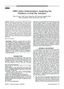
Fig. 4. Algorithm showing proposed method for evaluating for HER2 status in breast cancers.
variability for positive and negative results by immunohistochemistry displayed high kappa values (0.87; 0.81; 0.81; 0.74) for 4 observers. The results obtained by the fifth panelist showed poor agreement (kappa = 0.3). In contrast, kappa values for the numerical scores (0, 1+, 2+, or 3) were low (kappa = 0.31; 0.17; 0.43; 0.40; 0.25), demonstrating poor/ fair agreement in scoring the staining intensity. Incongruent interobserver immunohistochemistry scores that lead to different positive and negative interpretations were made on 5 slides (7%). As shown in Fig. 3, borderline staining (1-2+) was reported by at least one observer on 18 slides (26%). Four panelists used the borderline 1-2+ range for scoring. This designation was given by one observer to 17 slides (24%). The majority (82%) of slides with borderline staining were categorized as consensus negative by immunohistochemistry and were FISH-unamplified. The remaining 3 slides were categorized as consensus positive by immunohistochemistry and were FISHamplified. Observers who used the 1-2+ designation showed fewer incongruities with FISH. Many of the 1-2+ scores were rendered by one observer, but this observer had a 100% correlation with FISH results in the cases that scored frankly negative (0) or frankly positive (2-3+, or 3+).
Discussion Determination of HER2 status is an important independent clinical test for stratification of breast cancer patients. HER2 assessment is useful for prognosis, chemotherapy responsiveness, and selection for targeted monoclonal antibody therapy (Herceptin®) [5-10]. Because of the clinical significance of positive or negative HER2 results, pathologists and oncologists need accurate testing to determine the most appropriate therapy for breast cancer patients. Of the two FDA-approved methodologies for HER2 assessment, FISH testing is the most sensitive and specific [10]. However, the immunohistochemistry methodology is less expensive and more widely available. Our study demonstrated good correlation of immunohistochemistry with FISH results in a 24-patient cohort, particularly with results that were obviously positive or negative. Since amplification of the HER2 gene appears to be the primary mechanism responsible for protein overexpression [19], it follows that FISH and immunohistochemistry results should correlate. FISH is a quantitative assay, whereas quantitation of the HercepTest® immunohistochemistry results relies on the pathologist’s judgement in the interpretation of staining intensity. Therefore, it is important to
HER2 testing algorithm for breast cancer
understand the potential variables associated with this method. To assess possible variability in interlaboratory interpretation of immunostaining, the 24 immunohistochemistry slides initially analyzed at the reference laboratory were blindly reviewed by our in-house panel of pathologists. One of 12 FISHamplified cases was scored as 1+ (negative) by the reference laboratory pathologist but was scored as consensus positive by the in-house panel. In our limited study of 24 cases, interpretations of HercepTest® staining intensity between laboratories appeared most consistent in negative (unamplified) cases with no or little immunohistochemical staining. When the same 24 cases were prepared in-house and blindly scored in a side-by-side analysis of HercepTest®, no consistent interlaboratory variability of staining was detected (similar results were reported by Jacobs et al [20]). The panel’s average scores for HercepTest® slides prepared inhouse were comparable to the reference laboratory’s results for the same tumor blocks. To study intermethod variability, we compared results by the FDA-approved HercepTest® with those obtained using an automated stainer with another commercially available antibody (Ventana). As reported in previous studies [21-23], the HER2 antigen detection with the different polyclonal antibodies was quite variable. The NexES®prepared slides had a higher average score than either the in-house or reference laboratory HercepTest® results in ≥70% of cases; as a result, at least one observer interpreted the NexES® stain as borderline or positive in all FISH-unamplified cases. In addition, borderline staining was more common in the NexES®-prepared slides, compared to in-house or reference laboratory HercepTest®. Interobserver differences were largely responsible for variability of immunohistochemistry results. Kappa values showed good correlation between observers in overall positivity/negativity of results, but poor inter-observer agreement on individual scores of 0-1+, 2+, and 3+. Interpretation differences did not seem to be related to the observer’s level of experience with immunohistochemistry. Although the overall kappa was good, differences of interpretation of overall positivity/ negativity occurred in nearly 10% of cases.
9
We found concordance in the clearly negative (0) and clearly positive (3+) immunochemistry assays, but less correlation in the mid-range staining region. The majority (82%) of our borderline staining (1+ or 2+) cases were not amplified by FISH. Our data and previous studies [10,24-27] suggest that many 2+ immunohistochemistry results are false positives and that these patients may not benefit from treatment with trastuzumab. In addition, recent clinical trials have shown that only patients with FISH amplification respond to the targeted drug therapy (trastuzumab, Herceptin®) [12-15]. Some discordant cases could represent overexpression without amplification, although this has been reported to be a rare event–occurring in only ≥3% of all immunohistochemistry positive patients [19,24,28-30]. There were no cases in our study that demonstrated consensus overexpression without amplification. Recent reports suggest overexpression without amplification may not be as prognostically poor as overexpression with amplification [31]. Immunohistochemistry assay by HercepTest® is cost-effective as a first-line HER2 methodology and is more feasible in community-based laboratories. Furthermore, immunohistochemistry assay for the protein target of the monoclonal antibody is currently approved by the FDA for consideration for trastuzumab (Herceptin®) candidacy. Our study shows that immunohistochemistry is most sensitive and specific at low-level and high-level staining, but is less consistent with mid-range staining. Using immunostaining only for low-level and high-level results appears not to compromise quality, but can lead to significant cost savings for many patients. Problems with incongruent interpretations can be improved by using a mid-range category (1-2+) and further evaluating those cases with FISH. Our strategy is to evaluate patients with borderline immunohistochemistry results by FISH to clarify HER2 status (Fig. 4). Having both methodologies available also allows for ongoing quality assurance correlation and the study of outcomes data for both tests. An integrated approach maximizes the benefits of each test, while counterbalancing their limitations. Other recent studies have also suggested the benefits of an integrated approach [24-25,32-34].
10
Annals of Clinical & Laboratory Science
In conclusion, we have determined that the immunohistochemical test for HER2 can be unreliable when staining is of intermediate (1-2+) intensity. We developed an algorithm that initially performs immunohistochemistry for HER2 assessment. If results are within the 1-2+ staining range, repeat testing is performed by FISH. Our study suggests that this combined strategy may increase our ability to identify patients with HER2 amplification and/or protein over-expression in a more accurate and cost-effective manner. Ongoing clinical outcomes studies of large patient populations will determine the clinical efficacy of this testing algorithm for evaluating HER2 status .
7.
8.
Acknowledgements
9.
The authors thank Hurshell Hunt, Ph.D., for statistical review of the data and Marilyn Hegel for secretarial assistance. This study, supported by the Department of Pathology and Laboratory Medicine, Medical University of South Carolina, Charleston, SC, was presented in part at the Academy of Clinical Laboratory Physicians and Scientists’ meeting in Birmingham, AL, on 4 June 1999.
10.
References
11.
1. Akiyama T, Sudo C, Ogawara H, Toyoshima K, Yamamoto T. The product of the human c-erbB-2 gene: a 185-kilodalton glycoprotein with tyrosine kinase activity. Science 1986;232:1644-1646. 2. Slamon DJ, Clark GM, Wong SG, Levin WJ, Ullrich A, McGuire WL. Human breast cancer: correlation of relapse and survival with amplification of the HER-2/neu oncogene. Science 1987;235:177-182. 3. Press MF, Bernstein L, Thomas PA, Meisner LF, Zhou J-Y, Ma Y, Hung G, Robinson RA, Harris C, ElNaggar A, Slamon DJ, Phillips RN, Ross JS, Wolman SR, Flom KJ. HER-2/neu gene amplification characterized by fluorescence in situ hybridization: poor prognosis in node-negative breast carcinomas. J Clin Oncol 1997;15:2894-2904. 4. Xing WR, Gilchrist KW, Harris CP, Samson W, Meisner LF. FISH detection of HER-2/neu oncogene amplification in early onset breast cancer. Breast Cancer Res Treat 1996;39:203-212. 5. Ross JS, Fletcher JA. HER-2/neu (c-erb-B2) gene and protein in breast cancer. Am J Clin Pathol 1999;112 (Suppl.1):S53-S67. 6. Gusterson BA, Gelber RD, Goldhirsch A, Price KN, Säve-Söderborgh J, Anbazhagan R, Styles J,
12.
13.
Rudenstam C-M, Golouh R, Reed R, MartinezTello F, Tiltman A, Torhorst J, Grigolato P, Bettelheim R, Neville AM, Bürki K, Castiglione M, Collins J, Lindtner J, Seen H-J. Prognostic importance of c-erbB-2 expression in breast cancer. J Clin Oncol 1992;10:1049-1056. Têtu B, Brisson J. Prognostic significance of HER2/neu oncoprotein expression in node-positive breast cancer: the influence of the pattern of immunostaining and adjuvant therapy. Cancer 1994;73: 2359-2365. Leitzel K, Teramoto Y, Konrad K, Chinchilli VM, Volas G, Grossberg H, Harvey H, Demers L, Lipton A. Elevated serum c-erbB-2 antigen levels and decreased response to hormone therapy of breast cancer. J Clin Oncol 1995;113:1129-1135. Berns EMJJ, Foekens JA, van Staveren IL, van Putten WLJ, de Koning HYWCM, Portengen H, Klijn JGM. Oncogene amplification and prognosis in breast cancer: relationship with systemic treatment. Gene 1995;159:11-18. Pauletti G, Godolphin W, Press MF, Slamon DJ. Detection and quantitation of HER-2/neu gene amplification in human breast cancer archival material using fluorescence in situ hybridization. Oncogene 1996;13:63-72. Ravdin PM, Green S, Albain KS, Boucher V, Ingle J, Pritchard K, Shepard L, Davidson N, Hayes DF, Clark GM, Martino S, Osborne CK, Allred DC. Initial report of the SWOG biological correlative study of c-erbB-2 expression as a predictor of outcome in a trial comparing adjuvant CAF T with tamoxifen (T) alone. Proc Am Soc Clin Oncol 1998;17:19a (abstract 374). Mass RD, Sanders C, Charlene K, Johnson L, Everett T, Anderson S. The concordance between the clinical trials assay (CTA) and fluorescence in situ hybridization (FISH) in the herceptin pivotal trials. Proc Am Soc Clin Oncol 2000;19:75a (abstract 291). Seidman AD, Fornier M, Esteva F, Tan L, Kaptain S, Bach A, Arroyo CD, Currie V, Gilewski T, Theodoulou M, Moynahan ME, Moasser M, D’Andrea G, Sklarin N, Dickler M, Chin J, Denton C, Bacotti D, Willey J, Frye D, Hortobagyi G, Norton L, Hudis C. Final report: weekly (W) herceptin (H) and taxol (T) for metastatic breast cancer (MBC): analysis of efficacy by HER2 immunophenotype [immunohistochemistry (IHC)] and gene amplification [fluorescent in-situ hybridization(FISH)]. Proc Am Soc Clin Oncol 2000;19:83a (abstract 319).
HER2 testing algorithm for breast cancer 11 14. Vogel C, Cobleigh M, Tripathy D, Harris L, Fehrenbacher L, Slamon D, Ash M, Novotny W, Stewart S, Shak S. First-line, non-hormonal treatment of women with HER2 overexpressing metastatic breast cancer with herceptin (trastuzumab, humanized anti-HER2 antibody). Proc Am Soc Clin Oncol 2000;19:71a (abstract 275). 15. Buehler H, Bangemann N, Evers K, Becker C, Schaller G. Effective HER-2/neu diagnosis in breast cancer by a combination of immunohistochemistry and FISH. Proc Am Soc Clin Oncol 2000;19:76a (abstract 294). 16. Cohen J. A coefficient of agreement for nominal scales. Edu Psychol Meas 1960;20:37-46. 17. Fleiss JL. Statistical Methods for Rates and Proportions, 2nd ed. Wiley, New York, 1981, pp 143-147. 18. Landis JR, Koch GG. The measurement of observer agreement for categorical data. Biometrics 1977;33: 159-174. 19. Pauletti G, Dankebar S, Rong HM, Ramos L, Peng HJ, Seshadri R, Slamon DJ. Assessment of methods for tissue-based detection of the HER-2/neu alteration in human breast cancer: a direct comparison of fluorescence in situ hybridization and immunohistochemistry.J Clin Oncol 2000;18:3651-3664. 20. Jacobs TW, Gown AM, Yaziji H, Barnes MJ, Schnitt SJ. HER-2/neu protein expression in breast cancer evaluated by immunohistochemistry: a study of interlaboratory agreement. Am J Clin Pathol 2000;113:251-258. 21. Press MF, Hung G, Godolphin W, Slamon DJ. Sensitivity of HER-2/neu antibodies in archival tissue samples: potential source of error in immunohistochemical studies of oncogene expression. Cancer Res 1994;54:2771-2777. 22. Vang R, Cooley LD, Harrison WR, Reese T, Abrams J. Immunohistochemical determination of HER2/neu expression in invasive breast carcinoma. Am J Clin Pathol 2000;113:669-674. 23. Gancberg D, Lespagnard L, Rouas G, Paesmans M, Piccart M, Di Leo A, Nogaret J-M, Hertens D, Verhest A, Larsimont D. Sensitivity of HER-2/neu antibodies in archival tissue samples of invasive breast carcinomas: correlation with oncogene amplification in 160 cases. Am J Clin Pathol 2000;113:675-682. 24. Hoang MP, Sahin AA, Ordòñez NG, Sneige N. HER-2/neu gene amplification compared with Her2/neu protein overexpression and interobserver reproducibility in invasive breast carcinoma. Am J Clin Pathol 2000;113:852-859. 25. Ridolfi RL, Jamehdor MR, Arber JM. HER-2/neu
26.
27.
28.
29.
30.
3l.
32.
33.
34.
testing in breast carcinoma: a combined immunohistochemical and fluorescence in situ hybridization approach. Mod Pathol 2000;13:866-873. Lebeau A, Deimling D, Kaltz C, Sendelhofert A, Iff A, Luthardt B, Untch M, Löjrs U. HER-2/neu analysis in archival tissue samples of human breast cancer: comparison of immunohistochemistry and fluorescence in situ hybridization. J Clin Oncol 2001;19:354-363. Salmon DJ, Leyland-Jones B, Shak S, Fuchs H, Paton V, Bajamonde A, Fleming T, Eiermann W, Wolter J, Pegram M, Baselga J, Norton L. Use of chemotherapy plus a monoclonal antibody against HER2 for metastatic breast cancer that overexpresses HER2. N Engl J Med 200l;344:783-792. Persons DL, Borelli KA, Hsu PH. Quantitation of HER-2/neu and c-myc gene amplification in breast carcinoma using fluorescence in situ hybridization. Mod Pathol 1997;10:720-727. Jacobs TW, Gown AM, Yaziji H, Barnes MJ, Schnitt SJ. Comparison of fluorescence in situ hybridization and immunohistochemistry for the evaluation of HER-2/neu in breast cancer. J Clin Oncol 1999;17:1974-1982. Couturier J, Vincent-Salomon A, Nicolas A, Beuzeboc P, Mouret E, Zafrani B, Sastre-Garau X. Strong correlation between results of fluorescent in situ hybridization and immunohistochemistry for the assessment of the erbB2 gene status in breast carcinoma. Mod Pathol 2000;13:1238-1243. Farabegoli F, Ceccarelli C, Santini D, Baldini N, Taffurelli M, Marrano D, Treré D, Derenzini M. c-erbB-2 over-expression in amplified and nonamplified breast carcinoma samples. Int J Cancer (Pred Oncol) 1999;84:273-277. Wang S, Saboorian MH, Frenkel E, Hynan L, Gokasian ST, Ashfaq R. Laboratory assessment of HER-2/neu protein and oncogene in breast cancer specimens: comparison of immunohistochemistry assay with fluorescence in situ hybridization assays. J Clin Pathol 2000;53:574-581. Onody P, Bertrand F, Muzeau F, Bièche I, Lidereau R. Fluorescence in situ hybridization and immunohistochemical assays for HER-2/neu status determination: application to node-negative breast cancer. Arch Pathol Lab Med 2001;125:746-750. Tubbs RR, Pettay JD, Roche PC, Stoler MH, Jenkins RB, Grogan TM. Discrepancies in clinical laboratory testing of eligibility for trastuzumab therapy: apparent immunohistochemical falsepositives do not get the message. J Clin Oncol 2001;19:2714-2721.