Dodel, S. âStatistische Methoden zur Lösung des Inversen Problems in der Elektrodynamik.â Master of. Science Thesis. University of Tübingen, Germany, 1996.
Computers and Biomedical Research 33, 172–185 (2000) doi:10.1006/cbmr.1999.1538, available online at http://www.idealibrary.com on
Accuracy of Two Dipolar Inverse Algorithms Applying Reciprocity for Forward Calculation Päivi Laarne,* Jari Hyttinen,* Silke Dodel,† Jaakko Malmivuo,* and Hannu Eskola* *Ragnar Granit Institute, Tampere University of Technology, P.O. Box 692, FIN-33101 Tampere, Finland; and †Max-Planck-Institute for Fluid Dynamics, D-37073 Göttingen, Germany
Received September 1, 1999
Two inverse algorithms were applied for solving the EEG inverse problem assuming a single dipole as a source model. For increasing the efficiency of the forward computations the lead field approach based on the reciprocity theorem was applied. This method provides a procedure to calculate the computationally heavy forward problem by a single solution for each EEG lead. A realistically shaped volume conductor model with five major tissue compartments was employed to obtain the lead fields of the standard 10–20 EEG electrode system and the scalp potentials generated by simulated dipole sources. A leastsquares method and a probability-based method were compared in their performance to reproduce the dipole source based on the reciprocal forward solution. The dipole localization errors were 0 to 9 mm and 2 to 22 mm without and with added noise in the simulated data, respectively. The two different inverse algorithms operated mainly very similarly. The lead field method appeared applicable for the solution of the inverse problem and especially useful when a number of sources, e.g., multiple EEG time instances, must be solved. © 2000 Academic Press
1. INTRODUCTION The measurement of the electroencephalogram (EEG) provides noninvasively obtained data for localizing the electric sources generating the scalp potentials. During the past few years the EEG inverse problem has been widely studied. In brief, the solution consists of determining the equivalent source configuration generating the measured EEG within a defined volume conductor that preferably accurately reflects the patient’s head. The volume conductor model is used for obtaining the forward solution, i.e., the relationship between the equivalent sources and the scalp potentials. This provides the means to estimate the sources of the EEG. In general, mathematically there is no unique solution for the inverse problem. A physiologically meaningful solution can be found by applying anatomical, physiological, and mathematical constraints and adequate volume conductor and source models. Fast analytical methods can be applied in the calculation of geometrically simple volume conductor models, such as concentric and eccentric spheres (1–9). The shape and the tissue resistivities are major characteristics of the head as a volume conductor. This stresses the importance of applying realistic models for improved accuracy at the cost of 172 0010-4809/00 $35.00 Copyright © 2000 by Academic Press All rights of reproduction in any form reserved.
ACCURACY OF TWO DIPOLAR INVERSE ALGORITHMS
173
increased computing requirement. Both homogeneous and inhomogeneous models have been used (10–15). In inhomogeneous models the amount of the tissue layers usually varies from two to five. However, the number of included tissue compartments is increasing as computing capacity increases. For the forward solution boundary element (BEM) (11, 12, 16–18), finite element (FEM) (19, 20), finite difference (FDM) (21–23), and finite volume element (FVM) (10) methods have been applied. Each of these methods have their advantages and drawbacks in terms of the simplicity of the model construction, modeled anatomical details, the efficiency, and the required computer resources. Lately, models with several hundreds of thousands of elements can be processed in reasonable time using ordinary workstations. The inverse solutions can be divided into two rough groups, imaging and dipole localization methods (24). Inverse algorithms for single dipole localization have been generally based on the least-squares approach using different optimization techniques (e.g., 1, 2, 4). Also probabilistic methods have been applied (e.g., 25–26). The general scheme for the dipolar source localization has been to assume one source configuration, to calculate the corresponding forward solution, and then to compare the calculated potentials with the measured potentials until the optimal fit has been found. Since there are numerous possible source locations and directions, the solution to the forward problem must be efficient to find the solution in realistic time. When accurate and detailed realistic models are employed, the forward solution can be time-consuming, especially if the source of more than one EEG time instant is required (27 ). Therefore, the development of fast forward solution methods as well as inverse algorithms are equally important. The main objective of this study was to demonstrate the usability of the reciprocally calculated lead fields in the EEG inverse solution using an individually shaped inhomogeneous volume conductor model. In this respect the performance of two inverse algorithms using simulated potentials caused by artificial single dipole sources at different locations and varying directions were compared. The lead field approach based on the reciprocity theorem provides a procedure to calculate the computationally heavy forward problem by a single solution for each EEG lead.
2. MATERIALS AND METHODS A. The Lead Field Concept The determination of the forward solution can be based on the direct calculation of the scalp potentials due to the equivalent dipoles or on reciprocal approach by calculating the field at dipole location caused by current injected to the measuring electrodes. The reciprocity theorem states that the electric field inside the volume conductor, e.g., the head, generated by a unit current applied to the surface electrodes expresses how the same electrodes record potentials caused by dipole sources at any location within the volume conductor (28). Thus, the locations of the current source and the voltage measurement can be swapped and the relation between these two factors can be obtained. Thus, the lead field L F is determined by applying a reciprocal current Ir into the measurement electrodes. This current generates an electric potential field Φ in the volume
174
LAARNE ET AL.
conductor. The lead field is the negative gradient of the electric potential field normalized by the injected current (29): LF =
−∇Φ Ir
[1]
The scalp potential Φs (i.e., the potential difference between the surface electrodes) generated by a single dipole can be calculated by taking the dot product of the lead field and the dipole moment d: Φs = L F z d
[2] The lead field must be recalculated if the properties of the volume conductor, e.g., tissue resistivities, are altered, but the source location, direction, or the point of time do not affect the lead field. B. Construction of the Volume Conductor Model In this study the volume conductor model was based on the T1-weighted magnetic resonance (MR) images of an anatomically normal male, 28 years of age. One hundred transverse slices of 2-mm thickness were obtained using a 3D gradient-echo technique and GE Vectra 0.5T MR device. The images were stored as a set of 250 × 250 pixel matrices. The images were segmented using the semiautomatic IARD (image enhancement, amplitude segmentation, region growing, and decision tree) method (32). The scalp, skull, cerebrospinal fluid (CSF), gray matter, and white matter as well as the anatomical cavities (such as auditory meatus) were determined from the MR images. The segmentation procedure assigns a tissue code for every voxel in the original image. These data are as such applicable to the finite difference calculations (31). For the evaluation of the real electrode locations the EEG electrodes were replaced by oil capsules before the MR imaging. The electrode coordinates were defined from the MR images with the manual option of the segmentation program. Thus, the electrode locations were obtained in the coordinate system of the volume conductor model without any image-matching operations. In total 23 EEG electrodes were used, their positions being chosen according to the international 10–20 electrode system (Fp1 not used) and two additional electrodes were placed on the cheekbones. Bipolar recordings were considered between these electrode locations and a common ground electrode Fcz. Its position was between the Cz and Fz electrodes. The resistivity values for the different tissues were adopted from the literature (33,34). The resistivities of the scalp, skull, CSF, gray matter, and white matter, were set to be 2.3, 177.6, 0.65, 2.5, and 5.0 Ωm, respectively. All tissues were considered isotropic. The original image resolution was downscaled to achieve reasonable computing times. Each slice was converted to a 125 × 125 element matrix by superposition of four pixels to one. The resulting volume conductor model consisted of about 7-mm2 pixels and 430,201 nodes. C. Calculation of the Lead Fields The FDM model based on the segmented data was employed to calculate the lead fields (21, 30, 31). The current source (i.e., source nodes) was defined by selecting the
ACCURACY OF TWO DIPOLAR INVERSE ALGORITHMS
175
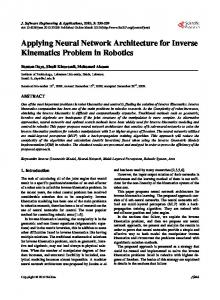
closest node to the actual electrode location and eight closest surface nodes. For the calculation of the lead field L F the reciprocal source current was set to 1 A which can be done due to the linearity of the problem by scaling the potential field by the applied current (Eq. [1]). This current varies in our model because the current source is set up using constant voltage between the source nodes and the Ohm’s law. The lead field procedure appoints the three-dimensional lead vector for each node in the volume conductor model by a single energization of the measurement lead. Thus, the forward calculation was repeated 23 times to obtain the lead fields of all EEG recording channels. D. Calculation of the Surface Potentials—Simulated Data The simulated potential data were generated by inserting a dipole source into the constructed volume conductor model and calculating the scalp potential with the FDM solver. Thus, to avoid trivial solutions due to reciprocity, the simulated potentials were not based on the same forward solution as the lead fields employed in inverse solution. Fifteen different cortical dipole locations at varying depths in the upward z direction (Fig. 1) were considered. Three different dipole directions (x, y, and z according to the rectangular grid used for modeling) for each location were used. In all, 45 cases were considered as simulated potential sets all scaled to correspond the potentials due to a 1 mA current source. For
FIG. 1. Dipole locations for simulated data. Three areas (temporal, central, and posterior) and five levels (1 to 5, 5 being the lowest) spaced by 1 cm were used. Three different dipole directions (x, y, and z) according to the Cartesian coordinate system shown in the figure were considered for each dipole location.
176
LAARNE ET AL.
a few cases (Temporal 1, Central 3, and Posterior 4) random noise (1, 5, 10, 20, and 30%) was added to the potentials. The sensitivity of the dipole localization to the changes in the tissue resistivities was demonstrated by setting the resistivity values of the CSF to 0.56 Ωm (35) and the skull to 160.0 Ωm when calculating the surface potentials of three dipoles (Temporal 3, Central 5, and Posterior 2). The calculation of the lead fields and simulated potentials were carried out on a 160-MHz Sun Ultra 1 workstation with 128 MB CPU, running the SunOS 5.5 operating system. The termination accuracy for the iterations was a 10−8 V difference in the absolute potential values. D. Source Localization Algorithms A single current dipole was assumed as source model. Thus, six dipole parameters (three location parameters r = (r1, r2, r3)T and three moment parameters d = (d1, d2, d3)T were to be determined. Two methods to obtain the inverse solution were applied: a leastsquares method and a probability based method. In the least-squares method the cost function K must be minimized with respect to the six dipole parameters: K(r, d) = Φ − L F d
2
[3]
Here Φ and L F d are the measured and calculated potentials at m electrodes, respectively. L F is the m × 3 lead matrix, containing the lead fields of the m electrodes at the location r under consideration. Note that the location dependence of K is completely determined by L F. The d-dependence of K can be removed by the so-called locally optimal dipole principle: For each location there is one dipole moment d that fits the measured potentials Φ best. This locally optimal dipole moment d is given by: T T d = (L F LF ) LF Φ −1
[4]
Φ is a vector in an m-dimensional space R. The three columns of L F span a threedimensional subspace S of this m-dimensional space. The cost function K is the squared Euclidean distance between Φ and vector L F d lying in S. If Φ also lies in S, there is a vector d for which L F d = Φ and the minimum of K becomes zero. If Φ does not lie in S, then K is minimized for the vector d, for which L F d is an orthogonal projection of Φ onto S. Inserting [4] into [3] makes the cost function K independent of the dipole moment parameters d: K(r) =
(I
)
T T − L F (L F LF ) LF Φ −1
m
2
[5]
Here Im is the m × m identity matrix. The solution of the inverse problem is taken to be the location r´, where K has its minimum, together with the locally optimal dipole at r'. In the probability-based method, an exponential probability density p(r) for the source location is assumed, which has its maximum at the minimum of the cost function K: p(r) =
1 N
− K (r)
e
2
[6]
ACCURACY OF TWO DIPOLAR INVERSE ALGORITHMS
177
N is a normalization factor
N=
∫
− K (r)
e
2
[7]
dV
G
where G is the region taken up by the volume conductor and dV indicates the volume integral. N is needed to define the probability density p. Note that p is symmetric in the m-dimensional space, but not necessarily in the three-dimensional location space. The expectation value r′
r′ = =
1
N∫
− K (r)
re
2
dV
[8]
G
together with the locally optimal dipole moment at r′ is taken to be the solution of the inverse problem. By using the expectation value one expects the solution being less sensitive to noise. The probability based dipole localization method was first introduced by Scholz and Schwierz (26) for magnetic measurements, but the application to electric potentials is straightforward and has been done in (36). In fact, the least-squares method can also be considered as a probability-based method; it is just the maximum likelihood estimation for probability densities like p, which have their maximum at the minimum of K. E. Measures of the Accuracy for Dipole Localization The performance of the source localization algorithms was estimated by the two measures in comparison with the known dipole source parameters: —the error of the spatial location, εl, [mm]
ε l = rsim − rcalc
[9]
—the moment direction error εd, [°] d sim z d calc ε d = acos d sim z d calc
[10]
The dipole magnitudes were not compared. In the lead field approach an actual point dipole is used, while in the simulated dipoles two nodes with finite grid distance formed the source and sink of the dipole. Thus, the magnitude values are not directly comparable. 3. RESULTS A. Forward Calculation The lead fields were calculated with an iterative FDM solver for all the 430,201 nodes in the volume conductor model. The lead field calculations for different leads required 44,601 to 81,807 iterations, the average being 62,570 iterations. The simulated surface potentials were obtained from the potential data at the nodes closest to real EEG electrode locations. The calculations for simulated potential data required 38,037 to 145,147 iterations to terminate. The average was 79,773 iterations.
178
LAARNE ET AL.
The iteration time for the forward computations with this volume conductor model and processor was 0.57 s per iteration. B. Inverse Solution The inverse solution using the lead fields and the simulated potential data was obtained for both algorithms in a couple of minutes when the search was restricted to the brain. The solution was thus found among the 177,868 nodes describing either the gray or white matter. The localization accuracy for dipoles of different directions is from 0 to 8.8 mm in a volume conductor model in size of an adult head (Fig. 2). The errors were in average 4.1 and 4.2 mm, respectively. The averages of different areas are given in Table 1. The moment direction error (Fig. 3) describes the angle between the calculated and the simulated dipole source moments. The angles vary from 0.5° to 27° using the leastsquares algorithm and from 2° to 50° using the probability-based algorithm. The averages of all dipoles are 10.5° and 11.8°, respectively. The averages of different areas are given in Table 2. The two algorithms gave otherwise very similar results, except in the case of x-directed dipoles where the performance of the least-squares algorithm was clearly better. Noise (1, 5, 10, 20, and 30 %) was added to the potentials of three dipoles at different locations and directions. The results are very similar for both algorithms and, thus, in Fig. 4 there are only the results for the least-squares algorithm. The localization error increased up to 22 mm and the moment direction error up to 30° while the noiseless values were less than 3 mm and 5°, respectively. A few simulations were conducted for demonstrating the effects of the resistivity values on the localization accuracy and dipole moment direction. The demand for accurate tissue resistivity data is obvious from the results given in Table 3. An extensive change in both the localization accuracy and moment direction due to 14% change in the CSF resistivity and 10% change in the skull resistivity was seen in the posterior dipole location. 4. DISCUSSION In this study a single equivalent dipole source was used in testing two inverse algorithms using simulated potential data. The localization accuracy is consistent with other studies (23, 37 ). It should be noted that the results were obtained using the minimum amount of measurement electrodes—the standard 10–20 electrode system—and thus, with poor spatial resolution. Obviously, increasing the number of recording channels the localization accuracy improves (37, 38). Our results demonstrate that neither the direction nor the depth of the dipole source plays an important role in localization accuracy. Thus, the deeper sources and cortical sources can be localized within the same error limits, if good signal-to-noise ratio is achieved and the tissue resistivities are well defined. The latter seems to be an elementary requirement if clinically obtained EEG data will be analyzed. The about 2-cm change in the dipole location due to 14% change in the CSF resistivity value on the brain–skull border (the CSF layer may totally vanish using 2.6-mm grid spacing) is dramatic. We have studied the effects of the tissue resistivities on the lead fields (39). The results were given as the average changes of the length and direction of the lead vectors in the brain. For the CSF the average for three EEG leads was 3.4% due
ACCURACY OF TWO DIPOLAR INVERSE ALGORITHMS
179
FIG. 2. Dipole localization accuracy in millimeters for different dipole directions: (a) x-directed dipoles, (b) y-directed dipoles, and (c) z-directed dipoles. The units in x axis correspond to the definitions given in Fig. 1 (i.e., “Temporal 3” applies to dipole location in the temporal area at the level 3).
180
LAARNE ET AL. TABLE 1 The Localization Error Averages
Area Temporal Central Posterior Average
Least, x
Prob, x
Least, y
Prob, y
Least, z
Prob, z
4.2 2.1 1.7 2.7
4.6 3.0 1.7 3.1
4.6 5.5 5.1 5.1
4.6 5.5 5.2 5.1
3.5 4.3 5.8 4.5
3.6 4.1 5.5 4.4
Note. Averages are calculated from the results of the five levels for each dipole area (refer to Fig. 1). The bottom line is the average of these averages for each dipole direction (x, y, and z). The values are given in millimeters. “Least” refers to least-squares algorithm and “Prob” refers to probability-based algorithm; x, y, and z refer to dipole directions.
to 10% decrease in the resistivity. The local changes especially at the tissue surfaces can be remarkably greater. This must be observed while the EEG sources often lie on the surface of the cortex. The localization errors in Fig. 2 are mainly due to the discretization of the volume conductor and the different source definitions used in the simulations and in the inverse procedure (dipole source vs point source). Otherwise the problem setup is ideal; the simulated data and the lead fields were obtained using same resistivity data and potential data can be considered noiseless (possibly some rounding errors can exist). The inverse solution includes in addition to the dipole location the estimate of the dipole moment, i.e., the direction and the magnitude of the source. The magnitudes were not used in this study due to the different definitions for the dipole source in the lead field approach and simulated data. For clinical applications, the location of the source seems to be the most important parameter. The lead field approach itself can be used for dipole magnitude estimation for point dipoles. However, the question arises if the EEG sources are volume sources rather than ideal dipoles. In (26 ) it is stated that the probability-based method avoids the drawback of being trapped in a local minimum, which can happen using iterative inverse methods (e.g., a gradient descent). The reason for the former is simply that to evaluate the expectation value r′ all nodes need to be considered. This may become impractical for larger volume conductors and so usually iterative methods in the least squares optimization method have to be used. The iterative methods are not straightforward applicable to the probabilitybased method without decreasing the spatial resolution. In this paper no iterative inverse methods were used but all nodes were considered, especially as in the lead field approach the solution is obtained by nature throughout the volume conductor model. This allowed comparing the performance of the two methods for volume conductors of reasonable size. In practice, both methods gave in general similar results. A great deal of research effort has been put on developing the EEG inverse procedures. The single equivalent dipole source has few clinical applications, and thus, more complex source models in terms of the number and type of the sources have been applied. Another aspect of development concerns including the temporal dimension in the solution. The source imaging methods have also aroused interest during the past few years (40).
ACCURACY OF TWO DIPOLAR INVERSE ALGORITHMS
181
FIG. 3. Errors in dipole directions in degrees between the known and calculated dipole moments for (a) x-directed (b) y-directed, and (c) z-directed dipoles.
A common problem to all these approaches is to solve the forward problem efficiently to obtain the inverse solution. The computing time of the lead fields is still remarkable in our system. Since there has not yet been demand for fast processing of the data, the forward calculation has been optimized by no means. The FDM calculation can be speeded up at least with a factor of 9 (41) and the modern PCs have more computing efficiency
182
LAARNE ET AL. TABLE 2 The Moment Direction Error Averages
Area Temporal Central Posterior Average
Least, x
Prob, x
Least, y
Prob, y
Least, z
Prob, z
11.5 9.8 4.3 8.5
28.7 13.2 5.2 15.7
9.7 20.9 13.9 14.8
9.7 15.0 13.0 12.6
9.4 5.6 9.6 8.2
9.1 5.0 7.4 7.2
Note. The values are given in degrees. “Least” refers to least-squares algorithm and “Prob” refers to probability-based algorithm, x, y, and z refer to dipole directions.
than the workstation used in this study. Therefore, calculating the lead fields of 64, 128, or even higher amount of EEG electrodes is possible in practice. We have demonstrated the usability of the lead field approach based on the reciprocity theorem in the EEG inverse problem by finite difference method using a realistically shaped volume conductor model. On the basis of the lead fields the inverse solution is
FIG. 4. Effects of the noise added to the simulated potentials on (a) the localization accuracy and (b) dipole moment directions using the least-squares algorithm. Results for three dipoles of different directions and depths are shown. “Temporal 1” is y-directed, “Central 3” is z-directed, and “Posterior 4” is x-directed dipole. Note that the x axis is not linear.
183
ACCURACY OF TWO DIPOLAR INVERSE ALGORITHMS TABLE 3 The Localization and Moment Direction (in Parentheses) Errors of the Three Simulated Dipoles When Changing the Resistivity Values of the CSF and the Skull Errors for altered resistivities CSF=0.56, Ωm
Reference values of errors Dipole Temporal 3, z Central 5, x Posterior 2, y
Skull=160, Ωm
Original least
Original prob
Least
Prob
Least
Least
4.8 (13.0) 3.3 (18.6) 0.0 (24.2)
4.3 (12.5) 3.3 (33.5) 0.0 (22.3)
5.8 (23.4) 5.8 (13.1) 19.7 (84.5)
3.8 (24.4) 5.7 (23.2) 19.7 (86.4)
5.6 (13.1) 8.4 (11.9) 15.3 (4.1)
5.4 (13.2) 8.4 (12.2) 15.3 (89.97)
Note. The values are given in millimeters and degrees, respectively. “Least” refers to least-squares algorithm and “Prob” to probability-based algorithm. The original resistivity values were 0.65 Ωm for the CSF and 177.6 Ωm for the skull.
straightforward and fast to calculate using alternative source models within the brain region. Thus, the lead field approach allows a wide spread of brain functions to be studied. ACKNOWLEDGMENTS The authors acknowledge the contribution of P. Ryymin, Tampere University Hospital, and T. Heinonen, Ragnar Granit Institute, for providing the anatomical data. The study was financially supported by the Academy of Finland (Grant 34171), the Foundation of Technology, Finland, and the Ragnar Granit Foundation
REFERENCES 1. Gaumond, R., Lin, J-H., and Geselowitz, D. Accuracy of dipole localization with a spherical homogeneous model. IEEE Trans. Biomed. Eng. 30(1), 29 (1983). 2. Cuffin, N. A comparison of moving dipole inverse solutions using EEG’s and MEG’s. IEEE Trans. Biomed. Eng. 32(11), 905 (1985). 3. Meijs, J., and Peters, M. The EEG and MEG, using a model of eccentric spheres to describe the head. IEEE Trans. Biomed. Eng. 34(12), 913 (1987). 4. Stok, C. The influence of model parameters on EEG/MEG single dipole source estimation. IEEE Trans. Biomed. Eng. 34(4), 289 (1987). 5. De Munck, J., Van Dijk, B., and Spekreijse, H. An analytic method to determine the effect of source modeling errors on the apparent location and direction of biological sources. J. Appl. Phys. 63, 944 (1988). 6. Salu, Y., Cohen, L., Rose, D., Sato, S., Kufta, C., and Hallett, M. An improved method for localizing electric brain dipoles. IEEE Trans. Biomed. Eng. 37(7), 699 (1990). 7. Mosher, J., Spences, M., Leahy, R., Lewis, P. Error bounds for EEG and MEG dipole source localization. Electroenceph. Clin. Neurophysiol. 86, 303 (1993). 8. Zhang, Z., and Jewett, D. Insidious errors in dipole localization parameters at a single time-point due to model misspecification of number of shells. Electroenceph. Clin. Neurophysiol. 88, 1 (1993). 9. Radich, B., and Buckley, K. EEG dipole localization bounds and MAP algorithms for head models with parameter uncertainties. IEEE Trans. Biomed. Eng. 42(3), 233 (1995). 10. Homma, S., Musha, T., Nakajima, Y., Okamoto, Y., Blom, S., Flink, R, Hagbarth, K-E. and Moström, U. Location of electric current sources in the human brain estimated by the dipole tracing method of the scalp–skull–brain (SSB) head model. Electroenceph. Clin. Neurophysiol. 91, 374 (1994). 11. Zanow, F., and Peters, M. Individually shaped volume conductor models of the head in EEG source localisation. Med. Biol. Eng. Comp. July, 582 (1995).
184
LAARNE ET AL.
12. Leahy, R., Mosher, J., Spencer, M., Huang, M., and Lewine, J. A study of dipole localization accuracy for MEG and EEG using human skull phantom. Electroenceph. Clin. Neurophysiol. 107, 159 (1998). 13. Cuffin, N. EEG localization accuracy improvements using realistically shaped head models. IEEE Trans. Biomed. Eng. 43(3), 299 (1996). 14. Rosenfeld, M., Tanami, R., and Abboud, S. Numerical solution of the potential due to dipole sources in volume conductors with arbitrary geometry and conductivity. IEEE Trans. Biomed. Eng. 43(7), 679 (1996). 15. Yvert, B., Bertrand, O., Echallier, J. F., and Pernier, J. Improved dipole localization using local mesh refinement of realistic head geometries: An EEG simulation study. Electroenceph. Clin. Neurophysiol. 99, 79 (1996). 16. He, B., Musha, T., Okamoto, Y., Homma, S., Nakajima, Y., and Sato, T. Electric dipole tracing in the brain by means of the boundary element method and its accuracy. IEEE Trans. Biomed. Eng. 34(6), 406 (1987). 17. Oostendorp, T., and Van Oosterom, A. The potential distribution generated by surface electrodes in inhomogeneous volume conductors of arbitary shape. IEEE Trans. Biomed. Eng. 38(5), 409 (1991). 18. Fletcher, D., Amir, A., Jewett, D., and Fein, G. Improved method for computation of potentials in a realistic head shape model. IEEE Trans. Biomed. Eng. 42(11), 1094 (1995). 19. Yan, Y., Nunez, P., and Hart, R. Finite-element model of the human head: Scalp potentials due to dipole sources. Med. Biol. Eng. Comp. Sept., 475 (1991). 20. Gevins, A., Le, J., Martin, N., Brickett, P., Desmond, J., and Reutter, B. High resolution EEG: 124-channel recording, spatial deblurring and MRI integration methods. Electroenceph. Clin. Neurophysiol. 90, 337 (1994). 21. Laarne, P., Eskola, H., Hyttinen, J., Suihko, V., and Malmivuo, J. Validation of a detailed computer model for the electric fields in the brain. J. Med. Eng. Tech. 19(2–3), 84 (1995). 22. Lemieux, L., McBride, A., and Hand, J. Calculation of electrical potentials on the surface of a realistic head model by finite differences. Phys. Med. Biol. 41, 1079 (1996). 23. Vanrumste, B., Van Hoey, G., Boon, P., D’Have, M., and Lemahieu, I. Inverse calculations in EEG source analysis applying finite difference method, reciprocity and lead fields. Proc. 20th Annu. Int. Conf. IEEE Eng. Med. Biol. Soc. 20(4), 2112 (1998). 24. Mosher, J., Baillet, S., and Leahy, R. EEG source localization and imaging using multiple signal classification approaches. J. Clin. Neurophys. 16(3), 225 (1999). 25. Greenblatt, R. Probabilistic reconstruction of multiple sources in the bioelectromagnetic inverse problem. Inv. Prob. 9, 271 (1993). 26. Scholz, B., and Schwierz, G. Probability-based current dipole localization from biomagnetic fields. IEEE Trans. Biomed. Eng. 41(8), 735 (1994). 27. Mosher, J., Leahy, R., and Lewis, P. EEG and MEG: Forward solutions for inverse methods. IEEE Trans. Biomed. Eng. 46(3), 245 (1999). 28. Malmivuo, J., and Plonsey, R. Bioelectromagnetism. “Principles and Applications of Bioelectric and Biomagnetic Fields.” Oxford Univ. Press, London, 1995. 29. Nunez, P., and Katznelson, R. “Electric Fields of the Brain.” Oxford Univ. Press, London, 1981. 30. Laarne, P., Hyttinen, J., Malmivuo, J., and Eskola, H. Application of reciprocity theorem to EEG using realistic head model. Med. Biol. Eng. Comp. 34(Suppl. 1, Part 2), 247 (1996). 31. Kauppinen, P., Hyttinen, J., Laarne, P., and Malmivuo, J. A software implementation for detailed volume conductor modelling in electrophysiology using finite difference method. Comp. Methods Prog. Biomed. 58, 191 (1999). 32. Heinonen, T., Eskola, H., Dastidar, P., Laarne, P., and Malmivuo, J. Segmentation of T1 MR scans for reconstruction of resistive head models. Comp. Methods Prog. Biomed. 54, 173 (1997). 33. Geddes, L., and Baker, L. The specific resistance of biological material—A compendium of data for the biomedical engineer and physiologist. Med. Biol. Eng. 5, 271 (1967). 34. Rush, S., and Driscoll, D. EEG electrode sensitivity—An application of reciprocity. IEEE Trans. Biomed. Eng. 16(1), 15 (1969). 35. Baumann, S., Wozny, D., Kelly, S., and Meno, F. The electrical conductivity of human cerebrospinal fluid at body temperature. IEEE Trans. Biomed. Eng. 44(3), 220 (1997). 36. Dodel, S. “Statistische Methoden zur Lösung des Inversen Problems in der Elektrodynamik.” Master of Science Thesis. University of Tübingen, Germany, 1996.
ACCURACY OF TWO DIPOLAR INVERSE ALGORITHMS
185
37. Krings, T., Chiappa, K., Cuffin, N., Cochius, J., Connolly, S., and Cosgrove, R. Accuracy of EEG dipole source localization using implanted sources in the human brain. Clin. Neurophys. 110, 106 (1999). 38. Malmivuo, J., Suihko, V., Eskola, H. Sensitivity distributions of EEG and MEG measurements. IEEE Trans. Biomed. Eng. 44(3), 196 (1997). 39. Laarne, P., Kauppinen, P., Hyttinen, J., Malmivuo, J., and Eskola, H. Effects of tissue resistivities on EEG sensitivity distribution. Med. Biol. Eng. Comp. 37(5), 555 (1999). 40. Pascual-Marqui, R. Review of methods for solving the EEG inverse problem. Int. J. Bioelectromagn. 1(1), 75 (1999). 41. Sachse, F. “Modelle des menschlichen Körpers zur Berechnung von physikalischen Feldern.” Dissertation. University of Fridericiana Karlsruhe, Germany, 1997.