Adhesion-dependent Cell Cycle Progression Linked to the Expression of Cyclin D1, Activation of Cyclin E-cdk2, and Phosphorylation of the Retinoblastoma Protein Xiaoyun Zhu, Motoaki Ohtsubo,* Ralph M. B6hmer, James M. Roberts,* and Richard K. Assoian Department of Cell Biology and Anatomy and Cancer Center, University of Miami School of Medicine, Miami, Florida 33101; and the *Department of Basic Sciences, Fred Hutchinson Cancer Center, Seattle, Washington 98104
Abstract. Growth factors and cell anchorage jointly regulate transit through G1 in almost all cell types, but the cell cycle basis for this combined requirement remains largely uncharacterized. We show here that cell adhesion and growth factors jointly regulate the cyclin D1- and E-dependent kinases. Adhesion to substratum regulates both the induction and translation of cyclin D1 mRNA. Nonadherent cells fail to phosphorylate the retinoblastoma protein (Rb), and enforced expression of cyclin D1 rescues Rb phosphorylation and entry into S phase when G1 cells are cultured in the absence of substratum. Nonadherent cells also fail to activate
'ITH the exception of some cells in the hematopoietic lineage, adhesion to substratum is required for cell cycle progression through G1 and into S phase. Cell adhesion is largely mediated by the interaction of extracellular matrix proteins with integrins, a heterodimeric family of cell surface matrix protein receptors (Hynes, 1987, 1992; Albelda and Buck, 1990; Hemler, 1990). Like growth factor receptors, the display of integrins varies in different cell types. Well studied anchoragedependent cells, such as fibroblasts, express integrins that bind to collagen, fibronectin, and vitronectin. Although less well studied, syndecans, a distinct class of cell surface adhesion molecules, also play an important role in mediating cell adhesion and focal contact formation (Woods and Couchman, 1994). lntegrins act as signaling receptors and transmit growth regulatory signals from the extracellular matrix to the cell. They lack the intrinsic kinase activities characteristic of growth factor receptors, but signal-transducing molecules such as focal adhesion kinase (FAK) 1 and IRS-1 can asso-
the cyclin E-associated kinase, and this effect can be linked to an increased association of the cdk inhibitors, p21 and p27. These data describe a striking convergence in the cell cycle controls used by the two major signal transduction systems responsible for normal and abnormal cell growth. Taken together with our previous studies showing adhesion-dependent expression of cyclin A, they also establish the cell cycle basis for explaining the combined requirement for growth factors and the extracellular matrix in transit through the Rb checkpoint, entry into S phase, and anchorage-dependent growth.
1. Abbreviations used in this paper: CAK, cyclin-activating kinase; cdk, cyclin-dependent kinase; CKI, cdk inhibitor; FAK, focal adhesion kinase; Rb, retinoblastoma.
ciate with integrin cytoplasmic tails (Vuori and Ruoslahti, 1994; Lewis and Schwartz, 1995; Miyamoto et al., 1995), These interactions likely explain the result that integrins, like growth factor receptors, can activate MAP kinase (Chen et al., 1994; Schlaepfer et al., 1994; Morino et al., 1995; Zhu and Assoian, 1995) and a number of other signal transduction events (Woods and Couchman, 1992; Vuori and Ruoslahti, 1993; Miyamoto et al., 1995). As opposed to these G0/G1 regulatory events that seem to be activated independently by integrins and growth factor receptors, the turnover of phosphoinositides (a hallmark of the G0/G1 transition) requires the coordination of signals from integrins and growth factor receptors. Integrin signals lead to the production of PIP2, and signals from growth factor receptors lead to the activation of phospholipase C and the turnover of PIP2 (McNamee et al., 1993; Chong et al., 1994). Indeed, the cooperative regulation of phospholipid turnover by integrins and growth factor receptors provides a paradigm for explaining the fact that soluble mitogens and the extracellular matrix have nonredundant roles in cell cycle progression through G1. Adherent cells irreversibly commit to cell cycle progression at a point in late G1 known as the restriction point (Pardee, 1989). Transit through the restriction point was originally defined as the switch from mitogen-dependent to -independent cell cycle progression. But the growth factor and adhesion requirements for proliferation are typically detected or lost in parallel; nontransformed cells are
© The Rockefeller University Press, 0021-9525/96/04/391/13 $2.00 The Journal of Cell Biology, Volume 133, Number 2, April 1996 391403
391
W
Please address all correspondence to R.K. Assoian, University of Miami School of Medicine, Department of Cell Biology and Anatomy, P.O. Box 01690 (R-124), Miami, FL 33101. Tel.: (305) 243-6423. Fax: (305) 2434431. e-mail:
[email protected]
mitogen and anchorage-dependent, and most transformed ceils are mitogen- and anchorage-independent (Pardee, 1989). This overlap in growth requirements raises the possibility that adhesion-dependent signals may also be involved in cell cycle progression through the restriction point. The molecular basis of restriction point regulation is not fully understood, but it correlates with the G1 hyperphosphorylation of the retinoblastoma protein (Rb). These phosphorylations are catalyzed by the cyclin-dependent kinase (cdk) family (Hinds et al., 1992; Ewen et al., 1993; Dowdy et al., 1993; Kato et al., 1993; Matsushime et al., 1992, 1994; Hatakeyama et al., 1994). Although several cyclin-cdk complexes can phosphorylate Rb in vitro, the timing of Rb phosphorylation in vivo indicates that cyclin D-cdk4/6 and cyclin E-cdk2 are likely to be the principal Rb kinases (Koff et al., 1992; Dulic et al., 1992; Meyerson and Harlow, 1994; Matsushime et al., 1994). Growth factors stimulate the expression of cyclins D and E and thereby activate the cdks that phosphorylate Rb (Matsushime et al., 1991; Lew et al., 1991; Koff et al., 1992; Ohtsubo and Roberts, 1993). The phosphorylation of Rb in G1 phase inactivates its growth inhibitory effects, presumably by allowing for the release of E2F (for reviews see Nevins, 1992; Sherr, 1994; Johnson et al., 1993). Rb also binds to other proteins, including c-abl (Kim et al., 1992; Gu et al., 1993; Welch and Wang, 1993, 1995; Dunaief et al., 1994), and these interactions may also contribute to the control that Rb imposes on cell cycle progression. Cdk activity is negatively regulated by specific cdk inhibitors (CKIs). There are two families of CKIs: p21/p27/ p57 (which bind to and inactivate all cyclin-cdk complexes; EI-Deiry et al., 1993; Xiong et al., 1993; Harper et al., 1993; Polyak et al., 1994a,b; Toyoshima and Hunter, 1994; Lee et al., 1995; Matsuoka et al., 1995) and the INK4s (which only inhibit complexes containing cdk4/6; Serrano et al., 1993; Harmon and Beach, 1994; Guan et al., 1994; Hirai et al., 1995; Chan et al., 1995). Low levels of p21 and p27 can be found in association with active cyclin-cdk complexes, but increased association of p21 or p27 leads to the inhibition of cyclin-cdk activity (Zhang et al., 1994; Polyak et al., 1994a,b; Nourse et al., 1994; Harper et al., 1995). CKIs are regulated by mitogenic and anti-mitogenic signals (Hannon and Beach, 1994; Kato et al., 1994a; Polyak et al., 1994a,b; Nourse et al., 1994), thereby providing a clear link between mitogenic signal transduction pathways and the cell cycle. Activation of the cdks also requires phosphorylation by cyclin-activating kinase (CAK; Kato et al., 1994b; Fisher and Morgan, 1994; M/ikel~i et al., 1994), but this enzyme seems to be constitutively expressed in an active form throughout the cell cycle (Matsuoka et al., 1994). Although the molecular pathways that link mitogen action and cell cycle progression are beginning to be understood, there is relatively little insight into the pathways by which cell adhesion activates cell cycle progression. Nevertheless, our previous studies in NRK fibroblasts showed that the stimulatory effect of adhesion on cell proliferation can also be understood in terms of the cyclins and cyclindependent kinases. In this cell line, growth factors stimulate transit from GO until late G1, and cell adhesion stimulates transit from late G1 into S phase (Guadagno and Assoian, 1991). NRK cells show an adhesion requirement for the
The Journal of Cell Biology, Volume 133, 1996
expression of cyclin A mRNA, and infection with a cyclin A retrovirus allows for anchorage-independent expression of cyclin A--dependent kinase activity and anchorage-independent growth (Guadagno et al., 1993). We also found that the expression of cyclin D1 mRNA is dependent upon cell adhesion in normal human fibroblasts (Bohmer et al., 1996). We now report that NRK cells express only a subset of the controls that normally link cell adhesion to cell cycle progression. In both NIH-3T3 cells and normal human fibroblasts, but not in NRK cells, we find that cell anchorage is required for the phosphorylation of the retinoblastoma protein. This effect can be linked to the adhesion-dependent expression of cyclin D1 and the adhesion-dependent activation of cyclin E-cdk2. Since cyclin D- and E-dependent kinase activities are also dependent upon mitogens, our results show that proper regulation of the G1 cdks requires the convergence of signals from growth factors and the extracellular matrix.
Materials and Methods Cell Culture Confluent (NRK cells) or 50% confluent (NIH-3T3 cells) cultures were trypsinized and seeded in fresh 150-ram-tissue culture dishes at near confluence in medium containing 5% serum. After the cells had attached, the medium was removed, the cells were washed with DME and synchronized in GO by incubation in 20-ml serum-free DME for 24-36 h (NIH-3T3) or 4 d (NRK). Early passage explant cultures of human foreskin flbroblasts were grown to density arrest, and then incubated for 3-5 d in serum-free DME containing ITS + (Collaborative Research, Waltham, MA). NIH-3T3 cells overexpressing human cyclin D1 and human fibroblasts overexpressing human cyclin E were prepared by retroviral infection as described (Guadagno et al., 1993); pools of G418-resistant colonies (>100 colonies per infection) were selected and serum-starved using the conditions described for the parent. The GO-synchronized cells were trypsinized, suspended in medium containing mitogens (5% FCS, 2-3 nM EGF for NRK and NIH-3T3 cells and 10% heat-inactivated FCS, 2 nM EGF for human fibroblasts) and cultured in monolayer or suspension using procedures similar to those described (Guadagno and Assoian, 1991; Han et al., 1993). At selected times after seeding, the cells were collected by centrifugation, either directly (suspended cells) or after trypsinization (monolayer cells). The collected cells (typically 1-5 × 10⁶ cells per time point) were washed twice with HBSS and extracted for total RNA or immunoblotting (see below). In most experiments, N10% of each sample was suspended in 0.5 ml of a solution (25% ethanol in calcium-free PBS) containing 2 ixg/ml HOECHST 33258. The cells were stored (4°C, >16 h) before flow cytometric analysis of DNA content. In several experiments, G0-synchronized NIH-3T3 cells were preincubated with mitogens in monolayer for 9 h (G1 phase cells) or 16 h (S phase cells) before trypsinization and incubation of the cells in monolayer and suspension. Fresh mitogen was added to these cultures during the incubation period.
Antibodies and lmmunoblotting NRK and NIH-3T3 cells (5 x 10⁶) and human fibroblasts (106) were extracted in 0.t ml lysis buffer (50 mM Tris, pH 7.4, 250 mM NaCI, 2 mM EDTA, 1% NP-40, 1 mM PMSF, 10 p,g/ml aprotinin, 10 p,g/ml leupeptin, 50 mM sodium fluoride, and 0.1 mM sodium orthovanadate). Unless noted in the figure legend, either 100 or 200 Ixg of each extract (determined by Coomassie binding; BioRad [Hercules, CA] protein assay) were fractionated on reducing SDS gels (7.5-12% acrylamide), and electroeluted onto nitrocellulose filters. Immunoblotting was performed as described (Zhu and Assoian, 1995) using filters blocked in BSA (anti-Rb [Pharmingen, San Diego, CA or Ciba-Corning, Alemeda, CA]) or nonfat milk (anti-cyclin D1 [Pharmingen or Upstate Biotechnology, Lake Placid, NY], anti-cdk2 [Upstate Biotechnology], anti-cdk4 [Pharmingen], antip21 [Pharmingen or Santa Cruz Biotechnology, Santa Cruz, CA], anti-
392
cyclin A, anti-cyclin E, and anti-p27. A pan-cyclin D antiserum was also purchased from Pharmingen. Enhanced chemiluminesence (ECL, Amersham, Arlington Heights, IL) was used to visualize the immunoblot signals.
1--
I.
16 h in Mn 9hinMn "l
:
I
Immunoprecipitations and In Vitro Kinase Assays Cell extracts (0.5 }xg) were incubated in their lysis buffer with 5-10 ~1 of cyclin E antiserum (1 h, 4°C). The reaction volume was brought to 0.5 ml with fresh lysis buffer, and the immune complexes were collected by incubation (l h at 4°C with rocking) with protein A agarose (50 p,1, Life Technologies, Gaithersburg, MD). For subsequent immunoblotting, the collected imrnunoprecipitates were washed five times in ice-cold lysis buffer and fractionated on reducing SDS-gels as described above. For determination of cyclin E-associated kinase activity, the collected immunoprecipitares were washed three times with cold lysis buffer and twice with room temperature kinase buffer (50 mM Tris-HCl, pH 7.4, l0 mM MgCI2). Kinase reactions were started by adding 2 Ixg histone H1, 25 I~M ATP, and 10 p~Ci [.y-32p] ATP (3,000 Ci/mmol) in a final volume of 30 pJ. The kinase reactions proceeded for 30 rain at 30°C at which time they were stopped by addition of 2 × SDS-sample buffer (30 I.d). The extent of histone H1 phosphorylation (the measure of kinase activity) was determined by SDSgel electrophoresis (12% acrylamide) and autoradiography.
Biosynthetic Labeling NIH-3T3 cells in late G1 (see above) were trypsinized, washed in DME lacking methionine and cysteine, and incubated (~2 x 10⁶ cells per 100-mm dish with 5 ml Met/Cys-free DME) in monolayer and suspension with mitogens. After 4 h, the cells were pulsed for 60 min with Translabel (I mCi/dish; ICN Biomedicals, Costa Mesa, CA). The cells were collected and extracted in 0.3 ml using the procedures described above. Equal amounts of TCA-precipitable radioactivity (107 cpm) were incubated with 5 i~1 of a rat monoclonal antibody to murine cyclin D1 (gift of C. Sherr) or CD44 (gift of L. Bourguignon) using the procedures similar to those described above except that the immune complexes were recovered by incubation with 50 ~1 protein G-plus agarose (Santa Cruz Biotechnology, Santa Cruz, CA). The washed immtmoprecipitates were fractionated on a reducing SDS-gel, and the amount of cyclin D1 was determined by fluorography.
RNA Blot Hybridization Total RNA was isolated from cells (typically 2.5 x 10⁶ cells per time point), and equal amounts of RNA (~10 Ixg) were fractionated on denaturing agarose gels. The filters were incubated with a random-primed eDNA probe for murine cyclin D1 using standard conditions. The hybridized filters were washed with 0.2 x SSPE, 0.1% SDS at 65~8°C.
Results To explore the effects of anchorage on cell cycle transit, monolayer cultures of anchorage-dependent fibroblasts in GO, late G1, and in S phase (Fig. 1) were trypsinized and transferred to suspension or reseeded in monolayer. We determined the effect of anchorage (or loss of anchorage) on the activity of cell cycle proteins, in particular the G1 and S phase cyclin-cdk complexes.
GO
[
G1 phase
Mn. "--
[
~Mn,,..
S phase
t
> Mn
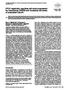
Figure1. Experimental design. Anchorage-dependent fibroblasts were synchronized in GO by serum-starvation in monolayer. These quiescent cells were trypsinized and incubated in monolayer (Mn) and suspension (Sp) before collection and analysis for adhesion-dependent cell cycle events. To obtain populations of late G1 and S phase cells, quiescent fibroblasts were preincubated with soluble mitogens in monolayer (for 9 and 16 h, respectively) before trypsinization and incubation in monolayer and suspension. When cultured with mitogens in monolayer, the G0to-S phase interval is N12-14 h for NRK and NIH-3T3 cells. (Guadagno et al., 1993) or protein during an 18-h incubation in suspension (Fig. 2 A). Expression of cyclin A was also anchorage-dependent in human primary foreskin fibroblasts (see Fig. 8 A). We observed a modest induction of cyclin A (Fig. 2 A) and DNA synthesis (not shown) after prolonged incubation (>18 h) of NRK cells in suspension. However, this muted induction was not sufficient to stimulate cell proliferation in suspension as assessed by colony formation in soft agar (Guadagno et al., 1993). The effect of cell anchorage on G1 progression in NIH3T3 and normal human fibroblasts was more profound than in NRK ceils. First, cyclin A was never induced during incubation in suspension (Figs. 2 A and 8). Second, growth factor-dependent hyperphosphorylation of the Rb protein was essentially anchorage-independent in NRK cells, whereas it was completely anchorage-dependent in NIH-3T3 (Fig. 2 B) and normal human fibroblasts (See Fig. 8 A). These results indicate that cell adhesion controls multiple cell cycle events during G1 and that the pathways causing Rb hyperphosphorylation had become largely anchorage-independent in NRK cells.
Adhesion-dependent Expression of Cyclin D1
Initial experiments tested the effects of anchorage on cell cycle progression from quiescence to S phase. Monolayer and suspension cultures of G0-synchronized NRK and NIH-3T3 cells were stimulated with soluble mitogenic growth factors (FCS/EGF). As previously reported, cells stimulated with soluble mitogens in suspension failed to complete G1 and enter S phase (Guadagno et al., 1993; Han et al., 1993). Expression of cyclin A is coincident with and necessary for the onset of S phase, and neither NRK nor NIH-3T3 fibroblasts synthesized cyclin A mRNA
The G1 cyclin-cdk complexes, cyclin D-cdk4 and cyclin E-cdk2, are thought to be the kinases that hyperphosphorylate Rb during G1 in fibroblasts (see Introduction). We compared the expression of cyclin D1 and cdk4 in monolayer and suspension cultures of G0-synchronized NRK and NIH-3T3 cells (Fig. 2 C) and found that cyclin D1 protein levels were growth factor-dependent in both cell lines. However, the growth factor-dependent induction of cyclin D1 protein was minimally affected by cell anchorage in NRK cells, consistent with the fact that Rb hyperphosphorylation was also anchorage-independent. In contrast, the induction of cyclin D1 was completely anchorage-dependent in NIH-3T3 cells, as was Rb hyperphosphorylation. Cyclin D1 is the predominant D-type cyclin expressed in both NRK and NIH-3T3 cells, as a monoclonal antiserum that recognizes all three D-type cyclins detected only cyclin D1 (not shown). Expression of cdk4 protein in both NRK and NIH-3T3 cells was neither anchorage- nor growth
Zhu et al. Cell Adhesion Regulates the G1 cdks
393
Adhesion-dependent Hyperphosphorylation of the Retinoblastoma Protein
Figure 2. Phosphorylation of the retinoblastoma protein is associated with adhesion-dependent expression of cyclin D1. NRK and NIH3T3 cells were synchronized in GO, and trypsinized and seeded with soluble mitogens (FCS/EGF) in monolayer and suspension. Cells were collected and extracted at the times shown. The extracts were fractionated on SDS gels and analyzed by immunoblotting with antibodies to cyclin A (A), Rb (B), cyclin D1 (C), and cdk4 (C). The upper and lower arrowheads in B, respectively, show the hyper- and hypo-phosphorylated forms of Rb. In D, an anti-cdk4 antibody was used to harvest cyclin D-cdk4 complexes from extracts (0.5 mg) of monolayer (Mn) and suspension (Sp) NRK and NIH-3T3 cells. The immunoprecipitates were fractionated on reducing SDS gels, and the amount of associated cyclin D1 was determined by Western blotting. In E, equal amounts of total RNA (assessed by ethidium bromide staining of rRNA) were isolated from extracts of monolayer and suspension NIH-3T3 cells and analyzed by Northern blot hybridization with a murine cyclin D1 cDNA. Extracts from monolayer and suspension cells were always analyzed in parallel and exposed to film for the same times. Approximate exposure times: A and B (30 s); C (1 min); D (3 min); E (4 d). factor-dependent, and cdk4 immunoprecipitations showed that cyclin D/cdk4 complex formation was unaffected by cell adhesion in N R K cells and strongly adhesion-dependent in NIH-3T3 cells (Fig. 2 D). Cyclin D1 m R N A was present in both monolayer and suspension cultures of NIH-3T3 cells (Fig. 2 E), and control studies (not shown) indicated that the m R N A was cytoplasmic in both conditions. Cyclin D1 m R N A levels were induced 3-5-fold by soluble mitogens in adherent 3T3 cells, and this induction was greatly reduced in the suspended cells (Fig. 2 E). We concluded that the decreased expression of cyclin D1 protein in suspended NIH-3T3 cells was at least partly due to the 3-5-fold lower levels of its mRNA, but that this effect might not be sufficient to completely account for the apparent absence of the cyclin D1 protein. Indeed, posttranscriptional control of cyclin D1 protein levels could be demonstrated by transferring NIH-3T3 cells to suspension late in G1, after growth factor-depen-
dent induction of the cyclin D1 m R N A had occurred. Quiescent NIH-3T3 cells were preincubated with soluble mitogens in monolayer for 9 h, at which time cyclin D1 m R N A (Fig. 3 A) and protein (Fig. 3 B) were almost fully induced. As determined by densitometric scanning, the subsequent incubation of these cells in monolayer and suspension had less than a twofold effect on the expression of cyclin D1 m R N A (Fig. 3 A), but the amount of cyclin D1 protein decreased almost fivefold and became almost undetectable within 5 h after the transfer of cells to suspension (Fig. 3 B). In contrast, expression of cyclin D1 was constant during the incubation of cells in monolayer (Fig. 3 B). Cycloheximide was added to the late G1 cells in order to compare the rates of cyclin D1 degradation in monolayer and suspension cells. The preaccumulated cyclin D1 protein (shown as 9 h) had a half-life of less than 1 hour in both culture conditions (Fig. 3 C). Thus, the turnover of cyclin D1 appeared to be anchorage-independent. Cyclin E levels were unaffected by cell adhesion, both in the ab-
The Journal of Cell Biology, Volume 133, 1996
394
Figure 3. Adhesion-dependent translation of cyclin D1 mRNA. G0-synchronized NIH-3T3 cells were preincubated with soluble mitogens (FCS/EGF) in monolayer for 9 h. These late G1 cells were trypsinized and reseeded in monolayer (Mn) and suspension (Sp) in the continued presence of mitogens. At the times indicated in the figure, the cells were collected and extracted for RNA blot and immunoblot analysis. A shows the expression of cyclin D1 mRNA (equal loading was confirmed by ethidium bromide staining of rRNA; shown as 28S). B and C show the expression of cyclin D1 and E (control) proteins. For the experiment shown in C, quiescent NIH-3T3 cells were preincubated with mitogen for 9 h to accumulate cyclin D1 protein. These preincubated cells were trypsinized, and then seeded in monolayer or suspension in the presence of cycloheximide (10 ~g/ml); collected cells were extracted at 1, 3, and 5 h, and the decay of cyclin D1 (accumulated during the preincubation) was determined by immunoblotting. For the experiments shown in D, the late G1 cells were pulsed (for the last 60 min of a 5-h incubation) with Translabel. As expected from the results of others (Benecke et al., 1978), total protein synthesis was decreased slightly by incubation in suspension (20% over four separate experiments as determined by TCA precipitation). Therefore, equal amounts of TCA-insoluble radioactivity were incubated with rat monoclonal antibodies to murine cyclin D1. Duplicate extracts from adherent cells were also incubated in parallel with monoclonal antibodies to cyclin D1 and CD44 (CON) to identify nonspecifically immunoprecipitated proteins. The collected immunoprecipitates were ffactionated on reducing SDS gels and analyzed by fluorography. E shows the results obtained when protein blots (prepared as described for B) were incubated with an antibody against Rb. The positions of hypo- and hyperphosphorylated Rb are shown by the lower and upper arrowheads, respectively. Approximate exposure times: A (18 h); B and C (2 rain); D (3 d); E (30 s). sence and presence of cycloheximide (Fig. 3, B and C, respectively). The rate of cyclin D1 synthesis was evaluated by immunoprecipitating cyclin D1 from extracts of late G1 cells that were incubated in monolayer and suspension and pulse-labeled with [35S]methionine. Minimal synthesis of cyclin D1 protein was detected in the late G1 cells after incubation in suspension whereas it was readily detected when the late G1 cells were incubated in monolayer (Fig. 3 D). Together, the results in Figs. 2 E and 3 indicate that both transcriptional and translational controls contribute to the adhesion-dependent expression of cyclin D1. Importantly, the extent of R b phosphorylation paralleled the
changes in cyclin D1 protein levels. Rb was hyperphosphorylated when late G1 cells were incubated in monolayer, and it dephosphorylated when these cells were transferred to suspension (Fig. 3 E). To determine if regulation of cyclin D1 protein was causally related to adhesion-dependent phosphorylation of Rb and entry into S phase, we studied the phenotype of cells that constitutively expressed cyclin D1 from an exogenous gene. W h e n NIH-3T3 cells were infected with a cyclin D1 retroviral expression vector, we found that expression of cyclin D1 protein became anchorage-independent. It is possible that the high level of cyclin D1 m R N A ex-
Zhu et al. Cell Adhesion Regulates the G1 cdks
395
Figure 4. Adhesion-dependent expression of cyclin D1 is linked to G1, but not S phase, phosphorylation of Rb. G0-synchronized NIH3T3 cells (A and C) or NIH-3T3 cells overexpressing human cyclin D1 (hD1/NIH-3T3; B) were preincubated with soluble mitogens in monolayer for 9 h (A and B) or 16 h (C). The cells were then trypsinized and incubated with soluble mitogens in monolayer (Mn) and suspension (Sp) for 1-5 h. Collected cells were extracted and the extracts were analyzed by immunoblotting with anti-Rb and anti-cyclin D antibodies. The hypo- and hyperphosphorylated forms of pRb are indicated by the lower and upper arrows, respectively. Exposure times: 30 s for each panel. Aiiquots of each sample were also analyzed by flow cytometry to assess the effect of adhesion and the expression of cyclin D1 on cell cycle progression. D shows the percent of S phase cells in the initial G0-sychronized cultures ("0"), the cultures after preincubation with mitogens in monolayer ("9" and "16" h), and the cultures after final incubation in monolayer and suspension for 5 h (Mn and Sp, respectively). pressed from the retroviral L T R compensated for the decreased translational efficiency in suspended cells or that the absence of the cyclin D1 untranslated regions in the retroviral vector evaded the adhesion controls on translation. Regardless of the specific molecular mechanism, the cyclin D1 protein was expressed at high levels in the quiescent and mitogen-stimulated transfectant, and this high level of expression was maintained even when late Gl-transfected cells were transferred to suspension (compare Fig. 4 A and 4 B). Flow cytometry and [3H]thymidine incorporation assays demonstrated that our NIH-3T3 cells and cyclin D1 transfectants had similar G0-to-S phase intervals when stimulated with both FCS and E G F in monolayer (data not shown). Cells constitutively expressing cyclin D1 differed from control cells in two important ways. First, the enforced expression of cyclin D1 allowed Rb to remain hyperphosphorylated when G1 cells were transferred to suspension (Fig. 4 B), indicating that cyclin D1 was necessary to maintain the hyperphosphorylated state of Rb during G1.
(Note that cyclin E-dependent kinase activity was induced during the 9-h preincubation with mitogens in these experiments, and the activity persisted throughout the subsequent incubation in monolayer and suspension [data not shown]. Thus, in this protocol the G1 phosphorylation of Rb is specifically controlled by the expression of cyclin D.) Second, the forced expression of cyclin D1 rescued entry into S phase. Aliquots of the cells used for the immunoblots above were processed for a flow cytometric analysis of cell cycle progression (Fig. 4 D). Consistent with the dephosphorylation of Rb, entry into S phase was inhibited when G1 control cells were incubated in suspension (Fig. 4 D, left) and expression of cyclin D1 overcame this G1 block (Fig. 4 D, middle). Since cyclin A is necessary for entry into S phase, these results also indicate that the forced expression of cyclin D1 affects the expression of cyclin A in nonadherent NIH-3T3 cells (see Discussion). In contrast to the results obtained with G1 cells, cyclin D1 was not necessary to maintain Rb hyperphosphorylation or cell cycle progression once cells had entered S
The Journal of Cell Biology, Volume 133, 1996
396
phase. Quiescent NIH-3T3 cells were preincubated with soluble mitogens in monolayer for 16 h to generate a population of S phase cells (refer to Fig. 1). The cells were then trypsinized and placed in suspension or returned to monolayer. Cyclin D1 protein still decreased when these S phase cells were incubated in suspension, but Rb remained hyperphosphorylated (Fig. 4 C). This result is in contrast to the behavior of Rb in late G1 cells, and it suggests that once cells have completed G1, other kinases maintain Rb in its hyperphosphorylated state. Cyclin A-cdk2, which remained active in the suspended S phase cells (not shown), is one candidate for the S-phase Rb kinase. The suspended S-phase cells were also able to progress into G2/M (Fig. 4 D, right) despite the fact that the expression of cyclin D1 remained adhesion dependent (Fig. 4 C). Overall, Fig. 4 shows that the expression of cyclin D1 protein is anchorage dependent throughout the cell cycle, but the biological consequence of this restriction (for both Rb phosphorylation and cell cycle progression) is evident only in G1 cells.
Cell Adhesion Regulates Cyclin E-cdk2 Activity Growth factor-dependent induction of cyclin E kinase activity was modestly delayed when N R K cells were cultured in suspension (Fig. 5 A), but it was completely blocked in suspended NIH-3T3 cells (Fig. 5 A) and primary human fibroblasts (see Fig. 8 B). The effects of adhesion on cyclin E-associated kinase activity could not be explained by changes in the levels of cyclin E or its catalytic partner, cdk2" the expression of these proteins was anchorage-independent in all three of these fibroblasts (Fig. 5 B and refer to Fig. 8 B). Thus, the activities of both G1 cyclin-cdk complexes (cyclin D-cdk4 and cyclin E-cdk2) are tightly regulated by cell adhesion in NIH-3T3 and human fibroblasts. In large part, both of these controls are absent in N R K cells. Cyclin E was immunoprecipitated from extracts of quiescent and growth factor-stimulated NIH-3T3 cells in monolayer and suspension, and all of the immunoprecipitates contained similar amounts of cdk2 (Fig. 5 C). Thus, formation of the cyclin E-cdk2 complex is unaffected by cell adhesion. Cyclin E-cdk2 complexes are activated by a CAK-mediated phosphorylation at threonine 160, and this phosphorylation can be detected as an increase in the electrophoretic mobility of cdk2 on SDS gels (Fesquet et al., 1993; Poon et al., 1993; Solomon et al., 1993). This CAKphosphorylated form of cdk2 was readily detected in the cyclin E complexes harvested from NIH-3T3 cells (compare total and cyclin E-associated cdk2 in Fig. 5 C), and the degree of CAK phosphorylation was unaffected by incubation of the cells in suspension (Fig. 5 C). Moreover, extracts of monolayer and suspended NIH-3T3 cells contained readily detectable C A K activity, and the amount of activity was largely unaffected by cell adhesion (Fig. 5 D). Thus, the strict adhesion requirement for cyclin E-kinase activity in NIH-3T3 cells could not be explained by differences in complex formation or by the regulation of CAK. C A K phosphorylation of cdk2 was also anchorage-independent in N R K cells (data not shown). Extracts from monolayer and suspension cultures of N R K and NIH-3T3 cells (Fig. 6) were analyzed by immunoblotting to determine whether cell adhesion controlled
Zhu et al. CellAdhesionRegulatesthe G1 cdks
Figure 5. Adhesion-dependent activity of cyclin E-cdk2. G0-synchronized NRK and NIH-3T3 cells were incubated in monolayer (&In) and suspension (Sp) in the presence of soluble mitogens (FCS/EGF). At the indicated time, cells were collected and extracted for the analysis of cyclin E-associated kinase activity using histone H1 as substrate (A). Identically prepared extracts were fractionated on reducing SDS gels and immunoblotted with antisera against cyclin E and cdk2 (B). To assess assembly of the cyclin E-cdk2 complex and its activation by CAK, extracts were prepared from quiescent NIH-3T3 cells (0) and cells that had been treated with soluble mitogens for 12 h in monolayer and suspension. Cyclin E complexes immunoprecipitated from the extracts were fractionated on SDS gels and immunoblotted with an antibody to cdk2 (C, left side). The migration of total cdk2 from the same extracts is shown by immunoblotting (C, right side). Note that the samples analyzed in C were run on one gel and exposed to film for the same time. For direct measurement of CAK activity (D), extracts (0.2 mg protein) from adherent and nonadherent cells were incubated with 1, 3, or 5 ixl of an antibody to MO15 (the catalytic subunit of CAK, Upstate Biotechnology Inc.) or normal serum (NRS, negative control), and the amount of CAK activity was determined by the ability of the immunoprecipitates to activate H1 histone kinase activity (H1) of a recombinant cyclin A-cdk2 complex (see Nourse et al., 1994 for procedures). The dose-dependent increase in CAK activity (with increasing amounts of anti-MO15) confirms that this analysis was performed in the linear range. Approximate exposure times: A and D (2 rain); B (1 min); C (30 s).
397
~d Q.
c) "5
Sp Mn p21
Sp Mn p27
$p Mn total
Figure Z Stoichiometry of CKI binding to cyclin E-cdk2 com-
Figure 6. Cell adhesion affects the association of p21 and p27 with cyclin E-cdk2 complexes in NIH-3T3 Cells. The figure shows immunoblots of total and cyclin E-associated p27 and p21 (as well as cyclin E and cyclin E-associated kinase activity) from quiescent (0) and mitogen-treated (12 h) NRK and NIH-3T3 cells in monolayer (Mn) and suspension (Sp). The levels of total and cyclin E-associated proteins were determined with 100 and 500 ~g extract, respectively. Approximate ECL exposure times for both NRK and NIH-3T3 cells: (30 s for total cyclin E, 30 s for total and cyclin E-associated p27, 5 min for total and cyclin E-associated p21). the expression of the cdk inhibitory proteins, p21 and p27, or their association with cyclin E-cdk2 complexes. In N R K cells, p21 was very low at quiescence and induced similarly when the cells were treated with soluble mitogen in monolayer or suspension, p27 was strongly expressed at quiescence and downregulated similarly during the incubation of NRK cells in monolayer and suspension. Similar amounts of p21 and p27 were associated with the cyclin E-cdk2 complexes isolated from monolayer and suspension cultures of N R K ceils. Thus, although mitogen regulated, the expression of total p21 and p27, and their association with cyclin E-cdk2 is anchorage-independent in N R K cells. The behavior of p21 and p27 in NIH-3T3 cells differed from that seen in N R K cells in two ways. First, the total levels of both p21 and p27 were slightly higher when the cells were cultured with mitogens in suspension (Fig. 6). Second, about threefold more p21 and about twofold more p27 were present in the inactive cyclin E complexes harvested from suspended NIH-3T3 cells (Fig. 6). Although there was some experiment-to-experiment variability in the levels of p21 and p27 that were associated with cyclin E-cdk2 complexes in nonadherent NIH-3T3 cells (e.g., refer to the error bars in Fig. 7), the increased association was reproduced in several independent experiments and the results shown in Fig. 6 are representative. We assessed the amounts of p21 and p27 that were associated with cyclin E-cdk2 by comparing signal intensities
The Journalof Cell Biology,Volume133, 1996
plexes. G0-synchronized NIH-3T3 cells were cultured in monolayer and suspension for 12 h in the presence of mitogens. Collected cells were lysed and equal aliquots (0.5 mg) of each sample were incubated with anti-cyclin E. The immunoprecipitates were thoroughly washed before fractionation on reducing SDS gels. Selected amounts of recombinant GST-murine p21, his-tagged murine p27, and GST-cdk2 were electrophoresed in parallel. The immunoprecipitates and standards were analyzed simultaneously by immunoblotting with antibodies that detect murine p21, p27, and cdk2. We visually matched band intensities to the standard curves and confirmed the results by densitometry (to correct for potential nonlinearity in the ECL signals). The recombinant standards were quantified by Coomassie blue staining (Bio-Rad assay) and purity was assessed on SDS gels. Quantification of GSTcdk2 and GST-p21 was confirmed with anti-GST immunoblots using a preparation of homogeneous GST as standard. From this analysis, we calculated the moles of cdk2, p21, and p27 in the cyclin E-cdk2 complexes from adherent and nonadherent cells. The results are presented as the moles of p21 and p27 per mol cdk2 and show the mean of two separate experiments. Error bars indicate the ranges observed. of components in the isolated complexes with standard curves for recombinant p21, p27, and cdk2 (Fig. 7). Individually, the moles of p21/cdk2 or p27/cdk2 were ~