ADVANCES IN BIOMEDICAL SCIENCE AND ENGINEERING Volume 1, Number 2, December 2014
ADVANCES IN BIOMEDICAL SCIENCE AND ENGINEERING
An Improved ASCII Character Encoding Method for Lossless ECG Compression Butta Singh1* , Dixit Sharma1 ,Manjit Singh2 ,Dilbag Singh3 1 Department
of Electronics and Communication Engineering, Guru Nanak Dev University Regional Campus, Jalandhar, India Scholar Punjab Technical University, Jalandhar, India and faculty Guru Nanak Dev University Regional Campus, Jalandhar, India 2 Department of Instrumentation and Control Engineering, National Institute of Technology Jalandhar, India *Corresponding author:
[email protected] 2 Research
Abstract: Storage and transmission limitations have made electrocardiogram (ECG) data compression an important aspect for ECG computerized systems. In this paper a lossless method based on modified American standard code for information Interchange (ASCII) character encoding for ECG data compression have been proposed. The Proposed method consists of compression algorithm comprising sign count; generation of array representing ECG sample’s signs (+ve, -ve alternatively), adaptive amplification factor; and grouping method of ECG samples and a reverse process for ECG reconstruction. The ability of the proposed compression algorithm has been investigated on the MIT-BIH Arrhythmia Database. The average percentage root mean square difference (PRD) of about 0.32, compression ratio (CR) of about 8.38, quality score (QS) of about 26.02, percent root mean square difference normalized (PRDN) of about 0.56, root mean square error (RMS) of about 0.0018 and SNR of about 45.46 was achieved on MIT-BIH data. The method is also compared with other compression algorithms and showed superior performance in term of PRD, CR, QS, PRDN, RMS and SNR. The novelty of proposed method is the nearly exact reproduction of the original signal (PRD=0.32) and a moderate CR. Keywords: ECG;Data Compression;Compression ratio;CR;PRD
1. INTRODUCTION Electrocardiogram (ECG) is an important non-invasive physiological signal for monitoring and diagnosing a patient’s heart. ECG is also used to diagnose different health disorders, including respiratory diseases, breathing disorders, and neuro-degenerative diseases. One of the largest drawbacks to the use of electrocardiography, however, is the huge storage capacity necessary to store vast amount of ECG signals. Some time transmission of the data from one place to another using line phone or mobile for right interpretation and diagnostic is required. In such situations the ECG data is become mandatory to compress effectively [1–3]. The objective of ECG compression is to reduce the information rate as well as to preserve the significant information in the reconstructed (decompressed) signal with low computational cost along with good compression ratio [4–7]. ECG compression methods proposed in literature to process transmit, and store the data efficiently can be classified into three categories. (i) The 1
ADVANCES IN BIOMEDICAL SCIENCE AND ENGINEERING
direct data compression method; preserves samples that contain important information about the signal by compressing data directly in the time domain [8–10]; (ii) The transform method; converts the time domain signal to other domains and analyzes the energy distribution [11–13]; (iii) The feature extraction compression method; extracts the parameters of the ECG signal [14, 15]. In recent years, several mobile telemedicine system techniques were proposed including, GSM (Global System for Mobile Communication) [16], Wireless Mesh Networks (WMN) [17] and Code Division Multiple Access (CDMA) [18]. In remote health care system, short message service (SMS) was also used to transmit compressed ECG signal [21]. The ECG data have been encoded using Coding Function, transformed to American standard code for information interchange (ASCII) codes, and then transmitted using a SMS emulator and mobile phone. The compressed file contains only ASCII characters and SMS system is used to transmit the compressed data. Mukhopadhyay et al. introduced an effective protocol for ECG compression by encoding essential information (sign bit, amplification factor, etc.) in their ASCII characters and average Percent root mean square difference (PRD) = 7.89, Compression ratio (CR) = 15.72 and Percent root mean square difference, normalized (PRDN) = 20.60 was achieved [7]. The encoding process was further modified by down-sampling, normalising inter-sample differences, grouping for sign and magnitude encoding, zero element compression and encoding in to ASCII characters and an improved average CR = 43.54 and PRD = 1.73 was obtained [19]. The problem with these compression methods is high PRD values, Therefore difference can be expected in morphological features of original and reconstructed ECG signals results in degradation of quality ECG Signal quality. Degrade ECG signal may produce a jitter in the estimation of the R wave fiducial point, which alters the spectrum considerably and produce inaccurate results and clinical misinterpretation. Although patterns of ECG hold considerable promise for clarifying issues in clinical applications, the inaccurate quantification and interpretation of these patterns may obscure critical issues or relationships and may impede rather than foster the development of clinical applications. In this article, we propose a lossless ASCII character encoding based improved compression method with low PRD between original and reconstructed ECG signals.
2. DATA In the emerging field of medical engineering, research subjects like cardiac arrhythmia detection, heart rate variability, ECG compression, cardiovascular and pulmonary dynamics and artificial intelligence based medical decision support, etc. are of major interest. The Massachusetts Institute of Technology (MIT) supplies some valuable resources for such research projects. These resources include databases containing recorded physiological signals and software for analyzing, viewing and creating such recordings. The MITBIH sinus arrhythmia database (http://ecg.mit.edu) is an extended collection of recorded physiological signals. In the present study, 17 ECG samples from Normal Sinus Arrhythmia database have been taken for investigation of the efficiency of the proposed method. The recorded samples were digitized at 360 samples per second per channel with 11-bit resolution over a 10 mV range
3. PROPOSED METHOD OF ECG COMPRESSION AND RECONSTRUCTION In the present work an effective protocol ECG data compression have been proposed. The proposed ECG compression method can be divided in to two parts the compression and decompression. All the steps used for compression and decompression are explained as follows. 2
An Improved ASCII Character Encoding Method for Lossless ECG Compression
3.1 Compression 3.1.1 Sign count The first step is to count the number of times the positive and negative samples occur in ECG array. For example if a(i) is an array containing ECG samples then sign count will be sgn a(j) as below a(i) = { 0.2350 0.4801 0.7202 0.5216 0.1578 0.0289 0.1641 } −0.0225 −0.1572 −0.3386 | {z } | {z } | {z } 3
5
2
sgn a(j) = {5 3 2}
3.1.2 Adaptive amplification factor In the next step, adaptive amplification factor was computed for a set of ten ECG samples to normalise them in a scale of 0 to 99. ECG samples of array a(i) ware rearranged as a1 a(1) a(2) . . . a(10) a a(11) a(12) . . . a(20) 2 ak = . = . . . . . aN/10 a(N − 9) . . . . a(N) where N is the total number of ECG samples. Largest number from that each array of ak is found out and using this largest number a adaptive amplification factor (amp ak ) for those ten ECG samples is generated in such a way that after amplification, integer part of each of the sample will be either less than or equals to nine 99. amp a(1) 99/ max(a(1), a(2)...a(10)) amp a(2) 99/ max(a(11), a(12)...a(20)) amp a(k)= . = . . . 99/ max(a(N − 9), a(N − 8)...a(N)) amp a(N/10) The ECG samples in each array of ak are normalised in a scale of 0 to 99 to form a new array bk b1 b(1) b(2) . . . b(10) b b(11) b(12) . . . b(20) 2 bk = . = . . . . .
b(N − 9)
bN/10
.
. . . b(N)
a(2) ∗ amp a(1) . . . a(10) ∗ amp a(1) a(1) ∗ amp a(1) a(11) ∗ amp a(2) a(12) ∗ amp a(2) . . . a(20) ∗ amp a(2) = . . . . a(N − 9) ∗ amp a(N/10) . . . . a(N) ∗ amp a(N/10)
3
ADVANCES IN BIOMEDICAL SCIENCE AND ENGINEERING
3.1.3 Grouping It is the most important compression logic throughout the algorithm. If all the samples of any array of bk are single digit then apply forward grouping as if b1 = [b(1) b(2) b(3) b(4) b(5) b(6) b(7) b(8) b(9) b(10)] then after forward grouping b1 = [10*b(1)+b(2) 10*b(3)+b(4) 10*b(5)+b(6) 10*b(7)+b(8) 10*b(9)+ b(10)] for example if b1 =[3 6 1 9 8 6 7 1 1 1] then after forward grouping b1 =[36 19 86 71 11] If only first two or either of the two is double digit sample and left are single digit samples then, values at first two positions are remain unchanged and apply forward grouping to last eight samples of array bk results in an array having six samples. Similarly apply forward grouping for last six, four or two samples if first four, six or eight samples are single digit sample results in seven, eight or nine samples respectively of array bk . Now each row of bk is reconstructed into a single array with adding 100 to last sample of each row for example if first three rows of bk after forward grouping is
9 12 22 24 31 33 bk = 14 14 15 16 19 17 15 12 12 11 1 6 7 7 77 89 98 then reconstructed array (r) will be r = [9 12 22 24 31 133 14 14 15 16 19 17 15 12 12 111 1 6 7 7 77 89 198] also these grouped integers will be encoded in their corresponding ASCII characters to form a array r encoded. Encoded array of grouped samples ‘r encoded’ along with the allied information like sign count, ‘sgn a(j)’ and Amplification factor ‘amp a(k)’ will be transmitted in their corresponding ASCII characters.
3.2 Decompression After compression, transmission and reception, next step is to decompress or reconstruct the ECG signal for clinical classification and analysis purpose. Reconstruction is developed using just the reverse logic of compression process. This module takes encoded ASCII character from the compressed ECG file and the decompression logic is applied on ASCII characters to reconstruct the original ECG signal. The ASCII value contains the Sign count ‘sgn a(j)’, Amplification factor ‘amp a(k)’ and encoded ECG samples ‘r encoded’. All these are necessary to get back the original ECG samples. Depending on the information stored in compressed file original ECG samples are generated using just the reverse logic of sign count, adaptive amplification factor and grouping respectively.
4. RESULTS 4.1 Evaluation of Compression Algorithm In biomedical signal compression, the clinical appropriateness of the reconstructed signal is usually determined through visual inspection. But to evaluate an accurate and quantitative analysis of a compression algorithm, data compression algorithms uses the following six performance parameters [16], [17]. 4
An Improved ASCII Character Encoding Method for Lossless ECG Compression
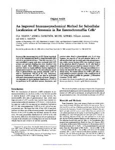
(a)
(b)
(c)
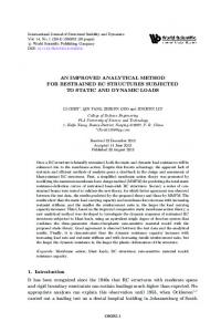
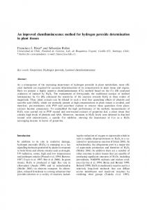
Figure 1. Original (a) Reconstructed (b) and Error Signal (c) of Data No. 100. CR=8.64; PRD=0.32
4.1.1 Compression Ratio (CR) Compression ratio (CR): defined as the ratio between the number of bits needed to represent the original and the compressed signal. No CR = Nc Where No and Nc are the number of bits in original and number of bits in the compressed ECG file respectively.
4.1.2 Percent Root Mean Square Difference (PRD) Percent root mean square difference (PRD): is the measure of acceptable fidelity s PRD% =
2
∑Ni=1 (yi − y¯i ) × 100% ∑Ni=1 y2i
Where yi and y¯i represents the samples in original signal and the samples in reconstructed signal and N is the total number of ECG samples
5
ADVANCES IN BIOMEDICAL SCIENCE AND ENGINEERING
(a)
(b)
(c)
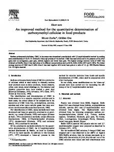
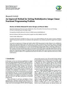
Figure 2. Original (a) Reconstructed (b) and Error Signal (c) of Data No. 117. CR=9.69; PRD=0.30
4.1.3 Percent Root Mean Square Difference, Normalized (PDRN) Percent root mean square difference, normalized (PDRN): normalized version of PRD, PRDN, which does not depend on the signal mean value ym . s PRDN% =
2
∑Ni=1 (yi − y¯i ) × 100% ∑Ni=1 (yi − ym )2
4.1.4 Signal-to-noise Ratio (SNR) Signal-to-noise ratio (SNR): can be represented as s SNR = 10 log
6
2 ∑Ni=1 (yi − ym ) ∑Ni=1 (yi − yi )2
An Improved ASCII Character Encoding Method for Lossless ECG Compression
(a)
(b)
(c)
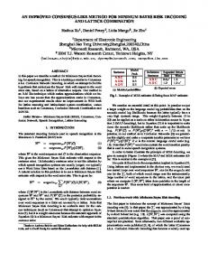
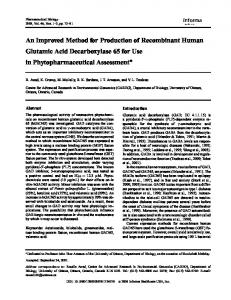
Figure 3. Original (a) Reconstructed (b) and Error Signal (c) of Data No. 119. CR=9.30; PRD=0.30
4.1.5 Root Mean Square Error (RMS) Root mean square error (RMS): The CR and PRD are global performance indicators. For local effects, the RMS is found more logical s 2
RMS =
∑Ni=1 (yi − y¯i ) (N − 1)
4.1.6 Quality Score (QS) Quality score (QS): is the ratio between the CR and PRD. The QS is a very rational performance indicator when it is difficult to estimate the best compression method while taking into account the reconstruction errors as well. A high score represents a superior compression method. QS =
CR PRD
4.2 Performance of Proposed ECG Compression Method To analyze the performance of proposed method 17 MIT-BIH arrhythmia datasets were selected as test signals. Each data file includes lead II ECG data, each lead comprises 6,50,000 data samples. The 7
ADVANCES IN BIOMEDICAL SCIENCE AND ENGINEERING
Table 1.
Performance of Proposed Algorithm on MIT-BIH Database Data
CR
PRD
PRDN
RMS
SNR
QS
100
8.64
0.3175
0.5951
0.0011
44.5089
27.2255
101
8.33
0.3275
0.4840
0.0013
46.3034
25.4258
102
7.95
0.3215
0.5249
0.0009
45.5921
24.7141
103
7.90
0.3552
0.4375
0.0014
47.1794
22.2335
104
7.47
0.3415
0.4592
0.0011
46.7601
21.8790
105
7.56
0.3281
0.3754
0.0015
48.5095
23.0400
106
7.23
0.3467
0.3851
0.0014
48.2893
20.8567
116
9.23
0.3066
0.5300
0.0035
45.5148
30.1182
117
9.69
0.3015
1.1164
0.0027
39.0436
32.1399
118
8.52
0.3103
0.6952
0.0030
43.1574
27.4460
119
9.30
0.3031
0.5740
0.0031
44.8223
30.6815
121
10.30
0.2949
0.8454
0.0025
41.4584
34.9285
122
9.69
0.3022
0.7596
0.0028
42.3879
32.0658
201
7.71
0.3273
0.4243
0.0008
47.4471
23.5448
205
8.52
0.3134
0.5790
0.0011
44.7457
27.1929
208
7.25
0.3389
0.3583
0.0017
48.9158
21.3981
210
7.19
0.3397
0.3962
0.0010
48.0409
21.1626
Table 2. Comparison of Proposed Method with Other Algorithms for Data no. 100, 117 and 119
8
Method
Data
CR
PRD
QS
Lee et al. (20% window size) [20]
100
23
1.94
11.86
Lee et al. (50% window size) [20]
100
9.6
0.44
21.82
Lee et al. [22]
100
24
8.10
2.96
Chou et al. [23] App. 1
100
24
5.21
4.61
Chou et al. [23] App.2
100
24
4.06
5.91
Fira et al. [24]
100
8.14
0.44
18.50
Filho et al. [25]
100
24
3.95
6.08
Proposed
100
8.64
0.32
27.23
Lee et al. (20% window size) [20]
117
24.4
1.17
20.85
Lee et al. (50% window size) [20]
117
10.4
0.42
24.76
Chou et al. [21] App. 1
117
10
0.98
10.20
Chou et al. [21] App. 2
117
13
1.18
11.02
Fira et al. [22]
117
5.17
0.61
8.48
Filho et al. [23]
117
8
0.75
10.67
Bilgin et al. [24]
117
10
1.03
9.71
Proposed
117
9.69
0.30
32.14
Lee et al. (20% window size) [20]
119
19.31
2.05
9.42
Lee et al. (40% window size) [20]
119
10.3
0.59
17.46
Lee et al. (50% window size) [20]
119
8.5
0.44
19.32
Bilgin et al. [24]
119
21.6
3.76
5.74
Fira et al. [22]
119
7.60
1.03
7.38
Chou et al. [21] App. 1
119
21.6
2.81
7.69
Chou et al. [21] App. 2
119
20.9
1.81
11.55
Filho et al. [23]
119
20.9
1.92
10.89
Proposed
119
9.30
0.30
30.68
An Improved ASCII Character Encoding Method for Lossless ECG Compression
Table 3. Comparison of Different ECG Compression Methods Algorithm
PRD
CR
QS
AZTEC [8]
28
10
0.36
m-AZTEC [25]
25.5
5.6
0.22
Hilton [13]
2.6
8
3.08
DJohan [26]
3.9
8
2.05
SPHIT [27]
1.18
8
6.78
Perceptual masks [28]
1.24
3.5
2.82
Shrouf [14]
5.3
11.6
2.19
Fira [22]
0.61
12.74
20.89
USZZQ and huffman coding [29] 2.73
11.06
4.05
Mukhopadhyay [7]
0.023
7.18
312.17
Lee(30% window size) [20]
0.61
16.5
27.11
Filho [23] JPEG2000(1)
0.86
8
9.3
JPEG2000(2)
1.03
10
9.71
Proposed
0.32
8.38
26.02
proposed method was programmed and implemented using Matlab. After the implementation, the average CR, PRD, PRDN, RMS, SNR, and QS based performance indicators were calculated and analyzed. Table 1 shows the values of the six performance indicators of 17 datasets of MIT-BIH arrhythmia database. The averages of the performance indicators as CR= 8.38, PRD= 0.32, PRDN= 0.56, RMS= 0.0018, SNR= 45.46, and QS= 26.02 were achieved on MIT-BIH dataset by proposed method. To evaluate the effectiveness and efficiency of method, performance indicators of are compared with performance of other compression method. Table 2 demonstrate the comparison of performances of other methods with the proposed method on three different normal and abnormal records data no. 100, 117, and 119 of MIT-BIH arrhythmia database. These three data sets have been chosen because their compression results are reported in the literature [20–23, 30]. The CR = 8.64, 9.69, 9.30, PRD = 0.32, 0.30, 0.30 and QS = 27.23, 32.14, 30.68 was achieved for data sets 100, 117 and 119 respectively. The advantage of the proposed method is its superior PRD and QS performance, although the method shows similar or slightly inferior performance in terms of CR compared to other methods. In agreement with wide consensus “QS is a better performance indicator while taking into consideration the both aspect reconstruction distortions (PRD) as well as a quantitative description of the compression (CR)” [22], the proposed method performs excellent by obtaining better QS as compared to other methods. It is implicitly assumed that the ECG reconstructed signals have been validated through visual inspection by the physician. Therefore, for visual inspection of proposed method, the original, reconstructed and error signals are shown in Figure 1 to Figure 3 for 1500 samples of data sets 100, 117 and 119 respectively. The closer scan at the figures discloses that reconstructed signals are similar to the original signals (very low PRD of the order of 0.3) and high quality reconstructed signals are obtained. Average CR, PRD and QS of all data sets of sinus arrhythmia data is also compared with other compression methods in Table 3. Almost all the clinical features of signals are preserved during the compression and reconstruction process and the method is secure to be used for ECG compression. The results obtained in this brief reveal new directions in ECG signal compression and it is worthwhile to further develop methods.
9
ADVANCES IN BIOMEDICAL SCIENCE AND ENGINEERING
5. CONCLUSION New compression techniques that can be directly applied to both regular and irregular ECG signals have been presented. The Compression and decompression processes of the proposed method are fast and simple and easy to implement. Proposed method is suitable to use in portable and mobile ECG monitoring system and may be helpful for the design of efficient ECG compression system. The novelty of method is the nearly exact reproduction of the original signal with a moderate CR. The method achieves good CR with low distortion and preservation of morphological features of ECG signals. The proposed method is shown to outperform some existing method in the literature by achieving high QS. The proposed method performs well in term of PRDN, SNR and RMS also.
References [1] Z. Arnavut, “ECG signal compression based on Burrows-Wheeler transformation and inversion ranks of linear prediction,” Biomedical Engineering, IEEE Transactions on, vol. 54, no. 3, pp. 410–418, 2007. [2] A. Bendifallah, R. Benzid, and M. Boulemden, “Improved ECG compression method using discrete cosine transform,” Electronics Letters, vol. 47, no. 2, pp. 87–89, 2011. [3] V. Kumar, S. C. Saxena, and V. Giri, “Direct data compression of ECG signal for telemedicine,” International Journal of Systems Science, vol. 37, no. 1, pp. 45–63, 2006. [4] M. B. T¨umer and M. C. Demir, “An adaptive signal compression system with pre-specified reconstruction quality and compression rate,” Computer Methods and Programs in Biomedicine, vol. 81, no. 2, pp. 99–105, 2006. [5] R. Borsali, A. Na¨ıt-Ali, and J. Lemoine, “ECG compression using an ensemble polynomial modeling: Comparison with the DCT based technique,” Cardiovascular Engineering: An International Journal, vol. 4, no. 3, pp. 237–244, 2004. [6] D. Tchiotsop, A. Tiedeu, and M. Kom, “Approaches for ECG data compression using orthogonal polynomials,” IRBM, vol. 31, no. 3, pp. 154–169, 2010. [7] S. K. Mukhopadhayay, S. Mitra, and M. Mitra, “An ECG compression technique using ASCII character encoding,” Measurement, vol. 45, no. 6, pp. 1651–1660, 2012. [8] J. Cox, F. Nolle, H. Fozzard, and G. Oliver, “AZTEC, a preprocessing program for real-time ECG rhythm analysis,” Biomedical Engineering, IEEE Transactions on, no. 2, pp. 128–129, 1968. [9] B. Furht and A. Perez, “An adaptive real-time ECG compression algorithm with variable threshold,” Biomedical Engineering, IEEE Transactions on, vol. 35, no. 6, pp. 489–494, 1988. [10] R. C. Barr, S. M. Blanchard, and D. A. Dipersio, “SAPA-2 is the Fan,” Biomedical Engineering, IEEE Transactions on, no. 5, pp. 337–337, 1985. [11] L. V. Batista, E. U. K. Melcher, and L. C. Carvalho, “Compression of ECG signals by optimized quantization of discrete cosine transform coefficients,” Medical engineering & physics, vol. 23, no. 2, pp. 127–134, 2001. [12] B. S. Kim, S. K. Yoo, and M. H. Lee, “Wavelet-based low-delay ECG compression algorithm for continuous ECG transmission,” Information Technology in Biomedicine, IEEE Transactions on, vol. 10, no. 1, pp. 77–83, 2006. [13] M. Manikandan and S. Dandapat, “Wavelet threshold based TDL and TDR algorithms for real-time ECG signal compression,” Biomedical Signal Processing and Control, vol. 3, no. 1, pp. 44–66, 2008. [14] A. Al-Shrouf, M. Abo-Zahhad, and S. M. Ahmed, “A novel compression algorithm for electrocardiogram signals based on the linear prediction of the wavelet coefficients,” Digital Signal Processing, 10
An Improved ASCII Character Encoding Method for Lossless ECG Compression
vol. 13, no. 4, pp. 604–622, 2003. [15] U. E. Ruttimann and H. V. Pipberger, “Compression of the ECG by prediction or interpolation and entropy encoding,” Biomedical Engineering, IEEE Transactions on, no. 11, pp. 613–623, 1979. [16] R. S. Istepanian and A. A. Petrosian, “Optimal zonal wavelet-based ECG data compression for a mobile telecardiology system,” Information Technology in Biomedicine, IEEE Transactions on, vol. 4, no. 3, pp. 200–211, 2000. [17] S. Pavlopoulos, E. Kyriacou, A. Berler, S. Dembeyiotis, and D. Koutsouris, “A novel emergency telemedicine system based on wireless communication technology-AMBULANCE,” Information Technology in Biomedicine, IEEE Transactions on, vol. 2, no. 4, pp. 261–267, 1998. [18] B. S. Kim and S. K. Yoo, “Performance evaluation of wavelet-based ECG compression algorithms for telecardiology application over CDMA network,” Informatics for Health and Social Care, vol. 32, no. 3, pp. 177–189, 2007. [19] M. Mitra, J. Bera, and R. Gupta, “Electrocardiogram compression technique for global system of mobile-based offline telecardiology application for rural clinics in India,” IET Science, Measurement & Technology, vol. 6, no. 6, pp. 412–419, 2012. [20] S. Lee, J. Kim, and J.-H. Lee, “A real-time ECG data compression and transmission algorithm for an e-health device,” Biomedical Engineering, IEEE Transactions on, vol. 58, no. 9, pp. 2448–2455, 2011. [21] H.-H. Chou, Y.-J. Chen, Y.-C. Shiau, and T.-S. Kuo, “An effective and efficient compression algorithm for ECG signals with irregular periods,” Biomedical Engineering, IEEE Transactions on, vol. 53, no. 6, pp. 1198–1205, 2006. [22] C. M. Fira and L. Goras, “An ECG signals compression method and its validation using NNs,” Biomedical Engineering, IEEE Transactions on, vol. 55, no. 4, pp. 1319–1326, 2008. [23] N. Rodrigues, E. da Silva, S. de Faria, V. da Silva, M. B. de Carvalho, et al., “ECG signal compression based on Dc equalization and complexity sorting,” IEEE Transactions on Bio-medical Engineering, vol. 55, no. 7, pp. 1923–1926, 2008. [24] A. Bilgin, M. W. Marcellin, and M. I. Altbach, “Compression of electrocardiogram signals using JPEG2000,” Consumer Electronics, IEEE Transactions on, vol. 49, no. 4, pp. 833–840, 2003. [25] V. Kumar, S. C. Saxena, V. Giri, and D. Singh, “Improved modified AZTEC technique for ECG data compression: Effect of length of parabolic filter on reconstructed signal,” Computers & Electrical Engineering, vol. 31, no. 4, pp. 334–344, 2005. [26] A. Djohan, T. Q. Nguyen, and W. J. Tompkins, “ECG compression using discrete symmetric wavelet transform,” in Engineering in Medicine and Biology Society, 1995., IEEE 17th Annual Conference, vol. 1, pp. 167–168, IEEE, 1995. [27] Z. Lu, D. Y. Kim, and W. A. Pearlman, “Wavelet compression of ecg signals by the set partitioning in hierarchical trees algorithm,” Biomedical Engineering, IEEE Transactions on, vol. 47, no. 7, pp. 849–856, 2000. [28] C. Rodrigo, M. Fabrizia, and V. Leonardo, “Near-lossless compression of ECG signals using perceptual masks in the DCT domain,” 2007. [29] M. S. Manikandan and S. Dandapat, “Wavelet threshold based ECG compression using USZZQ and Huffman coding of DSM,” Biomedical Signal Processing and Control, vol. 1, no. 4, pp. 261–270, 2006. [30] H. Lee and K. M. Buckley, “ECG data compression using cut and align beats approach and 2-D transforms,” Biomedical Engineering, IEEE Transactions on, vol. 46, no. 5, pp. 556–564, 1999.
11