Michele Migliore and Gordon M. Shepherd. Abstract | Characterizing the ...... St. John, J. L., Rosene, D. L. & Luebke, J. I. Morphology and electrophysiology of ...
PERSPECTIVES
OPINION
An integrated approach to classifying neuronal phenotypes Michele Migliore and Gordon M. Shepherd Abstract | Characterizing the functional phenotypes of neurons is essential for understanding how genotypes can be related to the neural basis of behaviour. Traditional classifications of neurons by single features (such as morphology or firing behaviour) are increasingly inadequate for reflecting functional phenotypes, as they do not integrate functions across different neuronal types. Here, we describe a set of rules for identifying and predicting functional phenotypes that combine morphology, intrinsic ion channel species and their distributions in dendrites, and functional properties. This more comprehensive neuronal classification should be an improvement on traditional classifications for relating genotype to functional phenotype.
The neurons of the nervous system are grouped into different types that were originally identified by Golgi, Cajal and their contemporaries on the basis of the shapes of their somatodendritic trees. These morphological phenotypes continue to provide the common nomenclature for different types of nerve cell, such as pyramidal neurons, basket cells and chandelier cells. However, to understand the role of a given morphological type of neuron in neural circuits, an understanding of the functional phenotype is needed. An important determinant of functional phenotype is the expression of specific types of ion channel. The distributions of different ion channels in the somatodendritic membrane are increasingly recognized as determining the characteristic firing patterns of a neuron and thereby defining its functional phenotype1. Therefore, the rules that underlie the expression of ion channels and their characteristic somatodendritic distributions are key to understanding how the functional phenotypes of neurons arise and are related to each other. An initial set of such rules was previously outlined2 on the basis of a limited set of neurons for which detailed data were available. As interest increases in dendritic functions and their roles in generating circuit properties, there is an urgent need to test the
810 | O CTOBER 2005
generality of these rules on larger populations of neuronal types, and to make predictions for those neurons of special interest for which dendritic ionic channel data are not yet fully available. The results of such a test support the emerging concept of a functional neuronal phenotype based on dendritic morphology, ion channel species, ion channel distributions and specific functional properties such as back-propagating action potentials (BP-APs) and somatic firing patterns. We discuss the advantages of this integrative framework compared with traditional methods of neuronal classification based on single morphological or functional properties. We then indicate how this approach can be combined with current studies in which physiological properties are correlated with differential gene expression3. The results highlight the need for more extensive exploration of the dendritic ion channel distributions and functional properties of important neuronal types. Overview of classification approach
Most of the data discussed here on membrane properties in different dendritic compartments of different neuronal types have been identified and integrated using NeuronDB, a database of neuronal properties that is part of SenseLab at Yale University, USA. The database shows the presence of different types of ion channel in a given canonical dendritic compartment, and allows researchers to carry out automated searches across corresponding compartments in all neurons in the database for a given property or arbitrary set of properties. This article is also coordinated with the ModelDB database of SenseLab, enabling the reader to download and use computational models to test the results for many of the main types of neuron covered here. For the purposes of this article, we begin the classification with the characterization of neurons as tending to have relatively thick or relatively thin dendritic trunks and initial branches. This is a relative rather than absolute distinction, which correlates roughly with
| VOLUME 6
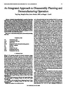
the current ability to patch onto a dendrite that is at least ~1 µm in diameter. As has previously been shown 2, these two morphological types are further characterized by a tendency for neurons with thick dendrites to have a high density of dendritic Na+ channels, and for those with thin dendrites to have few or no dendritic Na+ channels. Examples are shown in FIG. 1, in which the best studied representatives of the former are the large pyramidal neurons in layer 5 of the neocortex, mitral cells in the olfactory bulb and pyramidal neurons in area CA1 of the hippocampus. Representatives of the latter are the Purkinje cells of the cerebellum, thalamocortical relay cells and pyramidal neurons in area CA3 of the hippocampus. These characterizations apply to the overall organization of these dendritic trees, within which local differences can occur. For example, neurons with thick dendrites might have a significant proportion of thin branches with Na+ channels (such as the oblique dendrites of CA1 pyramidal cells4). Conversely, Purkinje and thalamocortical neurons with thin dendrites have relatively thick initial trunks with Na + channels, although their density falls off sharply with branching order and thin branches5,6. This initial characterization in terms of dendritic morphology and Na+ channel density has important implications for subsequent functional phenotypes. As is well known, the combination of thick dendrites and high Na+ would be predicted to be associated with extensive passive spread and active boosting of BP-APs, whereas thin dendrites and low Na+ would be predicted to be associated with limited passive spread and limited back-propagation. Below, we discuss these predictions in relation to BP-APs. Neurons with thick dendrites
The combination of dendritic morphology and dendritic Na+ channel expression is the first rule for phenotypic classification, leading to the subsequent rules for further differentiation within these classes. We begin by discussing neurons with thick dendrites. Dendritic Ca2+ channels. A possible next rule for defining neuronal subtypes would be the presence or absence of Ca2+ channels. However, experiments using direct or indirect recordings of dendritic electrical activity in all types of neural cells have consistently found Ca2+ membrane channels to be expressed7, although the relative densities and compositions of these channels have not been identified, especially for thin dendrites.
www.nature.com/reviews/neuro
© 2005 Nature Publishing Group
PERSPECTIVES
Relatively thick dendrites, high dendritic Na+
Neocortical L5 neuron
Mitral cell
CA1 hippocampal neuron
Thalamocortical neuron
CA3 hippocampal neuron
Relatively thin dendrites, low dendritic Na+
Purkinje cell
Figure 1 | Basic phenotypic categories of cells with thick dendrites and high dendritic Na+ channel density, and cells with thin dendrites and low dendritic Na+ channel density. Neurons with known dendritic Na+ channel density (green background) can be used to predict the presence of dendritic Na+ channels in neurons that show similar morphological properties, but for which no direct dendritic data are available (purple background). Neocortical layer 5 (L5) neuron reproduced, with permission, from REF. 70 © (1999) Society for Neuroscience. Mitral cell, Purkinje cell and thalamocortical neuron downloaded from the SenseLab public database. CA1 and CA3 neurons downloaded from the Duke/Southampton public archive (cells n128 and I51, respectively; see Further information).
Ca2+ channels are ubiquitous and are involved in all synaptic plasticity mechanisms, but the main role of these channels, especially low-voltage activated (LVA) channels, in specifying neuronal phenotypes probably involves modulating the ability of a neuron to generate calcium-dependent spikes and bursts. This ability is a common feature of very different cell types, such as deep neocortical8 and Purkinje neurons9. These possible functional phenotypes, which are based on details of different impulse firing patterns, are beyond the scope of this article. Fast, modulated integration phenotype. The presence or absence of Na+ channels correlates with the rapidity of synaptic integration
carried out by the neuron. Two crucial factors in this phenotype2 are the presence or absence of a BP-AP, and the presence or absence of the nonspecific cation current (Ih), which is activated by hyperpolarization and deactivated by depolarization. The presence of Ih seems to define a phenotype of a group of cells, which functionally would mean that in these cells the time window of integration could be modulated by this current10. For convenience, we refer to these neurons as having a ‘fast, modulated integration’ phenotype (FIG. 2). There is direct (patch clamp) or indirect (calcium imaging) evidence for a BP-AP in the dendrites of many types of neuron11, especially neurons with relatively thick dendrites. The best-studied examples are CA1
NATURE REVIEWS | NEUROSCIENCE
pyramidal neurons12 and deep neocortical pyramidal neurons13 (FIG. 2a). The actively supported back-propagating spike provides a narrow time window for enhancing rapid synaptic integration. Most of the cells that support BP-APs also show significant Ih, at least in the soma. As noted above, the main role of this current is to modulate the temporal window for synaptic integration (FIG. 2a, right). Interestingly, it seems that in all cases in which it has been tested, whenever Ih is expressed in the soma it is also expressed in the dendrites13–16. In the only two cases in which direct dendritic recordings were carried out (CA1 neurons10 and deep neocortical neurons14,15), the density of Ih was substantially higher in the dendrites than in the soma. This could be a general feature. A somatic Ih has been found in neurons that support dendritic BP-APs (FIG. 2b), and in neurons for which there are no indications of their dendritic properties (FIG. 2c). These include the nigral dopaminergic neurons17 and pyramidal neurons from the perirhinal cortex18, layers 2 and 5 of the entorhinal cortex19,20, and the olfactory cortex21. For all of these neurons the rules under discussion would predict a higher Ih in the dendrites than in the soma. A further hypothesis is that this property could be extended to those nonpyramidal neurons that show BP-APs and somatic Ih, such as globus pallidus neurons22, bitufted interneurons in cortical layers 2 and 3 REF. 23, oriens-alveus layer hippocampal interneurons24 and neostriatal cholinergic interneurons25. This rule (a somatic Ih associated with a higher dendritic Ih) also seems to apply to neurons that cannot be easily classified in terms of thick or thin dendrites, such as amacrine cells26. Fast, unmodulated integration phenotype. By contrast, a lack of Ih seems to define a phenotype of cells that are limited in their ability to modulate a time window of integration2 (FIG. 2a). For convenience, we refer to these neurons as having a ‘fast, unmodulated integration’ phenotype (FIG. 3). The prediction of a somatic Ih associated with a higher dendritic Ih (see previous section) would, therefore, not apply to neurons that lack Ih in the soma (FIG. 3). These neurons have relatively thick dendrites, and mainly show regular firing behaviour. A well studied example of this group is the olfactory mitral/tufted cells (FIG. 3a), for which there is electrophysiological evidence of BP-APs27 and lack of hyperpolarization-activated cyclic nucleotidegated potassium channel 1 (HCN1)28, one member of a gene family that encodes Ih. VOLUME 6 | O CTOBER 2005 | 811
© 2005 Nature Publishing Group
PERSPECTIVES
a Neurons with known somatic and
c Neurons with known somatic properties
dendritic properties
and unknown dendritic properties
ZD7268
Control
Dendrite Soma
CA1
Neocortical L5
Neostriatal cholinergic interneuron
Entorhinal cortex L2
b Neurons with known somatic properties and partially known dendritic properties
Nigral dopaminergic neuron
CA1 interneuron
Perirhinal cortex
DCN pyramidal neuron
Figure 2 | Cells with thick dendrites and high dendritic Na+ channel density are further subcategorized by back-propagating action potentials and modulation by Ih currents. a,b | Show the electrophysiological properties of neurons with several known somatic and dendritic properties (a) and neurons for which only a few dendritic properties are known (b). c | Predictions of the dendritic back-propagating action potentials and nonspecific cation current (Ih) of neurons that show similar morphological and somatic electrophysiological properties, but for which no direct dendritic data are available. All panels show typical recordings. Note, in all cases, the regular firing behaviour and the typical Ih-dependent ‘sag’ for hyperpolarizing currents. DCN, dorsal cochlear nucleus; L, cortical layer. Panel a reproduced, with permission, from REF. 71 © (1992) American Physiological Society; REF. 70 © (1999) Society for Neuroscience; and REF. 72 © (1999) The Physiological Society. Panel b reproduced, with permission, from REF. 14 © (2000) Elsevier Science and REF. 73 © (1996) American Physiological Society. Panel c reproduced, with permission, from REF. 74 © (1999) Society for Neuroscience; REF. 18 © (2001) Wiley-Liss; REF. 20 © (2004) Wiley-Liss; and REF. 75 © (1990) Society for Neuroscience.
This group would be predicted to have active support of action potential back-propagation, but no or little somatic or dendritic I h . Neurons with these properties would include pyramidal neurons from layers 2 and 3 of the neocortex29,30, (FIG. 3b), the lateral amygdala31 and layers 3 and 6 of the entorhinal cortex19,20 (FIG. 3c). Fast, time-dependent integration phenotype. In hippocampal CA1 pyramidal neurons, the dendritic density of KA — a fast, transient potassium current that is involved in the repolarization phase of the action potential — increases with distance from the soma12. Because of this distribution, the action potential amplitude could be modulated during back-propagation, with a dynamic dependence on the instantaneous activation or inactivation state of the KA channels32. So far, this is the only case in which this effect has been clearly observed, although it might also be present in the lateral dendrites of mitral cells33. As the overall calcium influx depends on the properties of the action potential34, the spike-time-dependent synaptic plasticity that is observed in these neurons35 might arise if a synaptic input that is activated a few milliseconds before the arrival of the BP-AP boosts the calcium influx through voltage-dependent
812 | O CTOBER 2005
calcium channels. For this reason we refer to this category as having a ‘fast, time-dependent integration’ phenotype. Neurons with thin dendrites
Next, we consider the class of neurons with thin dendrites. It is difficult to obtain direct electrophysiological recordings of BP-APs, different ionic currents, and physiological synaptic and firing properties in these neurons, and there have been few electrophysiological dendritic recordings beyond ~50 µm from the soma. Notable exceptions are the work of Martina et al.36, who investigated potassium channels in Purkinje neurons up to 250 µm from the soma, and Velte and Masland37, who were able to obtain dendritic recordings (although these were limited to the larger dendrites of 1–3 µm diameter) from retinal ganglion cells up to 320 µm from the soma. Most of the information about the possible phenotypes must, therefore, be inferred by correlating passive and morphological features, somatic recordings and indirect data (such as calcium imaging experiments). Slow integration, complex firing pattern phenotype. Using the same approach as for neurons with thick dendrites, we begin characterization of neurons with thin dendrites
| VOLUME 6
with the key property of BP-APs. As noted above, active, full-amplitude back-propagation of an action potential depends on the expression of dendritic Na+ channels. In the only neurons with thin dendrites for which there is some direct experimental evidence (FIG. 4a,b) — thalamocortical neurons6 and Purkinje cells5 — the density of dendritic Na+ channels was low or decreased with distance from the soma, and, consequently, action potential back-propagation was limited. The relative lack or absolute absence of the fast Na+ channels combined with the presence of the slower dendritic Ca2+ channels could result in slow integration properties. For convenience, we refer to cells in this group as having a ‘slow integration’ phenotype. The low or decreasing dendritic density of Na+ channels with distance from the soma is supported by indirect experimental findings in several other types of neuron with (mainly) thin dendrites. For example, in CA3 neurons there is indirect evidence for limited active action potential back-propagation38. In neurons such as retinal ganglion cells, a subpopulation of which have larger dendrites39 (FIG. 4c), the dendritic action potential amplitude at ~100 µm from the soma seems to be much lower than at the soma37 (by about 30 mV). Indirect findings in olfactory bulb granule cells indicate that, in these neurons, action potentials could be initiated in the dendrites. However, these action potentials never propagate at full amplitude to the soma, where only small (