Dec 13, 2010 - and a minimum of 1 MByte of RAM (a Mac Plus,. Mac SE, Mac SE/30, Mac II and Mac IIc/x series). HyperCard and several introductory ...
Journal of Biological Education
ISSN: 0021-9266 (Print) 2157-6009 (Online) Journal homepage: http://www.tandfonline.com/loi/rjbe20
An interactive computer-based alternative to performing a rat dissection in the classroom Megan Quentin-Baxter & David Dewhurst To cite this article: Megan Quentin-Baxter & David Dewhurst (1992) An interactive computerbased alternative to performing a rat dissection in the classroom, Journal of Biological Education, 26:1, 27-33, DOI: 10.1080/00219266.1992.9655239 To link to this article: http://dx.doi.org/10.1080/00219266.1992.9655239
Published online: 13 Dec 2010.
Submit your article to this journal
Article views: 19
View related articles
Citing articles: 4 View citing articles
Full Terms & Conditions of access and use can be found at http://www.tandfonline.com/action/journalInformation?journalCode=rjbe20 Download by: [University of Tasmania]
Date: 03 February 2016, At: 15:43
Downloaded by [University of Tasmania] at 15:43 03 February 2016
An interactive computer-based alternative to performing a rat dissection in the classroom Megan
Quenfin-Baxter
and David
Dewhursf
Improvements in the power, speed and screen resolution of microcomputers are increasing the potential of computer-assisted learning programs to provide interactive, realistic alternatives
Introduction Interest in computer-assisted learning (CAL) materials as teaching alternatives to laboratory experiments in biology has heightened with increased publicity and awareness of environmental issues, widespread debates concerning the rights of animals and recognition of the microcomputer as a powerful teaching aid. The educational purpose of performing animal experiments (particularly animal dissections) at school has been criticized by several organizations, including Abstract This paper describes a highly interactive Hypermedia atlas of a rat dissection which allows a student to investigate the relationships between anatomical structures in the body and provides information on the physiological function of many of them. The program is based around a number of photographic images of a rat dissection over which the computer is used to lay interactive features such as animation, information via Hypertext and question/answer facilities. Different user modes allow students to either seek assistance and access further information about any part of the rat (Browse mode), or test their knowledge by answering computer-generated questions (Test mode). The program runs from an easy-to-use menu palette and guidance is also provided by a map from which the student can access any part of the program. The potential of this program in teaching biology is also discussed. Key words: Rat, Dissection, Computer alternative.
those interested in ensuring that a student's refusal to undertake animal experiments does not result in academic penalties (Graham, 1989). These organizations are placing pressure upon educational establishments to find and provide suitable alternatives for those wishing to avoid animal experiments. Biological groups trying to preserve mandatory dissection practical, where they form part of the course requirements, have indicated a willingness to compromise providing suitable alternatives can be obtained. The response to this challenge has seen an increased use of diagrams, models, videos and computer simulations, none of which adequately fulfil all of the teaching objectives of a laboratory dissection. Computer-based simulations are potentially the most suitable alternative as they can combine more conventional teaching resources with an investigative or practical environment. So they go part-way to portraying the hands-on approach to learning provided by a dissection. The response of software developers to the call for more computer-based alternatives to dissections has been slow. This is partly due to the high cost of software development (Reed, 1987), and to scepticism about the ability of a computer to provide a completely realistic, alternative learning environment. When performing a dissection in an experimental situation a student is expected to interpret the dissected material in relation to information given in textbooks and lecture notes. A student using a screenJournal of Biological Education (1992) 26 (1)
17
Downloaded by [University of Tasmania] at 15:43 03 February 2016
A computer-based alternative to performing a rat dissection
based alternative faces a level of abstraction due to the necessity of interpreting the dissected material on the computer screen. As advances in technology improve the resolution of visual displays and increase the storage power of microcomputers, animal experiments depicted on a screen become more realistic. Poor graphics clarity in early animal dissection CAL programs such as 'Operation Frog' (Kohn and Miller, 1984), did little to aid interpretation of the material being presented and less to refute the claim that computer-based learning has little to offer as an alternative to more traditional teaching methods. An interactive computer-based alternative to an animal experiment can provide an environment similar to performing an actual animal experiment (Dewhurst, Brown, and Meehan, 1988). The two methods of conveying information do not, however, overlap exactly. This incomplete overlap results in three identifiable learning objectives, which differentiate learning via a practical session at the dissecting table, from the experience provided by a computerbased alternative. The three areas are: a) The area of learning which is peculiar to performing an animal dissection and not usually addressed by a computer-based alternative, is the development of dexterity and practical skills necessary to investigate anatomical structures. b) The central overlapping objectives of both performing a real dissection or using a computerbased alternative, include the development of investigative techniques and a general familiarization and comprehension of the material being studied. c) The area unique to a computer-based alternative is the ability to provide supportive information, feedback and help in an interactive and potentially repetitive environment, often without requiring the immediate attention of the tutor. The net result of this imperfect overlap of information delivery on a student using a computer-based alternative is a shift in emphasis away from practising technical skills towards operating in a more responsive and informative environment. The importance of the subsequent shift in a student's development depends on the quality of the CAL material and on the emphasis the student or tutor places on the necessity of practical experience and the development of technical skills. The CAL package described here (The Rat Stack) aims to maximize a student's understanding of the functional anatomy of the rat by comprehensively addressing the areas outlined in b and c above, without attempting what is clearly an impossible task of teaching actual dissecting techniques. It has been developed on computer hardware with relatively good graphics clarity using an authoring system based on the Hypermedia concept. Hypermedia is a powerful environment for managing and presenting information because it combines associative linking 28
Journal of Biological Education (1992) 26 (1)
Quentin-Baxter and Dewhurst
techniques with computer multimedia (text, graphics, sound and animation). It allows users to follow links between nodes of related information in a network, thus escaping from the more common linear or treebranching forms of presentation. In a learning environment mouse-and-icon driven Hypermedia CAL packages give students control over the presentation and content of the material within the context of the package.
The program The Rat Stack is a computer-assisted learning package designed to teach the functional anatomy of the rat in a highly interactive way to college, high school and undergraduate biology students. It runs under HyperCard™ on the Apple* Macintosh® microcomputer. To run correctly HyperCard requires a Macintosh with either a hard disk or two floppy disk drives and a minimum of 1 MByte of RAM (a Mac Plus, Mac SE, Mac SE/30, Mac II and Mac IIc/x series). HyperCard and several introductory HyperCard stacks are currently distributed free with every new Macintosh sold. The program is based around 32 high-resolution photographic images and diagrams of rat anatomy. It offers four modes of operation which promote a flexible approach to the dissection; from complete structural identification and textual descriptions of the material being presented, through to testing a student's knowledge of anatomy. Clicking the mouse on buttons in a menu palette on the right side of the screen offers additional textual information, overlays diagrammatic outlines of structures on an image, or generates questions which test a student's ability to identify structures on an image. Each image in the program comprises a photograph or diagram which forms the basis over which an invisible 'map' is laid. This map is made up of numerous buttons which cover the area of the structures in the photograph/diagram. Each structure on an image has one or more invisible buttons associated with it which adopt that structure's name, allowing them to respond appropriately to the presence of the mouse (cursor) or to a mouse-click—for example, in 'Browse' mode, a label appears on the screen identifying a structure as the user moves the mouse over it on the image (figure 1). When the program is asked to identify a structure on an image, a pointer locates all the buttons with that name and highlights them, so that a student may get an impression of the entire structure. Some of the more complex photographs of the dissection have a 'structures' function which overlays a photographic image with a diagrammatic outline of the major structures visible in that image. It is activated by a click on the 'S' button which appears in the menu palette. Interchanging between a photograph and its diagrammatic outline visually clarifies that part of the dissection, which may be as difficult to
A computer-based alternative to performing a rat dissection
Quentin-Baxter and Dewhurst
Label appears giving details ojthe structure the cursor is pointing to
Downloaded by [University of Tasmania] at 15:43 03 February 2016
*gtf M
(jT) (T) CD CD
elp
button
ap
button
A or Answering button Q or Question button T or Tally button S or Structures button draws a diagram of the image
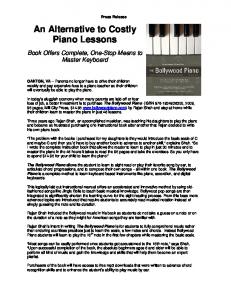
Cursor pointing to a structure of interest Figure 1 The third image in the abdominal dissection illustrating the labelling feature and showing the buttons available in the menu palette in 'Browse and Test' mode. As the mouse (cursor) is moved over the image, labels appear giving information about the organ or structure to which the mouse is pointing. 'Browse and Test' mode is indicated by the presence of information retrieval ('A' button) and questions ('Q' and 'T' buttons) in the menu palette. Clicking the mouse on a part of the image such as the thorax or abdomen will present the next image in that area. Help (?) is available and clicking on the small rat icon gains access to the map. Clicking the mouse on the 'S' button overlays a diagrammatic outline of the structures on the image which improves visual clarity. interpret when performing a real dissection as using an alternative. Whereas in practical dissection a student is expected to spend time cutting through layers of tissue, the program allows users to rapidly dissect the rat by clicking the mouse on a structure or area of interest on the image, such as the head, thorax, abdomen and feet. Clicking the mouse on one of the rat's feet in any of the images which show the whole animal, zooms to a close-up photograph of the appropriate foot, and clicking on a foot pad leads the user to a magnified diagrammatic cross-section through the skin. Using the features of Hypermedia, images are connected together so that a user might follow one path (e.g. clicking continuously on the abdominal region until the last picture in the series is reached), or jump around between different areas of the dissection (e.g. clicking on the thoracic region then on the dorsal aorta where it leaves the heart, which may take the student into the abdominal dissection to follow the descending path of the blood vessel). Using this nonlinear approach a user may jump forwards, backwards or laterally within the dissection in addition to following a step-wise refinement of a single line of interest. This ability to target a specific theme allows for repetition and hence reinforcement, offering an advantage over a dissection in which areas already exposed may be destroyed by further dissection.
A summary of the images in the dissection can be found in a map (figure 2) which may be obtained from anywhere within the program by clicking on the rat icon in the menu palette. Headings separate the dissection into eight areas: external anatomy, head and throat, thorax, abdomen, skeleton, intestines. reproduction and excretion. Clicking on a heading reveals icons of the images available in that area. A click of the mouse on an icon of an image takes the user to that image in the dissection. The map may help to orientate users within the program as it illustrates where a student has been within the dissection by highlighting (in inverse video) the icons of the images already viewed. Once all the images in a particular area have been observed the heading is also highlighted in inverse, indicating the completion of that section. Highlighting is intended as a guide for the user and headings or icons which are highlighted function similarly when not highlighted. The four modes of operation provide a challenging aspect to the program. Students may choose to look at (or ask about) fully-labelled images with additional information available, ('Browse' mode), or undertake the dissection by a more traditional method of identifying organs and structures without the benefit of labels or extra information ('Test' mode), the third mode, 'Browse and Test', combines the features of the first two modes. 'Input Text' mode is similar to Journal of Biological Education (1 992) 26 (1)
29
A computer-based alternative to performing a rat dissection
To change modes click and use as a menu by dragging downward
Quentin-Baxter and Dewhurst
A blackened heading indicates all of the images in inis section have been viewed by the current user
Click the Home icon to return to the Home stack The map 'help' is specific to the map only
Reproductive Organs-
Downloaded by [University of Tasmania] at 15:43 03 February 2016
^
^ EHcrelory Organs
-Click a heading to change the current view and to see small icons of the images available in that section
-Click an icon to view that image in the dissection The current view of small icons is from the 'Abdominal Dissection' section
When a user has visited an image its icon turns black
Figure 2 The map divides the dissection into eight areas. This example shows icons of the images available in the Abdominal dissection area. Clicking the mouse on another heading reveals icons of the images available in that area; clicking the mouse on an icon takes the user directly to that image in the dissection. The program illustrates a student's progress in inverse video. The user modes may be changed by clicking the mouse on the 'Change Browse/Test' button in the top left corner of the screen. A help (?) facility is also available and a user may exit to the 'Home' stack (quit the program). 'Browse and Test' but it allows a teacher with the correct password to alter text presented in the program. T h e modes control the choice of buttons available in the menu palette on the right hand side of the screen, and the first three modes may be easily changed from the 'Change Browse/Test' menu on the m a p screen (figure 2).
Browse mode In 'Browse mode' pop-up labels appear when the mouse (cursor) crosses the boundaries of structures on an image, so the area of a structure may be traced by moving the mouse over it on the screen. A student may locate and retrieve information about a structure via the 'A' (answer) button in the menu palette (figure 3).
Help button Map button (IT)
A or Answering button
Diagrammatic cross section through the small intestine.
Figure 3 An image from the Intestines section showing a diagrammatic view of magnified cross-sections through the small intestine. Clicking on the 'A' button in the menu palette produces a prompt for the name of a structure for which more information is sought. Some abbreviations and misspellings are accepted as valid entries. In response the organ or structure is indicated on the image (if it is present) by a 'finger pointer' and related textual information is presented. 'Browse' mode is denoted by the absence of the 'Q' and "T buttons. The map icon (small rat icon) is highlighted with a black background as this is the last image in this area and in order to proceed a user must return to the map to choose another image or area of the dissection. 30
Journal of Biological Education (1992) 26 (1)
Downloaded by [University of Tasmania] at 15:43 03 February 2016
A computer-based alternative to performing a rat dissection
A click on this button reveals a dialogue box which prompts for the name of a structure to be entered. Allowance is made for structures with more than one name and anomalies such as misspellings and abbreviations. If the input is recognizable as a structure or a set of structures which form another, (e.g. the intestines), it highlights on the current image the structure or structures with a 'finger pointer' and presents any related text. The 'finger pointer' identifies invisible buttons on the screen which correspond to the student's input and the pointer then flashes briefly over these buttons, effectively highlighting the area of the structure on the image. If the structure name entered by the student is not present on the current image, but is one for which textual information is available, then only text appears. This text is presented in a scrolling box on the left side of the screen (figure 4), and includes discussions of the function of all the major structures visible in the dissection in the program. Additional information which does not correspond to a structure name provides useful information about the rat, e.g. 'classification', 'weight', 'enzymes' and 'albino rat'. Words marked with an asterisk (*) in the text box have further information available and marked words may be clicked on to reveal the associated information. This HyperText linking facility allows for rapid navigation within the text in the database, which contains approximately 4700 words of text explaining the function of 120 structures in the rat. If the program cannot find information directly related to the input entered by the student, it looks to see if other information is sufficiently relevant to be offered in its place, to avoid returning an 'I don't know' message. Test mode When 'Test' mode is chosen students may navigate normally but structure-labelling and information retrieval features ('A' button) normally available in
Quentin-Baxter and Dewhurst
'Browse' mode are suppressed. This mode comes closest to mimicking a real dissection as students using it must rely upon normal support material such as textbooks and diagrams. Self-testing may be undertaken by clicking on the 'Q' button which randomly generates questions asking the student to identify an organ or structure on the current image (figure 5). To answer a question correctly a student must click the mouse on any part of the organ or structure on the image. If the student correctly identifies the structure another question is presented. If the structure is incorrectly identified the student may choose to attempt the question again or ask the program to highlight the structure with the 'finger pointer'. A dialogue box appears offering the choice of another attempt at answering the question, or asking the finger pointer to highlight the structure on the image. Up to five attempts at answering a single question are allowed. A score of correctly-answered questions is maintained for the student while the program is running and may be viewed by clicking on the T button (figure 1). The number of questions answered correctly after two attempts is also displayed along with the number of questions which have been cancelled. Print-outs of the score and any of the other screens may be obtained by pressing 'P' on the keyboard while depressing the command key.
Browse and Test 'Browse and Test' mode combines the features of both the 'Browse' and 'Test' modes described above. Images are fully annotated and all features of the 'A' button are available unless a series of questions is in progress. The features associated with 'Browse' mode become suppressed for the duration of a question series but resume after it is completed, or after the student terminates a question by clicking on the 'Cancel' button. elp
Blood enters the glomerulus In the kidney* under high pressure from the afferent * » orterloles end fluid Is filtered/forced out through pores In the thin epithelium of the capillaries, across the basement membrane and past the podocute* cells of
button
ap button Diagram of mole reproductive structures.
GQ CD
-A or Answering button Q or Question button T or Tally button
Figure 4 Screen display showing a diagram of the male reproductive structures (in •Browse and Test' mode) illustrating the text retrieval and HyperText facilities. The abbreviation 'glo' (for glomerulus) was entered at the prompt for a structure name after a click on the 'A' button. Clicking the mouse on words followed by an asterisk, such as 'kidney' in this example, returns additional textual information. The current mode is 'Browse and Test', indicated by the complete menu palette. Journal of Biological Education (1992] 26 (1)
31
A computer-based alternative to performing a rat dissection
Quentin-Baxter and Dewhurst
/Help
button
r*5i/
/Map
button
'(J[) CD
Q or Question button T or Tally button -S or Structures button draws a diagram of the image
V
Downloaded by [University of Tasmania] at 15:43 03 February 2016
d>
Please click on the spleen of the rat.
The question box while the program for a mouse-click
(Cancel}
-Click the cancel button at any time to terminate the question series
appears waits
Figure 5 An image from the Abdominal dissection showing the digestive system dissected out and displayed in 'Test' mode. The question presented would be answered correctly by clicking the mouse somewhere on the spleen. If a student responds incorrectly and opportunity is offered for another attempt, or the user may ask the program to highlight the area of the structure with the 'finger pointer'. Questions are randomly generated from the image on the screen after a click on the 'Q' button. The question series ceases when all the questions available have been presented, or when the series is terminated by a click on the 'Cancel' button. In 'Test' mode no labels appear and the 'A' button is not available from the menu palette.
Input Text The database of text which is available via the 'A' button may be altered by a person (such as a teacher) with access to the fourth user mode, 'Input Text'. It allows a teacher to add up to 5000 words of textual information to the database, or change or delete any of the existing text in the database. A password prevents a common entry to this mode. In all other aspects it is identical to the 'Browse and Test' mode. When 'Input Text' is in use an additional menu palette appears in the top left corner of the screen. Clicking on the 'Add Text' button invokes a series of steps which allow a user to enter a new keyword followed by the definition to be associated with it. If a new keyword already exists in the database the current definition is presented for alteration. Any new or changed entries are automatically saved and crossreferenced into the HyperText linking facility by adding asterisks to related information. This increases the likelihood that information will be seen by students. When keywords and their definitions are deleted all references to them are removed. This user mode enables the text in the program to be tailored to specific groups of students. In addition, tasks or instructions may be added. The program also has a comprehensive on-line facility and is supported by a manual which details 32
Journal of Biological Education (1992) 26 (1)
the technical aspects of using the program and contains several photographs of material which was used in the program's creation.
Discussion The program described aims to teach in a highly interactive way the functional anatomy of the rat, and provide a possible alternative to animal dissection for students where practical dissection experience and handling skills are considered to be of minor importance. However, if emphasis is placed on learning practical dissection skills, this program should not be considered as a replacement for an actual rat dissection as it cannot provide an environment for practising dissecting techniques. Operating the program is, however, likely to promote useful computer skills. Care was taken to ensure that throughout execution the program behaves in a non-intimidating manner. The computer acts as a 'tutor' who is available to return help and information at the user's request and who responds sensitively and anonymously to attempts at answering questions. The student-controlled approach to learning is a feature of Hypermedia and aims to promote investigative techniques as students must seek out information for themselves, as they would in a real dissection.
Downloaded by [University of Tasmania] at 15:43 03 February 2016
A computer-based alternative to performing a rat dissection
Estimates of how much time may be spent using the program depend on the student's response to the interactive environment and how much he/she wishes to learn. Some high school students may be advised to use the diagrams for reference only, as much of the information contained in them is detailed and may not be relevant to the course. A simple run-through of all the images in the dissection (without allowing time for attempting questions or retrieving additional information) may take a minimum of one and half hours if three minutes is spent with each image. Thus guidance from the teacher about which areas of the dissection are most relevant to the course (e.g. the thoracic region or the abdominal region) may be valuable, particularly where computer resources are scarce. The interactive nature of computer-based simulations as alternatives to animal vivisection allows them to escape from the one-way flow of information provided by diagrams, texts, models and videos. When used by groups of students simulations may help to promote discussion about the material being taught. This parallels a practical dissection situation where much more information is exchanged through student-student interaction. Situations in which CAL packages such as the one described form an alternative to an animal vivisection experiment, may be cost effective as a computer program may be a valuable teaching aid and re-used many times. If it is mandatory for students to learn dissection skills the program may be used to enhance interpretation efficiency and the confidence of students prior to performing a dissection, or used in retrospect to provide revision material and an opportunity to investigate specific areas of the dissection of further interest to the student. If a general view of functional anatomy is required then a demonstration of a dissected rat may be adequate live material when used in conjunction with the program. In many cases the object of a dissection is to impart an understanding of functional anatomy, and here the program is
Quentin-Baxter and Dewhurst
able to offer a large amount of supporting, repetitive and reinforcing information presented in an environment of discovery. Students are able to work at their own pace (within the constraints of access to hardware), and derive information relevant to their own interests as this program is self-explanatory and simple to use.
Availability The Rat Stack is available in either floppy or hard disk versions from Sheffield BioScience Programs, 11 Robinson Drive, Harrogate, North Yorkshire, HG2 9DJ, at the price of £80 for a multi-user licence.
Acknowledgement We would like to thank the Lord Dowding Fund (NAVS, UK) for their financial support of this project.
References Dewhurst, D. G., Brown, G. J., and Meehan, A. S. (1988) Computer simulation—an alternative to the use of animals in teaching? Journal of Biological Education, 22(1), 19-28. Graham, P. (1989) Non-animal alternatives for public school students. Humane Innovations and Alternatives in Animal Experimentation, 3, 94. Kohn, M. and Miller, P. (1984) Operation Frog. Scholastic Software, Scholastic Inc., 730 Broadway, New York, NY 10003, USA. Reed, M. J. (1987) Time and cost of producing high quality CBE materials. In Proceedings of the International Conference on Computer Aided Learning in Post-Secondary Education, pp. 185-187. Alberta, Canada: University of Calgary. The authors Megan Quentin-Baxter is an Analyst in the Computer Centre at Wolverhampton Polytechnic. Dr David Dewhurst is a Reader in the Faculty of Health and Social Care, Leeds Polytechnic, Calverley Street, Leeds LSI 3HE. (Reprint requests should be sent to Dr Dewhurst.)
Journal of Biological Education (1992) 26 (1)
33