W. Eric L. Grimson. 1. 1. Artificial Intelligence Laboratory. 2. Surgical .... Gibbs function G(T ,x) defines the relationship between x and its neighboring voxels [11].
Selected for Oral Presentation and Received Student Travel Award at ISBI 04: 2004 IEEE International Symposium on Biomedical Imaging: From Nano To Macro, Arlington, VA.
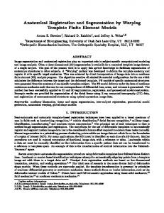
ANATOMICAL GUIDED SEGMENTATION WITH NON-STATIONARY TISSUE CLASS DISTRIBUTIONS IN AN EXPECTATION-MAXIMIZATION FRAMEWORK Kilian M. Pohl1 , Sylvain Bouix2,3 , Ron Kikinis2 , W. Eric L. Grimson1 1
Artificial Intelligence Laboratory MIT Cambridge MA, USA. {kpohl,welg}@ai.mit.edu
2
3 Surgical Planning Laboratory Department of Psychiatry Harvard Medical School Boston VA Healthcare System Boston, MA, USA. Boston, MA, USA {sylvain,kikinis}@bwh.harvard.edu
ABSTRACT High quality segmentation of brain MR images is a challenging task. To deal with this problem many automatic segmentation methods rely on atlas information of anatomical structures. We further investigate this line of research by introducing hierarchical representations of anatomical structures in an Expectation-Maximization framework. This new approach enables us to divide a complex segmentation scenario into less difficult sub-problems reducing the scenario’s statistical complexity. We demonstrate the method’s strength by segmenting a set of brain MR images into 31 different anatomical structures as well as comparing it to other methods. 1. INTRODUCTION Quantitative medical image analysis almost always involves segmentation to assess the volume and shape of anatomical structures. Even though automatic segmentation methods exist, labor intensive, manual delineation by expert anatomists is still common practice for high quality segmentations. Nevertheless, recent methods using prior knowledge of anatomical structures have shown promising results. This prior information is often represented as an atlas which can capture spatial, intensity and shape distributions of the anatomical structures of interest [1]. There are mainly two classes of atlas driven segmentation methods (see [1] for a detailed review). One class relies on deformable shape atlases which are defined by a deformable average model of a structure and principal modes of variations based on a sample population [2, 3, 4, 5]. When a new subject needs to be segmented, the model is deformed to best fit the subject’s image data and satisfy the prior knowledge given by the atlas. The second class of methods uses probabilistic atlases, modeling inter-subject This investigation was supported by the NIH (K02 MH 01110, RO1 MH 50747, R01 MH 40799, P41RR13218, and P01 CA67165), the NSF ERC CISST-973174-4, and the VA Merit Awards.
variability [6, 7, 8, 9, 10]. These methods first align the atlas to the patient and then use the atlas information as prior probabilities in the segmentation framework. This paper discusses an algorithm based on an implementation by Pohl et al. [11] which relates most closely to [9, 10]. The method first uses a non-rigid registration algorithm to align the atlas to the subject [12]. The images are then segmented using the atlas information in an Expectation-Maximization framework (EM) [13]. While this method generates very good results in general, we have found that the use of a probabilistic atlas also increases the risk of systematic biases, such as an over or underestimation of structures. For the specific problem of segmenting brain MR images in three different tissue classes (white matter (WM), gray matter (GM) and cortical spinal fluid (CSF)), we have found the following problems: (i) The subcortical gray matter is underestimated. In the subcortex, especially around the thalamus, the white matter fibers enter the gray matter structures, leading to poor contrast in the MR image. Accurate non-rigid alignment and segmentation are extremely difficult in this region. (ii) The cortical gray matter is overestimated. Segmenting the small folds of cortical gray matter correctly can be more complicated with a probability atlas. Its smooth borders increase the likelihood of bridges or cavities across adjacent banks of a sulcus and influence the segmentation towards an overestimation of cortical gray matter [6]. (iii) The method is difficult to scale. Increasing the number of structures in the segmentation problem also increases the problem’s statistical complexity and likelihood of misclassified voxels. We believe that incorporating a hierarchical representation of the tissues in the segmentation process provides a more powerful and more flexible framework not only for tissue classification but also for the segmentation of substructures of the brain. We will also show that this new framework is more robust than a number of other approaches and reduces the overestimation of cortical gray matter.
p(T (x)) := pT (x) · G(T , x) is the probability of the structure T being present at voxel x. pT (x) represents the spatial distribution of structure T defined by the atlas, and the Gibbs function G(T , x) defines the relationship between x and its neighboring voxels [11]. To integrate the anatomical tree into the E-Step, the atlas information from a sub-structure has to be properly permeated towards its parent structure. In brain MR images small anatomical structures, like the superior temporal gyrus, can be represented by global simple intensity distributions. Thus, the intensity distribution of a structure without any children can be represented by a normal Gaussian distribution GσT (I(x) − β(x) − µT ) where Gσ (X) ∼ N (0, σ). µT and σT define the mean and variance of the intensity distribution of T . For a structure with children, the intensity distribution is a spatially varying distribution defined by the weighted sum over the intensity distributions of its sub-structures. Let T be a parent structure with n children sub-structures T 0 with spatial distribution pT 0 . The spatially varying intensity distribution p(I(x)|T (x) = δT , B(x)) is then defined as : P T 0 ∈TSUB pTP0 (x)·p(I(x)|T (x)=δT 0 ,B(x)) , T 0 SUB 6= / T 0 ∈TSUB pT 0 (x) (2) G (I(x) − β(x) − µ ) , otherwise
2. METHOD To reduce the overestimation of cortical gray matter as well as increase the method’s robustness, we will divide the segmentation scenario into smaller problems. Before segmenting an image, the structures of interest will be ordered as a data tree based on their anatomical relationship (see figure 1). If a structure X, e.g. white matter, is part of another structure Y, e.g. brain, X will be located on a branch of Y. The segmentation is then divided into different levels starting at the top of the tree and ending at its leaves. For example (see figure 1), let us segment an MR image of a head into white matter (WM), gray matter (GM), cortical spinal fluid (CSF), and background (BG). Background and brain define the first level in the tree. Sub-Structures of the brain are CSF, WM, and GM, which define the second level of the data tree. Therefore, the method is a two step segmentation process. The head is first segmented in background and brain, which is then further parcellated into GM, WM and CSF.
σT
T
where TSUB := {T 0 |T is parent of T 0 } is a list of the substructures of T , e.g. BRAINSUB := {WM, GM, CSF}. The spatial distribution of a parent structure pT (x) is defined in a similar fashion. If TSUB = /0 for a structure T, the spatial distribution is defined by the aligned atlas. If TSUB 6= /0, the distribution of T is composed by the sum of its sub-structures. This definition is based on the assumption that all immediate children of a structure represent anatomical independent regions. As such, superior temporal gyrus and GM could not be immediate sub-structures of the structure BRAIN (see figure 1). Additionally, we introduce the parameter λ, which defines the influence of the atlas on the segmentation process. In our example, (see figure 1), the atlas is of great importance at the first level and therefore λ is set to 1. At the second level, the atlas caused mislabeling and thus λ is set close to 0 for each structure involved. The new definition for pT (x) is: (P 0 , TSUB 6= /0 T 0 ∈TSUB pT (x) (1 − λ) + λ · (3) as defined by the atlas , otherwise
Fig. 1. Anatomical structures ordered in a tree. In this example, background (BG) and brain (BRAIN) define the tree’s top level while gray matter (GM), cortical spinal fluid (CSF), and white matter (WM) compose the second level. The remainder of this section will discuss in detail the mathematical realization of the concept described above. Our implementation is based on the work by Pohl et al., [11]. It consists of two steps: the E-Step, which calculates the likelihood of each anatomical structure throughout the image, and the M-Step, which estimates the image inhomogeneities. The hierarchical model does not modify the MStep and we refer the reader to [11] for its derivation. The E-Step, however, needs to be redefined. Let p, the probability of a voxel of tissue class T , be defined as:
By substituting Eq. (3) and (2) in Eq. (1), the segmentation approach now incorporates the hierarchical anatomical dependencies. Based on those dependencies, the algorithm first segments the structures at the highest level and then works it way down the branches until it finally reaches the leaves of the anatomical tree. This procedure leads, as we will show in the next section, to more robust and accurate parcellations of the brain.
1 p(T (x)|I(x), B(x))= p(I(x)|T (x), B(x))p(T (x)) (1) Z Z is the normalizing term, T (x) is the indicator random vector representing the structures at voxel x (the hidden data), I is the input image (the observed data), and B(x) represents the image inhomogeneities (the parameters). 82
3. EXPERIMENTS AND VALIDATION Four MR datasets of the brain (256x256x124 SPGR and T2 volumes with 0.9375mm x 0.9375mm x 1.5 mm resolution) were segmented into BG, CSF, WM and GM, using four different methods: an EM approach without any atlas information (EM-Only) [13], an atlas dependent watershed approach (WS-Atlas) [8], an EM method using an atlas (EMAtlas) [11], and our new hierarchical method (EM-Hiery). The quality of each segmentation were assessed using STAPLE [14]. To evaluate a method, a ground truth is first estimated using the output of all methods except the one under evaluation. Afterward, the selected segmentation is compared to STAPLE’s ground truth using the Dice measure (see [11]). The results of our evaluation are shown in figure 2. Out of the four methods, EM-Only received the lowest score. Outliers, normally removed by an atlas, reduced its accuracy (see figure 4 (b)). EM-Atlas has good performance for WM and GM but performs poorly for CSF. WC-Atlas has overall good scores but is outperformed by EM-Hiery for all but one output (WM in case 4). As shown in figure 4 (c), EM-Atlas overestimates GM and underestimates CSF. The same trend can be observed in WS-Atlas (figure 4 (d)). EM-Hiery (figure 4 (e)) does well on all three tissue classes due to the new hierarchical definition of spatial and intensity distribution and the atlas influence parameter λ.
Fig. 3. Segmenting a brain into 31 different structures. A tree of the anatomical structure, consisting of three levels, guides the segmentation process. The complexity of the segmentation is thus divided into smaller problems that are more manageable by the EM algorithm. tion has very view outliers (figure 3). 4. SUMMARY AND DISCUSSION A new segmentation approach, guided by anatomical dependencies, has been presented. The main feature of the method is the use of a hierarchical data structure to decouple the complexity of the segmentation scenario from the number of structures to be segmented. Additionally, the influence of the atlas can be tailored at each level of the segmentation. As an example, atlas information plays an important role in segmenting an image into brain and background, as voxels in the skull and muscles can have very similar intensities to voxels inside the brain. However, when further parcellating the brain into WM, GM, and CSF the atlas is of less importance because large intensity differences between structures exist. As the results of our validation indicate, the new approach better coped with the GM overestimation and complexity issues addressed in section 1 than the other methods tested. Moreover, our new approach is also more flexible and allows for very complex segmentation scenarios. Parcellating a large number of structures can be elegantly handled by designing an appropriate data tree, reducing the stress on memory and computing requirements. If other methodology driven by anatomical dependencies exist, they are usually limited to specific scenarios and are difficult to scale or modify [15, 6]. We believe that the high quality results presented in this paper will instigate further applications in medical research. Nevertheless, our methodology can still suffer from a poor alignment of the atlas (a common problem in the subcortex). In future work, we plan to overcome this issue by rigidly aligning the atlas to the subject preserving the atlas’
Fig. 2. Comparison between four different segmentation methods. The Dice measure compares the segmentations to an approximated ground truth. Our new method (Hierarchy) achieves higher quality segmentation of the CSF. The method is more robust for the other two tissue classes. The method’s robustness and flexibility is further demonstrated by segmenting the brain into 31 structures. First, the image is segmented into brain and background. The background is segmented into skin and air. The brain is further separated into CSF, GM, and WM. On the lowest level we parcellate CSF into 7 and GM into 21 sub-structures. Currently we can only visually analyze the results due to a lack of experts’ segmentations. We note that the final segmenta83
(a) Image
(b) EM-Only
(c) EM-Atlas
(d) WS-Atlas
(e) EM-Hiery
Fig. 4. Segmentation of a MRI (a) by four different methods. The EM segmentation (b) is characterized by its many outliers, which are normally reduced by atlas information (see (c - e)). The atlas based EM approach (c) and the watershed method (d) smooth over small gyri resulting in an underestimation of CSF. This problem is less likely with the new approach (e). anatomical properties and then implement a non-rigid registration method within the EM framework.
image segmentation using prior information,” IEEE Trans Med Imag. In Press., 2003. [9] K. Van Leemput, F. Maes, D. Vanermeulen, P. Suetens, “Automated model-based bias field correction of MR images of the brain,” TMI, vol. 18, no. 10, pp. 885– 895, 1999.
5. REFERENCES [1] D.L. Pham, C. Xu, J.L. Prince, “Current methods in medical image segmentation.,” Annual Review in Biomedical Engineering, vol. 2, pp. 315 – 337, 2000.
[10] J. L. Marroquin , B. C. Vemuri, S. Botello, F. Calderon, A. Fernandez-Bouzas, “An accurate and efficient bayesian method for automatic segmentation of brain mri,” TMI, vol. 21, pp. 934–945, 2002.
[2] S. Ho, E. Bullitt, G. Gerig, “Level set evolution with region competition: Automatic 3-d segmentation of brain tumors,” Proceedings of the ICPR, 2002.
[11] K. M Pohl, W. M. Wells, A. Guimond, K. Kasai, M. E. Shenton, R. Kikinis, W. E. L. Grimson, S. K. Warfield, “Incoperating non-rigid registration into expectation maximization algorithm to segment mr images,” in MICCAI, 2002, pp. 564–572.
[3] A. Tsai, A. Yezzi, W. Wells III, C. Tempany, D. Tucker, A. Fan, W. Grimson, A. Willsky, “A shapebased approach to the segmentation of medical imagery using level sets,” IEEE Transaction on Medical Imaging, vol. 22, no. 2, pp. 137 – 154, 2003.
[12] A. Guimond, A. Roche, N. Ayache, J. Meunier, “Three-dimensional multimodal brain warping using the demons algorithm and adaptive intensity corrections,” IEEE Transactions in Medical Imaging, vol. 20, no. 1, pp. 58–69, Jan. 2001.
[4] M.E. Leventon, W.E.L. Grimson, O.D. Faugeras, “Statistical shape influence in geodesic active contours,” in CVPR, 2000, pp. 1316 – 1323. [5] J. Yang, L.H. Staib, J.S. Duncan, “Statistical neighbor distance influence in active contours,” in MICCAI, 2002, pp. 588 – 595.
[13] W.M. Wells III, W.E.L Grimson, R. Kikinis, F.A Jolesz, “Adaptive segmentation of MRI data,” IEEE Transactions on Medical Imaging, vol. 15, pp. 429– 442, 1996.
[6] B. Fischl, D. H. Salat, E. Busa, M. Albert, M. Dieterich, C. Haselgrove, A. van der Kouwe, R. Killiany, D. Kennedy, S. Klaveness, A. Montillo, N. Makris, B. Rosen, A. M. Dale, “Whole brain segmentation: Automated labeling of neuroanatomical structures in the human brain,” Neuron, vol. 33, 2002.
[14] S. K. Warfield, K. H. Zou, W. M. Wells, “Validation of image segmentation and expert quality with an expectation-maximazation algorithm,” in MICCAI, 2002.
[7] D. L. Collins, A. P. Zijdenbos, W. F. C. Barre, and A. C. Evans, “Animal+insect: Inproved cortical structure segmentation,” IPMI, vol. 1613, 1999.
[15] M. Kaus, S. K. Warfield, F. A. Jolesz,R. Kikinis, “Adaptive template moderated brain tumor segmentation in mri,” Bildverarbeitung fuer die Medizin, pp. 102–106, 1999.
[8] V. Grau, A.J.U. Mewes,M. Alcaniz, R. Kikinis, S.K. Warfield, “Improved watershed transform for medical 84