For example, the type I holoenzyme. (R2C2) and the dissociated regulatory subunit (R2) have fric- tional coefficients of 1.50 and 1.47, respectively, in contrast.
Vol. 262, No. 31, Issue of November 5, pp. 14961-14966,1987 Printed in U.S.A.
THEJOURNAL OF BIOLOGICAL CHEMISTRY 0 1987 by The American Society forBiochemistry and Molecular Biology, Inc
Antiparallel Alignment of the Two Protomers of the Regulatory Subunit Dimerof CAMP-dependent Protein Kinase I* (Received for publication, May 4,1987)
Jose BubisS, ThomasS . Vedvick, and Susan S.Taylor8 From the Department of Chemistry, Universityof California, Sun Diego, La Jolla, California 92093
native protein (Corbin et al., 1978; Potter and Taylor, 1978, 1979; Takio et al., 1980). The “hinge” region which is labile to proteolysis when the R-subunit is in its dissociated form is localized toward the NH2-terminal end of the polypeptide chain. Following proteolysis, the carboxy-terminal CAMPbinding domain retains most of the high affinity CAMPbinding properties of the native protein; however, this domain is monomeric. In some cases, the monomeric protein retains the ability to interact with the C-subunit forming an RC dimer which, like the holoenzyme, is activated by cAMP (Weldon and Taylor 1985; Weber and Hilz, 1979). Thus, the dimeric structure of the R-subunit is not essential for interacting with the C-subunit. The NH2-terminal fragment that results from limited proteolysis does not bind CAMP;however, it does remain as a dimer, thus, suggesting that the major sites of interaction between the two protomers of the native R-subunit are associated with this portion of the molecule (Potter and Taylor, 1979; Zick and Taylor, 1982; Reimann, 1986). The dimeric CAMP-dependent enzyme (RC) that is Although there are several distinct classes of CAMP-dependent protein kinase, each retains the same general struc- formed with proteolyzed R has a frictional coefficient of 1.35 ture. In all cases, the native protein is an inactive tetramer indicating that it is considerably less asymmetric than the which contains two regulatory (R)and two catalytic (C) holoenzyme or the R-subunit dimer (Taylor and Stafford, subunits which dissociates in the presence of cAMP into an 1978). Thus, it is likely that the NH2-terminal domain is the R-subunit dimer (R2) and two active C-subunits. Frictional major determinant in the R-subunit which leads to theassymcoefficients ( f / f o ) have established that the molecule has di- metry of the molecule. No evidence of cross-linking between mensional asymmetry. For example, the type I holoenzyme C-subunits hasbeen shown although cross-linking between R been (R2C2) andthe dissociated regulatory subunit (R2) have fric- subunits (R-R) andbetween R and C subunits (R-C) has tional coefficients of 1.50 and 1.47, respectively, in contrast described (First and Taylor, 1984;Zick and Taylor, 1982). to theC-subunit which is a rather typical globular protein (f/ This suggests that the two C-subunits in the tetramer are well segregated. fo = 1.18) (Zoller et al., 1979; Erlichmann et al., 1973). These In order to obtain some insight into the topological orienfrictional coefficients indicate a high degree of asymmetry in tation of the R-subunits, we have taken advantage of a unique the tetrameric enzyme and in the R2 dimer, and pose some interesting questions regarding the aggregation process and feature of the type I regulatory subunit. This molecule contains two sulfhydryl groups in the NH2-terminal domain the overall geometry of this protein. Each R-subunit has a well-defined domain structure. The which are involved in interchain disulfide bonding (Zick and COOH-terminal two-thirdsof the polypeptide chain includes Taylor, 1982). It is not clear whether the two protomers are two tandem CAMP-binding domains each of which is homol- disulfide bonded in the native protein or whether they simply ogous to the CAMP-binding domain of the catabolite gene are oxidized readily during thepurification procedure. Neveractivator protein in Escherichia coli (Weber et al., 1982; 1987). theless, it is apparent that both sulfhydryl groups from one Limited proteolysis also has provided some insight into the R-monomer are in close proximity to thesulfhydryl groups in structural organization of the R-subunit. In summary, the R- the adjacent chain such that interchain disulfide bonds can subunit can be cleaved proteolytically into two independent form readily. This observation, thus, provides two specific domains which each retain some of the properties of the points of contact between the polypeptide chains. Determining which specificdisulfide bonds are formed between the two * This work was supported by Public Health Service Grant GM- polypeptide chains should establish a general framework for 19301. The costs of publication of this article were defrayed in part the orientation of the two chains with respect to one another. by the payment of page charges. This article must therefore be hereby Since only two possibilities exist, we have identified the marked “aduertisement” in accordance with 18 U.S.C. Section 1734 specific sites of interchain disulfide bonding in this protein. solely to indicate this fact.
The purified type I regulatory subunit ofCAMPdependent protein kinase is a dimeric protein, and the two protomers of the dimer are linked by two interchain disulfide bonds. The disulfide linkages that join these two polypeptide chains have been identified in order to provide a structural basis for the orientation of the two chains in the asymmetric dimer. Disulfide bonds were found to exist exclusively between Cys-16 and Cys-37, and this assignment, thus, establishes a general antiparallel alignment of the two chains. Two other homologous proteins, the type I1 regulatory subunit and the cGMP-dependent protein kinase also are dimeric proteins. In all three proteins, a relatively small, nonhomologous, amino-terminal segment of the polypeptide chain is essential for maintaining the dimeric aggregation state.
$ Supported in part by the Consejo Nacional de Investigaciones Cientificas y Tecnol6gicas (CONICIT), Caracas, Venezuela. Present address: Dept. of Chemistry, Massachusetts Institute of Technology, Cambridge, MA 02139. § To whom reprints requests should be addressed.
EXPERIMENTALPROCEDURES
Materials-Reagents were purchased from the following sources: [‘4C]iodoaceticacid, ICN; iodoacetic acid, Aldrich; L-1-(tosy1amide)2-phenylethylchloromethylketone (TPCK)-treated trypsin, pepsin,
14961
14962
CAMP-dependent Protein Kinase: Orientation of R-Subunits in Dimer
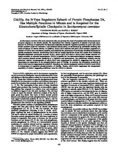
FIG. 1. HPLC elution profile of the tryptic digest of unreduced and fully reduced R’ alkylated with iodoacetic acid. R‘ isolated from porcine skeletal muscle was denatured and alkylated with [“C]iodoacetic acid. Tryptic peptides were separated by HPLC using aWaters Cls-pBondapak column and a gradient from 0.1% trifluoroacetic acid (pH 2.15) to 60% CH3CN in 120 min. The upper p a n e l shows the sample that was fully reduced with dithiothreitol prior to alkylation; the lower p a n e l shows the sample that was not reduced prior to alkylation. Four peptides designated A, B, C, and D were modified with [‘4C]iodoaceticacid in the fully reduced protein. Only two of these peptides, A and C, were “C-labeled in the nonreduced sample, and a new 219nm-absorbing peptide was observed.
2
0
’
40
a
a
m
1.D
IC Ij
II
a00
400
FR*cTIQI
TABLE I Amino acid compositions of cysteine- and cystine-containingpeptides The new tryptic peptide as well as peptides I11 and IV all indicated the presence of cystine in the amino acid composition. The numbers in parentheses indicate the expected composition for residues 12-61 in bovine R’ Tryptic peptides acid
A
B
CMC 0.30.30.20.4 ASP 3.3 0.9 Thr Ser 1.0 Glu 2.8 Pro GlY Ala Val 1.0 1.0 Met Ile Leu 1.4 2.1 TYr Phe His Lys1.0 1.0 1.0 Arg 0.7 0.9
c
New tryptic
D
0.9 3.0 0.8 1.0
1.0 0.8 2.9 0.6
0.6 0.7 1.7 1.8
1.4 2.0 1.0 0.8 1.0 1.8
0.9 2.2
peptide
(3) 0.5 (1) 0.8 (3) 13.5 4.5 (14) 4.5 1.7 (2) 1.2 (1) 4.61.0 (5) 1.5 (2) 0.4 (1) (3) 8.5 4.8 (8) 2.0 2.3 (2) 2.4 (2) 0.4 (1) 6.4 (6) 4.5 (5)
Peptic peptides
I
111
0.7 1.2
1.0 0.9 0.9
2.0 2.3 0.9 1.1 0.9 0.9 0.9
IV
0.9
1.8
1.0 0.5 1.0
1.0 0.8 0.9
1.0 1.0 1.1 1.0 1.0 1.0 3.02.0
1.0 2.0
United States Biochemical Corporation; thermolysin, Sigma; monobromobimane (Thiolyte Reagent), Calbiochem; trifluoroacetic acid (sequenal grade), Pierce Chemical Co.; acetonitrile (HPLC’ grade), Fischer Scientific; Cytoscint, West Chem. The abbreviations used are: HPLC,high performance liquid chromatography; dansyl, 5-dimethylaminonaphthalene-1-sulfonyl; PTH, phenylthiohydantoin.
-
Proteins-R1-subunit was purified from porcine skeletal muscle (Zick and Taylor, 1982). R-subunit also was purified from E. coli transformed with a pUC vector, pLST-2, which contains the complete coding region of bovine R’ (Saraswat et al., 1986). Labeling of Sulfhydryl Groups-R’-subunit (3-5 mg) was dialyzed against 200 mM Tris, 2 mM EDTA, pH 8.3. After bringing the solution to 6 M in guanidine HCI by the addition of solid guanidine HCI, the protein was incubated in the dark with 2 mM dithiothreitol for 1 h a t room temperature with stirring under nitrogen. In the case of the unreduced sample, this last stepwas eliminated. Finally, the protein solution was incubated with 10 mM monobromobimane for 3 h at 37 “C under nitrogen in the dark and then dialyzed exhaustively against 50 mM NH4HC03, pH8.3. Alkylation of sulfhydryl groups with [‘4C]iodoacetic acid was achieved according to the basic method of Crestfield et al. (1963) as described previously (Zick and Taylor, 1982). Isolation of Modified Tryptic Peptides-R-subunit that had been modified with either monobromobimane or with [“C]iodoacetic acid was digested with ~-l-(tosylamide)-2-phenylethylchloromethylketone-trypsin (1:50w/w) for 15h at 37 “C. The resultant peptides were resolved by HPLC using an Altex 332 system with either a Waters C1,-pBondapak reverse phase column (0.39 X 30 cm) or a Vydac Cla column (0.46 X 25 cm). The buffers employed were (a) 0.1% trifluoroacetic acid (pH 2.15) and ( b ) CH3CN. The particular gradients that were used are described in the figure legends. Further purification was achieved by rechromatographing the peptides on a Vydac Cls column using a different buffer system: ( a ) 10 mM sodium phosphate (pH 6.8) and (b) CH3CN. Prior to sequencing, the peptides were chromatographed again on a Vydac C18 column using a 20-min linear gradient from 0.1%trifluoroacetic acid to CH3CN (0-100%) to remove sodium phosphate which frequently interfered with the sequencing. Fluorescence was monitored using a Gilson Spectra/glo filter fluorometer equipped with fluorescein excitation and emission filter. Absorbance was monitored at 219 nm. Radioactive samples were counted in Cytoscint. Disulfide-bonded tryptic peptides were further treated with a variety of proteases. Proteolysis with thermolysin was performed in 100
CAMP-dependent Protein Kinase: Orientation
of R-Subunits Dimer in
14963
mM NH4HC03, 1 mM CaCl, (pH 8.0) at 45 “cfor 1 h using 2% thermolysin (w/w). This reaction was carriedout at room temperature when 1% sodium dodecyl sulfate was included in the mixture. Proteolysis with trypsin and chymotrypsin was carried out overnight at 37 “Cusing 1:50 protease to protein in 50 mM NH,HCOI, (pH 8.3) in the presence or absence of 2 M guanidine HCl. For digestion with pepsin, the peptide was lyophilized to dryness and then redissolved in99% formic acid. The peptide was then diluted 10-fold with a solution of pepsin (2%by weight of the substrate) in 1 mM HCl and was incubated with stirring at 25 “C for 4 h. The digestion was terminated by lyophilization. Amino Acid Analyses-Analyses were performed on a Beckman model 118C or a LKB Biochrom model 4400 automated amino acid analyzer. Samples were hydrolyzed in uacuo at 110 “C in 6 M HCl for 24 h. Sequencing-Manual dansyl-Edman degradation was carried out by two-dimensional thin-layer chromatography on polyamide sheets (8 X 8 cm) according to the method of Hartley (1970). Gas-phase sequencing was carried out on an Applied Biosystems model 470A protein sequencer. PTH-amino acids were identified by HPLC as described by Hunkapillar and Hood (1983) using an IBM Cyano Column.
(Kosower et al., 1978) which forms a chemically stable thiol adduct. The HPLC elutions of tryptic digests of the reduced (bottom panel)and unreduced (top panel)R-subunit areshown in Fig. 2. The fluorescent peptides obtained using monobromobimane are very similar to those obtained when [“CC] iodoacetic acid was used to modify the sulfhydryl groups of porcine R’. The denatured andfully reduced R-subunit yielded four fluorescent tryptic peptides. Two of these were contained in the NHz-terminalregion of the protein and were identified as Cys-16 and Cys-37, respectively (Fig.2, bottompanel). Both of these peptides were missing from the tryptic digest of the nonreduced protein (top panel). On the other hand, a new peptide was observed in the chromatogram of the unreduced R-subunit. This peptide eluted at 40% acetonitrile and contained both of the Cys residues, Cys-16 and Cys-37, that are involved in interchain disulfide bonds. The amino acid composition of the new peptide is shown in Table I, and the sequence of the first 17 residues of this new peptide, determined by gas phase methods, is shown in Table 111. The amino acid analysis and sequencing data established that this RESULTS tryptic peptide was accounted for by residues 12-61 in the R’In order to specifically identify all of the cysteine-contain- subunit. This large tryptic peptide which containedboth of the ing tryptic peptides, the regulatory subunit isolated from porcine skeletal muscle wasfully reduced, alkylated with [14C] interchain disulfide bonds was thentreated with various iodoacetic acid, and digested with trypsin. A similar reaction proteases in an effort to cleave between the cysteine residues, was carried out with protein that was not reduced prior to and the resultant proteolytic peptides were characterized. carboxymethylation in order to distinguish the disulfide- This peptide proved to be highly resistant to further proteobonded peptides. When these mixtures of tryptic peptides lytic cleavage. Neither trypsin,chymotrypsin, nor thermolysin were subjected to HPLC on a Waters C18-pBondapakreverse were capable of digesting the original peptide, even in the phase column and eluted as described under “Experimental presence of 2 M guanidine HC1 which was used in the case of Procedures,” the profiles shown in Fig. 1, A and B were trypsin andchymotrypsin or 1%sodium dodecyl sulfate which obtained for the reduced and unreduced protein, respectively. was used in the case of thermolysin. This resistance suggests When the fractions were measured for radioactivity, two that thedisulfide bonds are deeply buried. Only when pepsin peaks were identified in the case of the unreduced sample, was used, was the peptide cleaved further. The resulting peptic whereas four peaks were observed in the fully reduced sample. peptides were separated by HPLC as shown in Fig. 3 and The amino acid compositions and sequences of the cysteine- then characterized by gas-phase sequencing. Due to incomcontaining peptides shown in Fig. 1 are indicated in Tables I plete digestion, several of these peaks (111, IV, and V) conand 11. Additional peaks of radioactivity are accounted for by tained disulfide-linked peptides that each varied slightly. The sequence of the first 15 steps of peptide V is indicated in incomplete proteolysis. These sequences established that peptides A, B, C, and D Table I11 as well as the sequence of peptide I. The composicontained Cys 345, 16,360, and 37, respectively, based on the tions of some of the peptides are included in Table I. The amino acid sequence of bovine R’ (Titani et al., 1984). Three sequence of the original large tryptic peptide is summarized of the porcine peptides are identical to the bovine peptides, also in Fig. 4. and the changes in the fourth peptide as indicated in Table DISCUSSION I1 are all conservative replacements. Peptides B and D were Model building of the CAMP binding domains of the Rmissing from the tryptic digest of the nonreduced protein, and a new peptide was observed which was detected by ab- subunits on the basis of their homology with the catabolic sorbance at 219 nm and which eluted near the end of the gene activator protein and on the sites that are photolabeled with S-N,cAMP has provided some preliminary information gradient. A parallel experiment was performed using the expressed about the folding of each CAMP-binding domain and about bovine R-subunit isolated from E. coli transformed with a the potential orientation of the two tandem CAMP-binding plasmid, pLST 2, containing the cDNA for the bovine R’ domains relative to each other in the monomeric subunits subunit. Like the porcine protein, the two protomers of this (Weber et al., 1987). However, this modeling does not reflect dimer were covalently linked by interchain disulfide bonds on the overall symmetry of the two protomers in the R-dimer. (Saraswat et al., 1986). This R-subunit was modified with the The interchain disulfide bonds that join the two protomers of fluorescent sulfhydryl-specific probe, monobromobimane, the R’-subunit, therefore, were identified in order to map TABLEI1 Sequences of the cysteine-containing tryptic peptides from porcine R’ In positions where 2 residues are indicated, the upper residue refers to the porcine R’sequence and the lower to the bovine R‘ sequence. Peptide A Cys-Val-Lys Ser-Leu-Arg-Glu-Cys-Glu-Leu-Tyr-Val-Gin-Lys Peptide B Peptide C Leu-Leu-Gly-Pro-Cys-Ser-Asp-Ile-K-Lys-Arg Va 1 Leu Asp-Ser-Ile-Val-Gln-Leu-Cys-Thr-Ala-Arg-Pro-Glu-Arg-Pro-Met-Ala-Phe-Leu-Arg Peptide D
-
CAMP-dependent Protein Kinase: Orientation of R-Subunits in Dimer
14964
1 0.8.. A
c
0.4 -.
- 0.0 0.6
1
0.4
0.2 0.0 FRACTION NUYBER
FIG. 2. HPLC separations of the tryptic digest of unreduced and reduced R-subunit isolated from E. coli and treated with monobromobimane. The expressed R-subunit which was isolated from E. coli that had been transformed with pLST-2 was denatured, fully reduced with dithiothreitol (bottom panel) or untreated with reducing agent (top panel),and thenincubated with monobromobimane. After digestion with trypsin, the resultant peptides were separated by HPLC using a Vydac C,, column and a gradient of 0.1% trifluoroacetic acid (pH 2.15) to CH3CN. The following gradient was used 0-30% in 120 min, and then 30-60% in 60 min. In the case of the unreduced protein (top panel), a new absorbing peptide was observed at 40% CHaCN. Peptides A-Dwere the peptides that showed fluorescence.
TABLE 111 Sequences of the disulfide-bonded peptides from bovine R-subunits expressed in E. coli Sequences were determined by the gas-phase method. The tryptic peptide refers to the new peptide that is indicated in the upper panel of Fig. 2 and which was isolated from the unreduced R-subunit. The positions where cystine was located showed no identifiable PTH derivative and are indicated as X.In peptic peptide V, the location of the disulfide bond is indicated as a solid line. Step ~
2
1
Tryptic Peptide Peptic Peptide I Peptic Peptide V
3
4
5
6
7
8
9
10
11
12 14 13
15
~-
~
16
17
18
S e r - L e u - A r g - G l u - X -Glu-Leu-Tyr-Val-Gln-Lys-His-Asn-Ile-Gln-Ala-Leu-Leu Tyr-Val-Gln-Lys-His-Asn-Ile-Gln-Ala-Leu S e r - L e u - A r g - G l u - X -Glu-Leu-Tyr-Val-Gln-Lys-His-Asn-Ile-Gln c
X
-Thr-Ala-Arg-Pro-Glu-Arg-Pro-Met-Ala-Phe
specific points of contact between the monomers andto resolve the orientation of the two chains in the dimeric structure. The type I R-subunit contains a total of 4 cysteine residues (Titani et al., 1984). Two of these sulfhydryl groups lie close to theNHz-terminal endof the polypeptide chain, and, inthe purified protein, both of these cysteines are disulfide bonded to the adjacent subunit (Zick and Taylor, 1982), even in the presence of exogenous-reducingagents. The remaining sulfhydryl groups which lie in the second CAMP-binding domain are fully reduced. The fact that the R-monomers are linked by two disulfide bonds means that those two R-subunit monomers could potentially be aligned in either a parallel or an antiparallel manner. To differentiate between these two possibilities and definitively establish the orientation of both chains in the dimer, the peptide containing the disulfidebonded cysteines was isolated by HPLC and then digested further with pepsin. If the protomers are aligned in a parallel orientation, then a protease which cleaves between Cys-16 and Cys-37 in the linear sequence would yield two different peptides each yielding a single sequence. On the other hand, if the antiparallel orientationis found, cleavage with the same protease will produce one peptide which will contain a single disulfide bond between Cys-16 and Cys-37. Sequencing of this peptide would produce two different sequences, since two different amino termini wouldbe contained in the single
peptide. As was seen in Table 111, the two cysteine-containing peptides that were isolated each contained two different sequences corresponding to the regions flanking Cys-16 and Cys-37. This establishes definitively that theprotomers in the R*-subunit dimer are aligned inan antiparallelmanner, namely, Cys-16 of one monomer is disulfide bonded to Cys37 of the other. The localization of these disulfide bonds in the overall polypeptide chain is summarized in Fig. 4. Both disulfide bonds are located in the NHz-terminal 15% of the molecule. The localization in the primary sequence of R' of some of the peptides that were sequenced also are indicated in Fig. 4. As a general rule, disulfide bonding is associated with extracellular proteins. Incontrast,intracellularproteins which normally are exposed to an environment that favors reduction of disulfide bonds usually have their sulfhydryl groups in the reduced form. The regulatory subunit of CAMPdependent protein kinase I appearsto be an exception to this general rule in that the two protomers of this intracellular dimeric protein are covalently cross-linked by disulfide bonding in the purified protein. Interchain disulfide bonding has been demonstrated to occur in other proteins (Calissano et al., 1976; Flashner et al., 1972; Lehrer, 1975);however, in each of these cases, the disulfide bond was artificially generated in vitro and was not presentin the native purified protein. Other examples of disulfide bonds occurring in intracellular pro-
CAMP-dependent Protein Kinase: Orientation of R-Subunits in Dimer
24965
Peptlde V
0.6 0.5 0.4
0.3
I
0.2
I
0.1 0.0
1
AI 40
20
60
80
100
FRACTION NUMBER
FIG.3. HPLC separation of thepepsin-treated tryptic peptide containing the two interchain disulfide bonds. The new peptide observed after trypsin digestion of unreduced R-subunit (Fig. 2) was lyophilized, redissolved in 99% formic acid, and further proteolyzed with pepsin as described under “Experimental Procedures.” HPLC was carried out using the following 0.1% trifluoroacetic acid to CHSCN gradient: 0-40% CH&N for 120 min, and then40-100% C R C N in 30 min. Each of the resultant peptic peptides was rechromatographed by HPLC on a second column using a different gradient system of 10 mM sodium phosphate (pH 6.8) to CH3CN ( 0 4 0 % )in 80 min. As an example, the inset shows the rerun of peptide V which contained the interchain disulfide bonds. INTERCHAIN INTERACTION SITE
9
FIG.4. Antiparallel arrangement of the R’ monomers in the dimer showing some of the functional sites of R’ and including the sequence containing the interchain disulfide bonds. N - and C-refer to the amino- and carboxy-ends of the polypeptide chains, respectively. The disulfide bonds joining the NH2-terrninal fragments are indicated as -S-S-. The designated numbers on the sequence are based on the published sequence of bovine R’ (Titani et aL, 1984). Also indicated by horizontal arrows are the location in the primary structure of some of the peptic peptides sequenced by gas-phase procedures. teins, such as in glutathione reductase, lipomide dehydrogenase, and thioredoxin reductase, represent unique examples where the disulfide bond is an essential part of the catalytic mechanism (Williams, 1976). It is not clear yet whether the disulfide bonds in R’ are present in the native protein in the cell and, if so, whether they serve any function. If they are not, the ease with which they form spontaneously in vitro suggests that the residues are aligned in a veryclose and
precise manner and, furthermore, suggests that they are well shielded from the surface of the molecule that is exposed to the aqueous solvent. The fact that these disulfide bonds also form readily when the protein is expressed in E. coli further suggests that these cysteine residues are closely and precisely aligned and shielded from the cytoplasm since disulfide bonds are notin general formed in E. coli which has a good reducing environment surroundingthe intracellular proteins.
14966
CAMP-dependent ProteinKinase: Orientation of R-Subunits in Dimer Type I R-SUBUNlT
AC i - . m m ~ m m m . m # m -
Type II R-SUBUNIT
mmmmmmm*mmm-
INTERACTION SITES
e A L V T l C SUBUNIT
Myr,
Q-__
"_
e ___
~"
cGMP-dep. PROTEIN KINASE
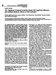
FIG. 5. Alignment of the R- and C-subunits of CAMP-dependent protein kinase with cGMP-dependent protein kinase. The primary region of contact between the two protomers in the R-subunits and in cGMPdependent protein kinase are indicated as a solidregion. The tandem cyclic nucleotide-binding domains are indicated by cross-hatching.Cysteine residues that are associated with interchain disulfide bonds are indicated as solid circles, whereas open circles indicate reduced sulfhydryl groups. The a-NH2groups of each protein are either acetylated (Ac) or myristylated (Myr) as indicated. The known phosphorylation sites in the C-subunit and the autophosphorylation site in R" also are indicated (E') as well as the conserved Lys (M that is part of the ATPbinding site.
The type I1 regulatory subunit shows extensive sequence homologies with the type I regulatory subunit (Takio et al., 1984a; Titani et al., 1984); however, most of these homologies are localized inthe two tandem CAMP-binding domains. Although the proteins are approximately the same size, the sequence homologies in the NH,-terminal portion are not significant even though this region contains the proteolytically sensitive hinge region and an essential recognition site for the C-subunit in both proteins. The type I1 regulatory subunit does not contain intersubunitdisulfide bonds and, in fact, does not even contain any cysteine residues in the aminoterminal portion of the molecule. Nevertheless, limited proteolysis has established that themajor contacts between the two protomers of the dimer are contained within the first 45amino acid residues since cleavage of the chain atresidue 45 yields a monomeric (42,000 dalton) CAMP-binding fragment (Reimann, 1986). A homologous example of interchain disulfide bonding in a purified protein is found in cGMP-dependent protein kinase. This cGMP-dependent protein kinase has a frictional coeffilike itsCAMP cient ( f l f o ) of 1.42 confirmingthatittoo, counterpart shows a high degree of asymmetry (Gill et al., 1977). In addition, Gill et al. (1977) demonstrated that this kinase also existedas a dimer with both protomers covalently cross-linked by interchain disulfide bonds. As in the case of R', the purified cGMP-dependent protein kinase was completely cross-linked despite the presence of 2-mercaptoethanol in all buffers throughout the purification. cGMP-dependent proteinkinase is homologous toboththe regulatory and catalytic subunits of CAMP-dependent protein kinase; however, it is a chimeric protein where the two domainsare covalently joined as part of a contiguous polypeptide chain (Takio et al., 1984b). The regulatory domain is localized in the amino-terminal portionof the protein andis followed by the catalytic domain. This relationship of the cGMP-dependR and C subunits of CAMP-dependent protein kinase to the ent protein kinase is summarized Fig.in5. Extensive sequence homologies are found throughout the catalytic domain and in the cyclic nucleotide-binding domains.Only the small region preceding the CAMP-binding site lacks sequence homology and is significantly shorter than the correspondingregion of both R-subunits. Nevertheless, limited proteolysis has established that the interchain disulfide bond is localized in this
region as itis in R' (Monken andGill, 1985). Since the cGMPdependent protein kinase containsa single Cys in this NH2terminal segment, this must represent a specific contact point between the two protomers in thisenzyme. In summary, allof these homologous proteins, R', R", and the cGMP-dependent protein kinase are asymmetric dimeric proteins which are retained in their dimeric state by either covalent or noncovalent interactions that are restricted toa relatively small segmentlocalized at the amino-terminal portion of the polypeptide chain. Although these proteins do not share sequence homologies in this portion of the molecule, this short segmentat the amino terminus apparently serves a common functionandis responsible formaintainingthe dimeric aggregation state in eachcase. REFERENCES Calissano, P., Mercanti, D., and Levi, A. (1976)Eur. J. Biochem. 71.45-52 Corbin, J. D.,Sugden, P. H., West, L., Flockhart, D. A,, Lincoln, T. M., and McCarthy, D. (1978)J. Biol. Chem. 253,3997-4003 Crestfield, A. M., Moore, S., Stem, W. H. (1963)J. Bud. Chem. 238, 622-627 Erlichman, J., Rubin, C. S., and Rosen, 0.M. (1973)J. Brol. Chem. 248,76077609 First, E. A., and Taylor, S. S. (1984)J. Biol. Chem. 259, 401174014 Flashner, M., Hollenberg, P. F., and Coon, M. J. (1972)J. E d . Chem. 247, 8114-8121 Gill, G. N., Walton, G. M., and Sperry, P. J. (1977)J. Biol. Chem. 252,64436449 Hartley, B. S. (1970)Biochem. J. 119,805-822 Hunkapillar, M. W., and Hood, L. E. (1983)Methods Enzymol. 91,486-493 Kosower, E. M., Pazhenchevsky, B., and Hershkowitz, E. (1978)J. Am. Chem. SOC. 100,6516-6518 Lehrer, S. S. (1975)Proc. Natl. Acad. Sci. U. S. A. 72, 3377-3381 Monken, C. E., and Gill, G. H. (1985)Arch. Biochem. Bwphys. 240,888-903 Potter, R. L., and Taylor, S. S. (1978)Arch. Biochem. Biophys. 90, 174-180 Potter, R. L., and Taylor, S. S. (1979)J. Biol. Chem. 254,2413-2418 Reimann, E. M. (1986)Biochemistry 25, 119-125 Saraswat. L. D.. Filutowics., M.., and Tavlor. ~. S. S. (1986) . . J. Biol. Chem. 261. 11091-iios6' Takio, K., Smith, S. B., Krebs, E. G., Walsh, K. A., and Titani, K. (1984a) Biochemistry 23,4200-4206 Takin. R. -D.. Smith. S. B.. Krebs. E. G.. Walsh. K. A.. and Titani. - ...... K.. Wade. ~. K. (&b) Biochemis~ry231.4207-4218 ' Takio K. Walsh K. A. Neurath, H. A,, Smith, S. B., Krebs, E. A,, and Titani, K. (19SO)FEBb Lett.' 114,8348 Taylor, S. S., and Stafford, P.H. (1978)J. Biol. Chem. 253, 2284-2287 Titani, K., Sasagawa, T., Ericsson, L. H., Kumar, S., Smith, S. B., Krebs, E. G., and Walsh, K. A. (1984)Biochemistry 23,4193-4199 Weber, I. T., and Hilz, H. (1979)Biochem. Bzophys.Res. Comm.,9O,1073-1081 Weber, I. T., Steitz,T. A,, Buhis, J., and Taylor, S. S. (1987)Blochemutry 26, 343-351 Weber, I. T., Takio, K., Titani, K., and Steitz, T. A. (1982)Proc. Natl. Acad. Sci. U. S. A. 79, 7679-7683 Weldon S. L. and Taylor S. S. (1985)J. Biol. Chem. 260,4203-4209 William;. C. H. (1976)in 'he Enzymes (Borer, P. D., ed) Vol. 113,pp. 89-173, Academic Press, New York Zick, S. K., and Taylor, S. S. (1982)J. Biol. Chem. 267,2287-2293 Zoller, M. J., K e r b a g e , A. R., and Taylor, S. S. (1979)J. Biol. Chem. 254, 240&2412