ACE angiotensin-converting enzyme. Ang-1 angiopoietin-1. AP1 activator protein 1 ... real-time microvisualization .....
Application of Collagen Matrices for Enhancing Cardiac Regeneration
Ali Ahmadi
Thesis submitted to the Faculty of Graduate and Postdoctoral Studies in partial fulfillment of the requirements for the Doctorate in Philosophy degree in Cellular and Molecular Medicine
Department of Cellular and Molecular Medicine Faculty of Medicine University of Ottawa
© Ali Ahmadi, Ottawa, Canada, 2014
Abstract Injectable biomaterials have emerged as a treatment for myocardial infarction (MI). They can be applied either as an enhancement for cell therapy or as a stand-alone treatment for MI. The main focus of this study was to apply circulating angiogenic cells (CACs) with or without an injectable collagen matrix for MI treatment in a mouse model. Furthermore, a collagen-chitosan matrix was tested for modulating the myocardial maladaptive remodeling post-MI. First, the in vivo thermo-gelling and retention properties of the collagen matrix were validated using positron emission tomography (PET) tracer and quantum dot (Qdot) labelled matrix in MI mouse hearts. The therapeutic potential of the matrix ± CACs was then tested in a mouse MI model. The results showed that CACs-only and matrix-only treatments were associated with cardiac function preservation. However, in combination, CAC + matrix therapy had a synergistic effect and significantly improved cardiac function (echocardiography), perfusion and viability (PET scan), increased cell engraftment and arteriole density, and reduced the infarct size. CAC-matrix interaction through the integrin 2 receptor was essential for the observed therapeutic effect. In a third study, the addition of chitosan (a polysaccharide) to the collagen matrix was shown to reduce maladaptive remodeling post-MI by limiting cardiac fibroblast-to-myofibroblast differentiation and scar formation. In conclusion, these collagen-based hydrogels hold promise to enhance cardiac repair as a delivery scaffold for therapeutic cells, and/or as a stand-alone treatment, which can actively modulate the environment including the fibrotic process after MI.
ii
Acknowledgments I would like to express my gratitude to my supervisor Dr. Erik Suuronen for giving me the opportunity to work in his lab and for providing excellent guidance and support over years. I also thank my co-supervisor Dr. Marc Ruel for his great help and support during my PhD. I would like to thank the members of my thesis advisory committee, Dr. Darryl Davis, and Dr. Maxwell Hincke for their input and advice. During my PhD, I have had the opportunity to work alongside some great people who not only helped me along the way, but also provided a very productive and pleasant working environment for all trainees including myself. Many thanks to Branka Vulesevic for her kind help and advice. I would like to thank Dr. Rob deKemp, Dr. Jean DaSilva and Dr. Rob Beanlands for their great advice and priceless help throughout my projects. Also, I would like to thank Suzanne Crowe, Brian McNeill, Rick Seymour, Joanne McBane, Donna Padavan and Eva Mathieu who helped me with many, many things. I would like to mention our Molecular Function and Imaging Program collaborators, Animal Care and Veterinary Services staff and also Drew Kuraitis, Mary Zhang, Pingchuan Zhang, Chao Deng, Tanja Sofrenovic and Celine Giordano. I would like to thank my family for their support over the years. I could not have done it without them.
iii
Table of Content ABSTRACT …………………………………………………………………………………..…ii ACKNOWLEDGMENTS ……………………………………………………………………...iii LIST OF FIGURES …………………………………………………………………………….ix LIST OF TABLES ……………………………………………………………………………...xi ABBREVIATIONS …………………………………………………………………………….xii CHAPTER 1: INTRODUCTION ………………………………………………………………1 1.1 Structure of the Heart ………………...……………………………………………….............2 1.2 Coronary Artery Disease: Myocardial Infarction and Remodeling ………………………......4 1.3 Conventional Treatment Strategies for Heart Failure …………………...……………………7 1.4 Clinical Challenge of Heart Failure ……………………………………………………...…...7 1.5 Endogenous Myocardial Regeneration in Humans …………………………………………..8 1.6 Role of Endogenous Stem/Progenitor Cells in Cardiac Regeneration ……….……..…….....9 1.7 Cell Therapy for the Infarcted Myocardium ………………………………………………...10 1.7.1 Stem Cells Applied in Clinical Trials ……………………………….…………….11 1.7.2 Endogenous Mobilization of Stem Cells ………………………………………….12 1.7.3 CAC Therapy ……………………………………………………………………...13
iv
1.8 Characterization of CACs …………………………………………………………………...14 1.9 Clinical Trials with CACs …………………………………………………………………...16 1.10 Limitations of Cell Therapy ………………………………………………………………..18 1.11 Biomaterial Enhancement Strategies for CAC Therapy …………………………………...18 1.12 Injectable Biomaterials ...…………………………………………………………………..21 1.12.1 General Considerations of Injectable Biomaterials ……………………...............21 1.12.2 Injectable Biomaterials as a Scaffold for CAC Transplantation ……....................23 1.12.3 Injectable Biomaterials as a Stand-alone Therapy …………………………….…24 1.13 ECM in Normal and Remodeling Heart …………………………………………………...25 1.13.1 Normal ECM Structure …………………………………………………………..25 1.13.2 Integrin Receptors …………………………………………………......................26 1.13.3 Role of Itgs in ECM and CAC Cross-Talk …………………………….…….......26 1.13.4 ECM Alterations after Infarction ……………………………………….………..28 1.14 Cardiac Fibroblasts: Key components of Cardiac Remodeling ………………………........30 1.14.1 Role of Cardiac Fibroblasts in Post-MI Repair ………………………….……….30 1.14.2 Cardiac Fibroblasts as a Therapeutic Target after Infarction ……………….........30 1.14.3 Applying Biomaterials to Target Fibroblasts ……………………………….........31
v
1.15 Summary …………………………………………………………………………………...31 1.16 Research Plan …………………………………………………………………………........32 1.16.1 Aims ……………………………………………………………………………...32 1.16.2 Hypotheses ……………………………………………………….……………....33 1.17 Role in Research ………………………………………………………...............................33 Chapter 2: PET Imaging Reveals Effective Injection and Targeted Retention of a Collagen Matrix in a Mouse Model of Myocardial Infarction ………………………………………..34 2.1 Notes on Chapter ………………………………………………………………………........35 2.2 Contributions of Co-authors ………………………………………………………………..36 2.3 Abstract ……………………………………………………………………………………..37 2.4 Introduction …………………………………………………………………………………38 2.5 Methods ……………………………………………………………………………………..40 2.6 Results ………………………………………………………………………………….........44 2.7 Discussion …………………………………………………………………………………...55 2.8 Supplementary Section ……………………………………………………………………...60 Chapter 3: The Role of Integrin α2 in Cell and Matrix Therapy that Improves Perfusion, Viability and Function of Infarcted Myocardium …………………………………………...63 3.1 Notes on Chapter …………………………………………………………………………….64 vi
3.2 Contributions of Co-authors ………………………………………………………………...65 3.3 Abstract………………………………………………………………………………………66 3.4 Introduction …………………………………………………………………………………66 3.5 Materials and methods ………………………………………………………………………68 3.6 Results ………………………………………………………………………………………73 3.7 Discussion …………………………………………………………………………………..90 3.8 Conclusion …………………………………………………………………………………..92 3.9 Supplementary Section ……………………………………………………………………...94 Chapter 4: A Collagen-Chitosan Injectable Hydrogel Improves Cardiac Remodeling in a Mouse Model of Myocardial Infarction ………………………………………........................98 4.1 Notes on Chapter …………………………………………………………..………………...99 4.2 Contribution of Co-authors ………………………………………………………………...100 4.3 Abstract …………………………………………………………………………………….101 4.4 Introduction ………………………………………………………………………………...103 4.5 Methods …………………………………………………………………………………….104 4.6 Results ……………………………………………………………………………………...108 4.7 Discussion ………………………………………………………………….........................123 Chapter 5: General Discussion ………………………………………………………………126 vii
5.1 Minimally Invasive Collagen Matrix Delivery ………………………………………….....127 5.2 Collagen Matrix as Enhancement Strategy for CAC Therapy ……………………………..128 5.3 Optimum Timing of Intervention after MI ………………………………………………...130 5.4 Collagen-Based Hydrogels as Cell Therapy Enhancement Strategy or Stand-alone Approach ……………………………………………………………………………………......................131 5.5 Future Directions …………………………………………………………………………..133 References ……………………………………………………………………………………..140 Appendices …………………………………………………………………………………….179 Appendix A- Methods for the Figures of Chapter 5 …………………………………………...179 Appendix B – Authorizations ………………………………………………………………….180
viii
List of Figures FIGURE 1.1: Cardiac remodeling after MI ………………………………………………….....6 FIGURE 1.2: Biomaterial application strategies for MI ……………………….........................20 FIGURE 1.3: Biomaterial delivery methods …………………………….….………………….22 FIGURE 1.4: Integrin-ILK pathway ….…………………………………….………………....29 FIGURE 2.1: Representative images of PET scans ………………………….………………...47 FIGURE 2.2: PET imaging of matrix retention and distribution properties ….………………..49 FIGURE 2.3: Biodistribution ………………………………………………….……………….51 FIGURE 2.4: Qdot labeling efficiency ………………………………………………………...53 FIGURE 2.5: Evaluation of Qdot-labeled matrix in MI heart …………………………………54 SUPPLEMENTARY FIGURE 2.1: Qdot-collagen matrix reaction scheme …………………62 FIGURE 3.1: Combined CACs+matrix therapy improves the perfusion, glucose uptake, and function of MI mouse hearts ……………………………………………………………………79 FIGURE 3.2: CACs+matrix therapy limits adverse remodeling and improves vascular density and transplanted cell retention …………………………………………………………………..81 FIGURE 3.3: Integrin α2β1 is required for the functional enhancement of CACs on collagen matrix ……………………………………………………………………………………………83
ix
FIGURE 3.4: The synergistic effect of CACs+matrix therapy in MI mouse heart is lost when integrin α2 is blocked in CACs …………………………………………………………………85 FIGURE 3.5: Collagen matrix-enhanced function of CACs is dependent on integrin α5 ……..87 FIGURE 3.6: Collagen matrix-enhanced integrin α5 expression involves Itgα2 signaling and the ERK pathway ……………………………………………………………………………………89 SUPPLEMENTARY FIGURE 3.1: Integrin α2 is required for increased ILK expression in matrix-cultured mouse BM-CACs ………………………………………………………………94 SUPPLEMENTARY FIGURE 3.2: The ability of CACs+matrix therapy to limit adverse remodeling is inhibited when itgα2 is blocked in CACs ………………………………………..95 FIGURE 4.1: Cardiac fibroblast culture ……………………………………………….…112-113 FIGURE 4.2: Left ventricular EF and FS in MI mice injected with different treatments …….114 FIGURE 4.3: Infarct size assessment 1wk and 3wks after treatment delivery ……………….115 FIGURE 4.4: Arteriole density in mouse MI hearts ………………………………………….117 FIGURE 4.5: CD68+ cells in mouse MI hearts ……………………………………………….119 FIGURE 4.6: MMP9 and TIMP2 levels in the treated hearts ………………………………...121 FIGURE 5.1: Ratio of C-kit+ cells in the infarcted myocardium to the entire ventricles …….135 FIGURE 5.2: Circulating serum levels of VEGF and G-CSF in MI mice 3wks after treatment delivery…………………………………………………………………………………………137
x
LIST OF TABLES SUPPLEMENTARY TABLE 3.1: Summary of qPCR primers ………………………………96
xi
Abbreviations 13
NH3
[13N]-ammonia
18
F-FDG
2-[18F]fluoro-2-deoxy-D-glucose
18
F-HFB
hexadecyl-4-[18F]fluorobenzoate
18
F-NaF
18
F- sodium fluoride
ACE
angiotensin-converting enzyme
Ang-1
angiopoietin-1
AP1
activator protein 1
ATP
adenosine 5’-triphosphate
BM
bone marrow
CACs
circulating angiogenic cells
CDCs
cardiosphere-derived cells
CMR
cardiac magnetic resonance
COX
cyclooxygenase
CSCs
cardiac resident stem cells
CXCR4
CXC chemokine receptor type 4
DAPI
4'6-diamidino-2'-phenylindole
xii
EBM-2
endothelial basal medium 2
ECM
extracellular matrix
EDC
ethyl(dimethylaminopropyl) carbodiimide
EF
ejection fraction
ELISA
enzyme-linked immunosorbent assay
eNOS
endothelial nitric oxide synthase
EPCs
endothelial progenitor cells
FACS
fluorescence activated cell sorting
FAK
focal adhesion kinase
FGF
fibroblast growth factor
FISH
fluorescence in situ hybridization
FOV
field-of-view
FS
fractional shortening
G-CSF
granulocyte colony-stimulating factor
GFP
green fluorescent protein
GSK3
glycogen synthase kinase 3
HGF
hepatocyte growth factor
xiii
HIF-1
hypoxia-inducible factor 1
HMG-CoA
5-hydroxy-3-methylglutaryl-coenzyme A
HPS
hematoxylin-phloxine-saffron
HSCs
hematopoietic stem cells
HUVECs
human umbilical vein endothelial cells
IGF
insulin-like growth factor
IL
interleukin
Itg
integrin
ILK
integrin-linked kinase
LDL
low density lipoprotein
LVEF
left ventricular ejection fraction
MBq
millibecquerel
MHC
myosin heavy chain
MI
myocardial infarction
MNCs
mononuclear cells
MMP
matrix metalloproteinase
MSCs
mesenchymal stem cells
xiv
mTOR
mammalian target of rapamycin
NF-B
nuclear factor kappa-light-chain-enhancer of activated B cells
NHS
N-Hydroxysuccinimide
NOS
nitric oxide synthase
PBS
phosphate buffered saline
PCI
percutaneous coronary intervention
PDGF
platelet-derived growth factor
PET
positron emission tomography
Qdots
quantum dots
RMV
real-time microvisualization
ROIs
regions of interest
ROS
reactive oxygen species
SCID
severe combined immunodeficiency
SD
standard deviation
SDF
stromal cell-derived factor
SEM
standard error of the mean
SM
skeletal myoblast
xv
SMA
smooth muscle actin
SPECT
single-photon emission computed tomography
TGF
transforming growth factor
TIMPs
tissue inhibitor of metalloproteinases
VEGF
vascular endothelial growth factor
VEGFR-2
vascular endothelial growth factor receptor 2
vWF
von Willebrand factor
xvi
Chapter 1: General Introduction
1
1.1 Structure of the Heart The heart is a fibromuscular cone-shaped organ situated in the middle thoracic mediastinum (Mahadevan, 2012). Its inferior surface lies on the diaphragm central tendon and its base is adjacent to the esophagus and descending aorta. The left atrium and a part of right atrium constitute the base. The heart’s left and right surfaces lie medial to a lung and a phrenic nerve. The sternum and the costal cartilages protect the anterior surface of the heart (Whitaker, 2010). There are four chambers in the heart: the right and left atria and the right and left ventricles. These are separated by atrioventricular valves: the tricuspid valve on the right and the mitral valve on the left. Each ventricle is separated from its great artery by a semilunar valve with crescent-shaped cusps: the pulmonary valve between the right ventricle and the pulmonary artery and the aortic valve between the left ventricle and the aorta (Katz, 2006). The heart wall is made up of three layers: epicardium, myocardium and endocardium. The epicardium is the visceral layer of the pericardium which is a double-walled enclosing sac around the heart. The outer pericardium is a fibrous protective connective tissue which anchors the heart to the diaphragm and great vessels; and the inner pericardium is a thin serous membrane composed of two layers: the parietal layer (lining the inner surface of the fibrous pericardium) and the visceral pericardium (epicardium). Between the two layers of the visceral pericardium there is a serous fluid that allows the two membranes to glide smoothly (Des Jardins, 2008). The myocardium is the muscular part of the ventricle walls and is made up of overlapping spiral sheets which sweep from the heart base to the apex. The muscle fibers at the outer surface of the left ventricle are arranged parallel to the base-apex axis of the heart. At the inner surface of ventricular myocardium, the muscle fibers are oriented circumferentially (Katz,
2
2006). As a result of this myocardial fiber pattern, when the left ventricle contracts it twists and turns toward the chest wall and to create the apical impulse (Opie, 2004). The oxygenated blood from the lungs flow into the left atrium and subsequently enters the left ventricle as the mitral valve opens. The mitral opening happens only during the left ventricle relaxation phase (diastole) that reduces the left ventricular pressure. During left ventricular contraction (systole), the two mitral valve cusps are forced to close which prevents the blood from flowing back to the left atrium (Opie, 2004). The ventricular cusps are tethered at the margins by thin fibrous structures (chordae tendinae) that attach to long muscular projections of the ventricular inner wall (papillary muscle) (Des Jardins, 2008). As the mitral valve is shut by left ventricular contraction, the aortic valve is forced to open by the increasing ventricular pressure and the blood travels throughout the circulation (Opie, 2004). The myocardium is irrigated by the coronary arteries which arise from the aorta. Sinus of Valsalva is located behind each of the three aortic valve cusps. The anterior and left posterior sinuses give rise to the right and left coronary arteries while no coronary artery originates from the right posterior sinus (Katz, 2006). The left coronary artery divides into the left anterior descending and the circumflex coronary arteries. The former runs on the anterior wall and supplies the apex and the interventricular septum and the latter irrigates the posterior wall of the left ventricle and also the left atrium. The right coronary artery supplies the right atrium and then divides into the posterior descending and marginal arteries (Des Jardins, 2008); in about 10% of human hearts the posterior descending artery originates from the circumflex artery (Katz, 2006). The posterior descending artery supplies the posterior wall of both ventricles and the marginal branch supplies the lateral wall of the heart (Des Jardins, 2008).
3
1.2 Coronary Artery Disease: Myocardial Infarction and Remodeling Atherosclerosis is the most prevalent cause of morbidity and mortality in the developed world. Briefly, formation of the atherosclerotic plaque is initiated by endothelial dysfunction, intimal accumulation of lipoproteins, leukocyte recruitment and accumulation of lipoprotein particles in monocytes (foam cell formation) (Strom, 2011). As the plaque progresses, smooth muscle cells migrate into the intima and extracellular matrix (ECM) synthesis and degradation is altered. Finally, calcification, fibrosis and smooth muscle cell death occur which may be accompanied by hemodynamic stresses leading to plaque disruption and thrombus formation (Libby et al., 2002). The clinical manifestation of coronary artery plaque rupture is an acute cardiac event such a myocardial infarction (MI) (Libby, 2002), which is characterized at the cellular level by myocyte necrosis due to severe impairment of blood flow to the myocardium. The progression of cell death from sustained ischemia often starts in the sub-endocardium (sub-endocardial infarct) (Rhee, 2011) and spreads toward the sub-epicardium in a wave front (transmural infarct) (Opie, 2004, Rhee, 2011). The wave front phenomenon is caused by increasing intraventricular pressure and progressive failure of left ventricle. Therefore, the larger the initial infarct zone is, the larger and faster the wave front is (Opie, 2004). Occlusion of a major coronary artery leads to cell death by apoptosis, necrosis, or a combination of both (Nadal-Ginard et al., 2003). Apoptosis induces acute modification in the myocardial structure and impairment of myocardial force development (Cheng et al., 1995). Necrosis causes inflammation, macrophage infiltration, fibroblast activation, and finally scar formation (NadalGinard et al., 2003). Different mechanisms have been proposed for the irreversible myocardial damage in the context of severe prolonged ischemia: (1) inhibition of the sodium pump; (2) substantial loss in the reservoir of adenosine 5’-triphosphate (ATP); (3) metabolically or 4
mechanically damaged cell membrane; (4) free radical formation; and (5) calcium overload. The amount of tissue which is irreversibly damaged by the severe ischemia is referred to as the infarct size. Therefore, upon occlusion of a major coronary artery, the whole area supplied by that artery is at risk of infarction. In a typical rabbit model, about 60% of the ischemic area will undergo infarction after coronary artery occlusion (Opie, 2004). The irreversible damage of functional myocardial cells quickly leads to impaired contractile function of the ventricle and results in systolic dysfunction, which is characterized by cardiac output decrease and loss of synchronous myocardial contraction. Therefore, the damaged myocardium becomes hypokinetic (reduced contraction), akinetic (no contraction at all) or dyskinetic (bulging out of the infarcted region) during contraction of the remaining functional ventricular tissue. Furthermore, diastolic dysfunction of the left ventricle develops because ventricular diastolic relaxation is an energy dependent process; the heart’s function is adversely affected by reduced ventricular compliance and elevated filling pressures. After MI, cardiac remodeling eventually occurs and the abnormal loading conditions change the geometry of both infarcted and non-infarcted regions (Rhee, 2011). Within days, the infarct area begins to expand and becomes thinner. Within days to months, global remodeling results in overall ventricular dilation, mitral valve dysfunction, augmentation of wall stress, formation of aneurysms and ventricular arrhythmias (Jessup and Brozena, 2003, Rhee, 2011). A recent study using contrast enhanced cardiac magnetic resonance (CMR) imaging showed that adverse cardiac remodeling in remote areas starts as early as 5 days post-MI (Chan et al., 2012). These structural and functional abnormalities ultimately lead to heart failure (Jessup and Brozena, 2003) (Figure 1.1).
5
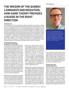
Figure 1.1 Cardiac Remodeling after MI. In the infarcted area of the myocardium, cardiomyocytes undergo cell death (apoptosis and necrosis) and the infarct area expands and becomes thinner within days. The remodeling continues over a period of weeks, which leads to morphologic and physiologic alteration of the entire LV. Reproduced with permission from (Jessup and Brozena, 2003); Copyright Massachusetts Medical Society
6
1.3 Conventional Treatment Strategies for Heart Failure Thrombolysis treatment has been shown to decrease the transmural MI mortality from 11% to less than 7%, if applied within a few hours of the onset of infarction (Bohula, 2012). Mortality and morbidity was even further reduced by the advent of acute percutaneous coronary intervention and also with the addition of anti-platelet agents (e.g. glycoprotein IIb/IIIa inhibitors) (Schomig et al., 2000). Conventional post-discharge therapy for MI patients targets controlling lipids (statins), blood pressure (angiotensin-converting enzyme (ACE) inhibitors), heart rate (beta blocker), and blood coagulation (anti-platelet therapy) (Bohula, 2012). Furthermore, rigorous attention to underlying risk factors like diabetes and smoking is essential (Rhee, 2011). The use of implantable automatic defibrillators has also been shown to further reduce the mortality rate by about 7% over five years (Bardy et al., 2005). Despite conventional treatment strategies, heart failure has increased in prevalence in North America and worldwide (Kannel, 2000, Mendez and Cowie, 2001). This is due to: (1) a significant decrease in mortality results in the survival of patients with a large MI who progress to heart failure; (2) an increase in the number of patients at risk of MI as the population ages; and (3) the development of cardiac risk factors at a younger age (Bohula, 2012). 1.4 Clinical Challenge of Heart Failure In spite of recent advances in conventional therapies for acute and chronic heart failure, the mortality rate remains significant: ~4% in hospital, ~10% in the first two months after discharge, and up to 30% after one year (Jong et al., 2002, Yancy et al., 2006). Although ACE inhibitors are proven to be efficient in reducing the mortality associated with heart failure (Shearer et al.,
7
2013), current therapies can only slow the progression of cardiomyocyte loss, but they fail to reverse the process of cardiac remodeling (Bohula, 2012). It has been shown that a left ventricular ejection fraction of ≤30% is associated with a high risk of sudden cardiac death (Rhee, 2011), which warrants heart transplantation as the definitive treatment (Bohula, 2012). Heart transplantation has a survival rate of 85% at the end of first year and 50% after ten years (Taylor et al., 2008). The major limitation of transplantation is that the demand largely surpasses the donor supply (Langone and Helderman, 2003). 1.5 Endogenous Myocardial Regeneration in Humans Myocardial regeneration has emerged as a potential treatment for heart failure since the myocardium of both non-mammalian and mammalian hearts has shown regenerative capacity (Bohula, 2012). Several studies suggest that the human heart has some degree of cellular turnover: (1) the measurement of carbon-14 (generated by nuclear bomb tests) integrated into human cardiomyocyte DNA has shown that cardiomyocyte renewal occurs at the rate of 1% at the age of 25 and decreases to 0.45% at the age of 75, which suggests a renewal of more than half of the cardiomyocytes during an average life span (Bergmann et al., 2009); (2) in human aortic stenosis, a small level of cardiomyocyte mitosis and division has been demonstrated (Urbanek et al., 2003); (3) in human end-stage heart ischemia the number of mitotic cardiomyocytes increases to 10 times that in the normal heart (Kajstura et al., 1998); and (4) biopsies of patients who underwent heart transplantation demonstrated that in nearly 30% of cardiomyocytes, DNA synthesis and replication occur in the context of heart failure (Beltrami et al., 1997).
8
1.6 Role of Endogenous Stem/Progenitor Cells in Cardiac Regeneration An experimental study used a double transgenic mouse model in which constitutive expression of -galactosidase was replaced by green fluorescent protein (GFP) expression in cardiomyocytes upon tamoxifen treatment. This study showed that MI induced the formation of new cardiomyocytes in the peri-infarct and non-infarcted areas from an immature endogenous myocyte or progenitor cell pool (Hsieh et al., 2007). Results such as these have led to greater research into understanding the potential for endogenous stem/progenitor cells to contribute to cardiac regeneration in response to injury or disease. In the human body, adult progenitor cells that exist in the circulation or within niches in the bone marrow (BM), home to damaged tissues in response to appropriate induction signals (Koyanagi, 2012). Hematopoietic stem cells (HSCs), endothelial progenitor cells (EPCs) and mesenchymal stem cells (MSCs) have all been reported to be mobilized and/or recruited by various signals such as granulocyte-colony stimulating factor (G-CSF) and stromal cell-derived factor-1 (SDF-1) (Koyanagi, 2012). The contribution of marrow-derived circulating cells to cardiomyocytes and endothelium formation was shown in a study of male recipients of female BM allograft by applying dual color fluorescence in situ hybridization (FISH) (X and Y chromosome-specific probes) (Thiele et al., 2004). A similar study indicated that in male recipients of female heart transplantation, the endothelial cells and vascular smooth muscle cells show a high degree of chimerism (24.3±8.2% and 11.2±2.1%, respectively) from extracardiac sources (Minami et al., 2005). Similarly, in another study, Y chromosome positive nuclei were detected in post-mortem transplanted hearts of male patients who had heart transplantation from female donors. This chimeric study
9
concluded that 18% of cardiomyocytes, 20% of coronary arterioles and 14% of capillaries originated from male cells, indicating recruitment from the male host into the female donor heart (Quaini et al., 2002). These findings have been challenged by a more recent study using HSCs carrying a fusion gene consisting of the -myosin heavy chain (MHC) promoter and a modified -galactosidase gene. The cells were injected to the peri-infarct myocardium in mice 5h after MI surgery. Tracking of the transplanted cells up to 28 days post-MI showed no cardiomyocyte transdifferentiation of HSCs (Murry et al., 2004). Although marrow-derived progenitor cells may not transdifferentiate to cardiomyocytes after MI, it is suggested that their main therapeutic effect is through the release of growth factors and induction of angiogenesis (Balsam and Robbins, 2005). 1.7 Cell Therapy for the Infarcted Myocardium Although the body mounts an endogenous regenerative response, it is insufficient to prevent progression of heart disease. Therefore, regenerative therapies are an attractive approach to restore tissues and cardiac function. Multiple clinical and experimental studies have focused on cell therapy for improvement of myocardial regeneration after infarction (Bohula, 2012). In this context, there are three main therapeutic aspects to be addressed: (1) the best cell or cardiac tissue to be targeted; (2) the most efficient method of stimulating regeneration; (3) the optimum timing for intervention (Kuraitis et al., 2010). Presumably, the best cardiac regeneration strategy involves the replacement of the lost cardiomyocytes (Bohula, 2012). However, experimental and clinical studies have also highlighted the importance of angiogenesis in the inadequately perfused myocardium (Suuronen et al., 2007, Erbs et al., 2007, Kawamoto et al., 2003).
10
1.7.1 Stem Cells Applied in Clinical Trials Different cell types including BM-derived HSCs, MSCs, resident cardiac stem cells (CSCs), skeletal myoblasts (SMs) and circulating angiogenic cells (CACs) have been tested and shown to improve heart function in humans (Segers and Lee, 2008). Undifferentiated marrow-derived mononuclear cells (MNCs) have been shown to improve left ventricular ejection fraction (LVEF) (Seeger et al., 2007). CD133+ cells, which constitute a primitive subtype of MNCs, were applied in a non-randomized clinical trial, and improved LVEF and enhanced myocardial glucose uptake were observed (Bartunek et al., 2005). MSCs are stromal cells which can be isolated from bone marrow and adipose tissue and have the capacity to exert paracrine effects to suppress inflammation and apoptosis (Amado et al., 2005). Several clinical studies are investigating the safety and feasibility of MSC delivery in MI patients. Current published data has indicated that a high dose (6×10¹⁰) but not a low dose (5×10⁶) MSC injection is associated with a significant improvement in LVEF (Tongers et al., 2011). CSCs, which are lineage- ckit+, have shown a capacity to generate cardiomyocytes (Beltrami et al., 2003). Since the total number of CSCs in the heart is low, CSCs have been expanded ex vivo from endomyocardial biopsies (Bearzi et al., 2007). The Marban group has identified a CSC population that can be isolated from self-adherent clusters (cardiosphere-derived stem cells (CDCs)), and which show cardiomyogenic potential (Davis et al., 2010). A recent randomized controlled trial of CDC therapy in 17 MI patients has showed promising results in terms of decreased scar tissue and increased viable myocardium (Malliaras et al., 2014). SMs are isolated from muscle biopsies, and have been expanded ex vivo and delivered to the infarcted heart (Murry et al., 1996). The most comprehensive SM Phase II clinical trial has indicated no improvement in LVEF after 6 months and even a significant LVEF deterioration in the high dose group (Menasche et al., 2008). Since the myoblast injection has
11
also been associated with increased arrhythmic events in patients (Menasche et al., 2003), SM application for MI patients is controversial (Tongers et al., 2011). The characterization and application of CACs, which are a heterogeneous population of angiogenic cells, will be discussed later in this chapter. These results indicate, for the most part, that cell therapy can be safe and effective at improving cardiac function in patients; however, improvements are still needed to further enhance the degree to which cell therapies can promote repair and regeneration. 1.7.2 Endogenous Mobilization of Stem Cells In addition to cell transplantation, strategies are being developed to attract greater numbers of endogenous stem cells to the injured or diseased tissue. G-CSF is the major mobilizing agent that has been used in clinical trials. One study showed a modest improvement in LVEF, wall motion and infarct zone wall thickness in patients that received G-CSF for 6 consecutive days following angioplasty. This effect was attributed to higher mobilization of CD34+ MNC into the systemic circulation (Ince et al., 2005). In contrast, a meta-analysis conducted in 2008, indicated no therapeutic benefit associated with G-CSF therapy in the context of MI (Zohlnhofer et al., 2008). Yet, a more recent meta-analysis on the clinical outcomes of G-CSF treatment indicated nonsignificant trends towards LVEF improvements in 3-6 months after injection (Zimmet et al., 2012). Another potential approach is to target the SDF-1 pathway, which is known to enhance progenitor cell trafficking. It has been shown that recruitment of angiogenic cells via the SDF-1 signaling pathway is required for myocardial regeneration; competitive inhibition of SDF-1 by a mutant form of the chemokine, delivered on a lentiviral plasmid, was associated with a decreased regenerative response and reduced cardiomyocyte repopulation in a fetal sheep model of MI (Allukian et al., 2013). In a recent clinical study, a non-viral DNA plasmid encoding human 12
SDF-1 was delivered to 17 ischemic cardiomyopathy patients via endomyocardial injection. Four months after treatment, the patients’ 6-minute walk distance and quality of life were reported to be improved. These therapeutic benefits persisted at 12 months post-treatment (Penn et al., 2013). Adult stem cells secrete a multitude of growth factors, chemokines, and enzymes that not only play an important role in different stages of tissue regeneration, angiogenesis and cardiac remodeling, but also recruit more progenitor cells to the ischemic site (Gnecchi et al., 2008). In fact, paracrine signaling is believed to be the main mechanism by which transplanted cells exert their positive effects on the MI heart, rather than direct tissue generation/replacement (Cho et al., 2007). In summary, the recruitment of endogenous progenitor cells constitutes an essential element of myocardial regeneration. The types of cells contributing to regeneration may vary depending on the pathophysiologic state of the heart. In the normal heart, resident CSCs are responsible for myocardial turnover. In the infarcted myocardium, a regenerative response arises from CSCs and circulating endogenous progenitor cells which are recruited to the damaged tissue (Malliaras et al., 2013). 1.7.3 CAC Therapy In this context, CACs have emerged as a promising cell source for myocardial repair and regeneration. They release growth factors with multiple positive effects including cardiomyocyte survival, neovascularization, the prevention of adverse remodeling, and the induction of endogenous progenitor cell mobilization (Shintani et al., 2001, Takahashi et al., 1999, Jujo et al., 2008). Furthermore, there is evidence that CACs have the potential for differentiation into endothelium, therefore directly contributing to new tissue formation (Cho et al., 2003). The
13
safety of CAC therapy has been demonstrated by multiple clinical trials (Tongers et al., 2011), and will be discussed in detail later in this chapter. 1.8 Characterization of CACs Endothelial precursors, positive both for hematopoietic stem cell marker (CD34) and endothelial markers (vascular endothelial growth factor receptor-2 (VEGF-R2)), were first isolated from human peripheral blood in 1997 by Asahara et al. The isolated CD34+ cells (first termed endothelial progenitor cells (EPCs)) showed potential for endothelial differentiation in vitro and also incorporation to newly formed blood vessels in vivo (Asahara et al., 1997). Several studies to identify putative markers for human EPCs have been conducted using hematopoietic marker CD133 (Gehling et al., 2000, Shi et al., 1998). EPCs are often characterized as CD133+ and VEGF-R2+ cells, which in the presence of appropriate growth factors, can differentiate to endothelial cells which express CD34 and von Willebrand factor (vWF) markers and incorporate acetylated low-density lipoprotein (LDL)(Shi et al., 1998). However, a study demonstrated that CD133+CD34+VEGF-R2+ cells isolated from umbilical cord or from peripheral blood after GCSF stimulation, do not differentiate into endothelial cells in vitro (Case et al., 2007). Concurrently, another group demonstrated that only CD34+CD45- cells and not the CD34+CD45+ cells show the potential to form endothelial cell colonies (Timmermans et al., 2007). Another study indicated that CD133+CD34-VEGR-2+ cells are precursors for CD133+CD34+VEGFR-2+ cells, but the CD34- cells show a higher potency with respect to homing and vascular repair in patients with limb ischemia and also in an artery injury mouse model (Friedrich et al., 2006). These findings are not consistent in identifying EPC specific markers. The complexity of EPC characterization is, to some degree, attributable to differences between in vitro culture conditions
14
and in vivo environments; the growth factors and chemokines secreted in the damaged tissue can alter cell differentiation and function in a fashion which is not reproducible in vitro. The transplanted cells, on the other hand, interact with other cells of the tissue by paracrine mechanisms which further adds to the complexity of in vivo environment (Koyanagi, 2012). These are some of the reasons why pro-angiogenic cells obtained from the culture of peripheral blood mononuclear cells are commonly referred to as circulating angiogenic cells (CACs), to reflect their heterogeneity and function, rather than their status as true endothelial precursors. Typically, CACs are generated from the culture of bone marrow-derived mononuclear cells (from BM or peripheral circulation) on fibronectin; the adherent cells after 4-7 days are referred to as early CACs. These CACs express endothelial markers (e.g. VEFG-R2, vWF, and CD31) (Kalka et al., 2000) and myeloid markers (e.g. CD45 and CD14). After 14 days of culture, late CAC cultures form large colonies on the plate. These cells express a higher level of VEGFR-2 and demonstrate a higher potential to incorporate in the capillaries; however, late CACs secretes less cytokines compared to early CACs (Koyanagi, 2012). The characterization and identification of CACs has been controversial. None of the current identifying and quantifying methods has shown a reliable capacity to predict the in vivo behavior of the cells. Moreover, it is unknown whether the culture-modified cells only represent an artificial phenotype or they naturally exist in the circulation or BM (Fadini et al., 2012). Overall, the main difference between the types of CACs is the culture time. The short-term protocols (4-7 days) yield myeloid/hematopoietic cells and the long-term protocols (more than 14 days) give a more homogenous population of CACs with a reduced cytokine release profile (Koyanagi, 2012). The in vivo capacity of CACs to enhance angiogenesis differs with respect to their culture protocol. It has been shown that short-term culture CACs mainly enhance tissue perfusion by
15
providing potent growth factors that promote angiogenesis (Di Santo et al., 2009, Urbich et al., 2005, Rehman et al., 2003). The cells gained by long-term culture contribute to the structure of new blood vessels by differentiating to endothelial cells (Yoon et al., 2005b, Hur et al., 2004). Despite the existence of different methods for CAC isolation and culture, defining CACs has been challenging because of the following reasons: 1) although it has been shown that a variable percentage of endothelial cells is derived from BM (Gunsilius et al., 2000), a common precursor for marrow-derived blood cells and endothelial cells has not been identified (Fadini et al., 2012); 2) growing evidence indicates a new concept that mature endothelial cells can dedifferentiate and enter the circulation with an overlapping endothelial-hematopoietic phenotype (e.g. CD34 expression) in case of tissue injury (Chao and Hirschi, 2010); and 3) many cell surface markers are expressed by both hematopoietic and endothelial cells which complicates the determination of lineage difference between the two (Fadini et al., 2012). 1.9 Clinical Trials with CACs Several clinical studies have demonstrated the efficacy and feasibility of CAC therapy. Autologous CD133+ cell injection in combination with bypass surgery resulted in improved cardiac function and perfusion in a group of myocardial ischemia patients (Stamm et al., 2003). Another clinical study has tested the therapeutic benefit of autologous CD133+ cell implantation in patients who were candidates for limb amputation due to critical limb ischemia. This treatment improved treadmill walking time and prevented leg amputation (Burt et al., 2010). A nonrandomized clinical trial (TOPCARE-AMI trial) indicated improved LV function in patients treated with a heterogeneous population of CACs (Schachinger et al., 2004). In the REPAIRAMI randomized trial, intracoronary injection of marrow-derived progenitor cells improved LV function significantly (Schachinger et al., 2006). In this context, not only CACs, but also 16
marrow-derived MNCs have shown the capacity for moderate but significant improvement of LV function (Assmus et al., 2006). Moreover, the efficacy of marrow-derived MNCs for moderately increasing LV function (~3%) and slightly improving the quality of life, has been shown in a randomized double-blind clinical trial (van Ramshorst et al., 2009). In the BOOST trial, intracoronary delivery of unselected marrow-derived progenitor cells after percutaneous coronary intervention (PCI) was associated with a transient increase in LV function at 6 month; however, this effect did not persist until month 18 post-injection (Meyer et al., 2006). A phase I double-blind randomized controlled trial was conducted using CACs in patients with severe inoperable coronary heart disease. The autologous CACs were collected from peripheral blood MNCs and injected directly to the ischemic myocardium using an electromechanical mapping system. This treatment was associated with improved clinical parameters such as reduced angina frequency and increased exercise duration and overall showed a trend favoring the CAC therapy (Losordo et al., 2007, Losordo et al., 2011). Overall, clinical studies reported an enhanced recovery in cardiac function after CAC therapy (Dimmeler et al., 2008). Despite these promising results, the long-term therapeutic effects of CAC therapy is not elucidated yet (Koyanagi, 2012); it has been suggested that the cell therapy benefit may be sustained for at least two years following treatment delivery (Assmus et al., 2010). However, the effect of CAC therapy is variable due to different subtypes of CACs used in clinical trials and also because the cells have been applied at different stages of myocardial ischemia (Koyanagi, 2012). Several clinical trials are underway to further study the prospect of cell therapy in patients with different types of ischemic disease, including coronary artery disease. As discussed before, the most important issue to be addressed in these second generation trials, is to maximize sustainable therapeutic benefits for the patients (Wollert and Drexler, 2010b). Given the variation in clinical outcomes
17
with similar cell isolation protocols in previous trials (Hirsch et al., 2008, Huikuri et al., 2008, Lunde et al., 2006, Schachinger et al., 2006), it is essential to establish a standard protocol for cell isolation, characterization and delivery to the target tissue (Wollert and Drexler, 2010b). This is also confirmed by a recent meta-analysis on marrow-derived stem cells indicating that clinical trials with more discrepancies in patient selection and study design yield larger variances in results compared to studies with fewer discrepancies (Nowbar et al., 2014). 1.10 Limitations of Cell Therapy Cell therapy is mainly limited by poor cell engraftment and survival after delivery, regardless of the cell type used (Wollert and Drexler, 2010b). Moreover, co-morbidities (e.g. diabetes), advanced patient age and heart failure have a negative impact on the functional activity of marrow-derived progenitor cells, including CACs. The cells isolated from patients with diabetes and heart failure demonstrate impaired activity in repairing denuded arteries when transplanted into mice (Dimmeler and Leri, 2008), which is associated with reduced in vitro functionality such as decreased colony formation and impaired migration capacity (Assmus et al., 2007). Cell therapy enhancement strategies are, therefore, required to augment the therapeutic potential of progenitor cells (Wollert and Drexler, 2010b). 1.11 Biomaterial Enhancement Strategies for CAC Therapy Strategies for enhancing the efficacy of transplanted progenitor cells are needed in order to overcome the challenges posed by the hostile environment of the infarcted myocardium, which include: (1) ischemia and reduced perfusion of the infarcted area; (2) inflammatory response due to oxidative stress and cytotoxic cytokines and (3) loss of normal ECM (Wollert and Drexler, 2010b). These conditions reduce cell engraftment and function, thus limiting the therapeutic 18
potential of cell transplantation. The use of biomaterials presents an opportunity to address all of these limitations. Biomaterials are being designed to improve the host environment into which the cells are being transplanted, to enhance the therapeutic potential of the transplanted cells, and to increase the engraftment of the transplanted cells. Theoretically, an ideal biomaterial will improve the secretion of growth factors and cytokines from the cells, promote their engraftment and survival and guide their phenotype and function (Wollert and Drexler, 2010b). Several different biomaterial approaches have been tested for cell delivery in the context of MI, including: (1) transplantable cell sheets generated in vitro from cells that have been stimulated to produce their own ECM; (2) culturing cells on a scaffold in vitro and then suturing the cultured tissue on the epicardial surface of the heart; (3) decellularized cardiac ECM; and 4) injectable biomaterials as delivery scaffolds (Christman and Lee, 2006) (Figure 1.2).
19
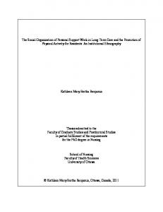
Figure 1.2 Biomaterial application strategies for MI. (A) A biocompatible mesh can be fixed around the infarted heart to prevent LV dilation. (B) Progenitor cells are cultured on biomaterials to form a tissue in vitro. The tissue is grafted on the epicardial surface of the heart. (C) Progenitor cells are delivered within an injectable matrix. (D) Injectable biomaterial can be used as a stand-alone therapy for MI. (E) Injectable scaffold have also shown the capacity to act as a vehicle for growth factors and/or chemokine delivery. Reprinted with permission from (Christman and Lee, 2006); Permission from Elsevier
20
1.12 Injectable Biomaterials 1.12.1 General Considerations of Injectable Biomaterials The injectable biomaterial approach is less invasive than implanting a polymer mesh or an epicardial patch and is therefore more favorable for clinical application (Christman and Lee, 2006); injectable materials have the potential to be delivered via catheter. This also allows for direct treatment delivery to the damaged myocardium. Injectable biomaterials are mainly hydrogels, which are made of cross-linked polymer networks from natural or synthetic sources. Within the body, the hydrogel is solidified by a variety of ways depending on how the hydrogel is cross-linked and also its chemical makeup. Hydrogel cross-linking may be achieved by covalent bonds, physical entanglements or ionic interactions which can respond to triggers like pH or temperature (Radisic and Christman, 2013). Possible non-invasive methods for hydrogel delivery are intracoronary infusion (Leor et al., 2009) and transendocardial injection (Seif-Naraghi et al., 2013) (Figure1.3). The disadvantages of both methods are: (1) risk of leakage to the blood stream at the time of injection; (2) risk of solidification due to exposure to the body heat during the time the hydrogel flows through the catheter to its target area at the infarcted myocardium. Therefore, a very quick gelling injectable material may not be compatible with these two delivery methods and also the challenge of being hemocompatible to reduce the risk of emboli is a major concern. The preferable biomaterial delivery method is via direct myocardial injection that reduces the exposure of injected material to the circulation (Johnson and Christman, 2013). Finally, the depth and quantity of injected material must be carefully monitored to minimize the risk of cardiac arrhythmia after injection (Radisic and Christman, 2013).
21
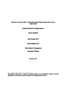
Figure 1.3 Biomaterial delivery methods. Injectable biomaterials can be injected in experimental models via 3 routes: 1) transendocardial injection; 2) intracoronary delivery by using an inflated percutaneous transluminal coronary balloon; and 3) direct injection to the infarcted myocardium. Reprinted from (Stamm et al., 2009); Permission from Elsevier
22
1.12.2 Injectable Biomaterials as a Scaffold for CAC Transplantation Many materials have been tested for the delivery of multiple cell types (Radisic and Christman, 2013). Of particular interest to the present work are biomaterials for CAC therapy. The injection of CACs with a fibrin matrix (used commercially as a sealant) resulted in improvement of cardiac function and angiogenesis (Chekanov et al., 2003). Another study investigated CAC delivery within a thermo-gelling collagen matrix in a rat ischemic hindlimb model; results showed that the CACs (labeled with a positron emission tomography (PET) tracer) were mostly retained at the injection site in the hindlimb, whereas the cell-only injection showed a poor cell retention after 4hrs (Zhang et al., 2008b). The injection of a collagen-based matrix to the ischemic hindlimb of rats increased arteriole density, and this effect was further improved by adding CD133+ cells to the collagen matrix. The same study also demonstrated the positive effect of the collagen matrix on the retention of transplanted cells in the target tissue compared to the cell-only group (Suuronen et al., 2006). CAC delivery within a macroporous alginate scaffold has been shown to be associated with augmented blood vessel density, improved perfusion and prevention of foot necrosis in a severe combined immunodeficiency (SCID) mouse model of hindlimb ischemia (Silva et al., 2008). Kim et al. have developed a biosynthetic material as a scaffold for CAC therapy in a murine dermal injury model. The scaffold increased the survival and retention of injected cells and improved their vascular repair potential (Kim et al., 2009). An injectable polylactic coglycolic acid-based injectable matrix has also been used as a vehicle for delivering CACs and angiogenic growth factors (VEGF, hepatocyte growth factor (HGF) and angiopoietin-1 (Ang-1)). The therapeutic benefits of CAC delivery in the scaffold (enhanced incorporation to blood vessels and higher capillary density) is improved when the pro-angiogenic growth factors are also delivered (Saif et al., 2010). In the same animal model, co-injection of
23
MSCs and EPCs within a thermo-gelling engineered peptide hydrogel (PuraMatrix) show synergistic effects in promoting angiogenesis compared to either element injected alone (Allen et al., 2011). The focus of another study was the fate of EPCs after injection within an injectable fibrin matrix; the cells were transfected with -galactosidase in vitro and the mixture of cells and matrix was injected subcutaneously at the dorsum of rats. The cells within the matrix formed blood vessels and also migrated along the intermuscular septa and differentiated to mature endothelial cells (Bleiziffer et al., 2011). A therapy consisting of CD133+ cells in a collagen type 1 patch was favorable for blood vessel formation, but it did not promote the formation of cardiomyocytes or vascular smooth muscle cells in a rat MI model (Pozzobon et al., 2010). Overall, the use of a biomaterial appears promising for improving the benefits that can be obtained from cell therapy. 1.12.3 Injectable Biomaterials as a Standalone Therapy Although injectable biomaterials were first developed to improve cell survival and engraftment after transplantation by providing a temporary ECM (Radisic and Christman, 2013), it was later discovered that they can also be utilized as a stand-alone therapy as well, to support the left ventricular wall and prevent the negative effects of cardiac remodeling (Johnson and Christman, 2013). For example, an injectable fibrin glue has been shown to preserve cardiac function and left ventricular geometry in a rat model of heart ischemia (Christman et al., 2004). In a rat model of hindlimb ischemia, a collagen-based matrix containing sialyl LewisX which binds L-selectin membrane receptor on progenitor cells, improved the recruitment of endogenous and exogenous L-selectin+ cells and improved arteriole density and perfusion in the ischemic tissue (Suuronen et al., 2009).
24
The exact mechanism by which certain hydrogel-only treatments in experimental models show therapeutic effects is still under debate. A recent study indicated that the myocardial injection of an alginate-chitosan hydrogel prevents adverse cardiac remodeling in a rat MI model potentially by improving angiogenesis, attenuating inflammation and reducing cardiac cell apoptosis (Deng et al., 2014). Although some studies suggest mechanical wall support by thickening the infarct wall as a potential mechanism involved in the therapeutic benefits conferred from myocardial injection of hydrogels, more recent studies indicated that bioactivity of injected materials plays an essential role in attenuating the negative cardiac remodeling (Radisic and Christman, 2013). For example, in one study, a bio-inert and non-degradable synthetic polymer was injected to the infarcted rat myocardium one week after MI surgery. The results indicated that passive reinforcement of myocardial structure could preserve infarct wall thickness, but it was not sufficient to prevent adverse post-MI remodeling and restore normal ECM (Rane et al., 2011). Therefore, bioactivity appears to be an important consideration in the development of injectable biomaterials to be used as stand-alone therapies; insight into the required functions of such biomaterials may come from a better understanding of the role of the native ECM. 1.13 ECM in Normal and Remodeling Heart 1.13.1 Normal ECM Structure Normal cardiac ECM is made up of basement membrane and stromal matrix. The former provides a support for tissues’ peripheral cells such as the outer layer of blood vessels and the latter maintains structural support for the cells within a tissue (Kuraitis et al., 2012b). The ECM key components include collagens (e.g. collagen type 1 and 4), non-collagenous glycoproteins (e.g. fibronectin, laminin, vitronectin), proteoglycans (e.g. transmembrane transforming growth
25
factor beta receptor), glycosaminoglycans (e.g. chondroitin sulfate) and matrix-bound growth factors (e.g. VEGF, interleukin-1 (IL-1)). The ECM provides essential structural support and also a regulatory system for the cells; it not only regulates cells by transducing signal through integrin (Itg) receptors but also act as a reservoir for growth factors and ligands (Friedman, 2010). 1.13.2 Integrin Receptors Integrins (Itgs) are adhesion receptors composed of non-covalently bound and subunits. The Itg family is made up of 18 and 8 subunits which form 24 different Itg heterodimers. The amount of subunits is generally the determinant of heterodimer formation since the subunits are more abundant in the cells. Upon ligand binding to a region at the intersection of two subunits, the subunit undergoes conformational changes which trigger multiple signaling cascades. The major ECM-integrin binding sites (and some of their corresponding integrins) are: (1) the RGD sequence (e.g. 51) on fibronectin, vitronectin and fibrinogen; and (2) the GFOGER sequence (e.g. 11, 21) on the fibrillar collagens. In summary, the Itg receptors interact actively with ECM and relay cell-specific signaling which can alter the conformation of the receptor itself, leading to cell functional changes (Barczyk et al., 2010). 1.13.3 Role of Itgs in ECM and CAC Cross-Talk CACs contribute to the angiogenic process through four inter-related phases: (1) mobilization from the BM reservoir to the blood stream in response to chemoattractant agents; (2) homing to the damaged tissue; (3) migration through the ECM; and (4) differentiation to endothelial cells and/or provision of paracrine signaling. During this process, CACs need to interact with adjacent cells and also the ECM (Caiado and Dias, 2012).
26
Itg receptor subunits 2, 1 and 2 have been shown to play a major role in CAC interaction with the ECM (Carmona et al., 2008, Chavakis et al., 2005). A recent in vitro study has shown that collagen type 1 induces the self-renewal of mouse embryonic cells through activation of 2 1 Itg which up-regulates integrin-linked kinase (ILK) (Suh and Han, 2011). ILK is a serine threonine kinase that has been shown to bind to the intracellular domain of Itgs (Hannigan et al., 2005) and its activation is stimulated by the interaction of cells with the ECM (Cho et al., 2005). ILK activation plays an important role in promoting pro-survival (Akt/NFB) and inhibiting pro-apoptotic signaling (Chiarugi and Giannoni, 2008) (Figure 1.4). In a rat MI model, the injection of adenoviral vector expressing ILK to the myocardial periinfarct areas one week post-MI was associated with reduced infarct size, LV mass preservation, and enhanced angiogenesis. However, ILK over-expression in this MI model did not result in significant improvement in cardiac function (Ding et al., 2009). Moreover, in a mouse model of hindlimb ischemia, CACs which were transfected with the ILK gene showed enhanced survival, reduced apoptosis and higher capacity to restore blood flow (Cho et al., 2005). It has also been shown that ILK is down-regulated in CACs isolated from patients with coronary artery disease. This is associated with CAC dysfunction which can be rescued by restoration of ILK expression in vitro (Werner et al., 2008). Itg 5 is also highly expressed in CACs and plays a pivotal role in the cell-ECM interaction which is required for homing to the injury site, regulation of gene expression and angiogenesis (Caiado and Dias, 2012). A study has indicated that statins improve endothelial repair in a rat model of arterial denudation by recruiting marrow-derived CACs to the injury site. Notably, the
27
CACs had increased expression of Itg 5 and 1 at the protein and the mRNA level (Walter et al., 2002). Blocking Itg 5 in marrow derived CACs has been shown to decrease the therapeutic capacity of CACs to repair blood vessel endothelium in a mouse model of pulmonary vascular injury (Wary et al., 2009). Knowledge of CAC-matrix interactions and the integrins involved is likely to help optimize biomaterials designed to enhance CAC therapy. 1.13.4 ECM Alterations after Infarction Disruption of the normal matrix after MI leads to LV geometry alteration and cardiac dysfunction. A severe inflammatory reaction is triggered by cardiomyocyte death and leads to activated leukocyte infiltration; the acute inflammatory phase lasts almost 4 days in large mammals. This activates matrix metalloproteinases (MMPs) which mediate matrix degradation. The matrix debris and dead cells are subsequently cleared by macrophages; myofibroblasts then accumulate and generate a large quantity of type I and type III collagen. As the reparative phase continues, a mature infarct scar develops and myocardial mechanical properties change, which results in LV dilation and sphericity. In this context, it has been shown that ECM proteins play a major role in modulating fibroblast/myofibroblast phenotype and gene expression (Dobaczewski et al., 2010).
28
Figure 1.4 Integrin-ILK pathway. Upon adhesion of collagen to Itg receptor, the β subunit undergoes a conformation change leading to ILK activation, which then phosphorylates Akt and GSK3. Akt phosphorylation increases its activity, which ultimately results in cell survival, angiogenesis and vascular protection. GSK3 phosphorylation relieves the negatively regulated MMP9 pathway. (AP1: activator protein 1; Casp3: caspase 3; COX2: Cyclooxygenase 2; MMP9: matrix metalloproteinase 9; GSK3: glycogen synthase kinase 3; HIF1: hypoxia-inducible factor 1; ILK: integrin linked kinase; mTOR: mammalian target of rapamycin; NF-B: nuclear factor kappa-light-chain-enhancer of activated B cells; VEGF: vascular endothelial growth factor)
29
1.14 Cardiac Fibroblasts: Key components of Cardiac Remodeling 1.14.1 Role of Cardiac Fibroblasts in Post-MI Repair As the post-MI inflammation subsides (mainly due to activation of IL-10 and transforming growth factor (TGF)-), fibroblasts proliferate and differentiate to myofibroblasts which secrete a substantial amount of ECM proteins. Simultaneously, IL-10 and TGF- induce the secretion of tissue inhibitor of metalloproteinases (TIMPs) that improve matrix deposition and preservation. This proliferative phase lasts 4-14 days which is succeeded by the maturation phase, characterized by matrix cross-linking and fibroblast quiescence (Dobaczewski et al., 2010). In the aging myocardium, fibroblasts respond poorly to growth factors; this leads to decreased collagen deposition, defective scar formation and ventricular dilation (Chen and Frangogiannis, 2013). 1.14.2 Cardiac Fibroblasts as a Therapeutic Target after Infarction Targeting fibroblasts may be a promising strategy for post-MI cardiac remodeling since cardiac fibroblasts are the main effector cells of the reparative phase (Chen and Frangogiannis, 2013). For example, targeting TGF-1 has been shown to be an effective approach to modify cardiac remodeling (Bujak and Frangogiannis, 2007). However, inhibition of fibroblast function may induce adverse cardiac remodeling by reducing the tensile strength of the scar (Chen and Frangogiannis, 2013). The time frame of intervention is also important since inhibition of TGF signaling at the early stages of infarction prolongs the inflammatory reaction (Ikeuchi et al., 2004). Late TGF- inhibition may be ineffective because fibrosis and advanced scar formation
30
have occurred. Thus, improved/preserved cardiac function depends on optimal anti-fibrotic intervention, which remains elusive (Chen and Frangogiannis, 2013). 1.14.3 Applying Biomaterial to Target Fibroblasts A scaffold-based 3D human fibroblast culture has been tested as an epicardial patch in immunodeficient mice after MI surgery; the patch delivery was associated with increased angiogenic response compared to control mice; however, the mechanisms of this angiogenic effect were not investigated (Kellar et al., 2001). A potential mechanism is the release of angiogenic factors from the transplanted fibroblasts, such as fibroblast growth factor (FGF). In another study, FGF was delivered within an injectable chitosan hydrogel to a rat MI model and resulted in improved cardiac function, reduced fibrosis and higher arteriole density (Wang et al., 2010). The animals injected with growth factor alone did not show a significant improvement compared to the control, indicating that biomaterials can provide a means to prevent rapid washout and sustain FGF release over the course of study (Wang et al., 2010). A similar study confirmed that FGF delivery within a pH- and thermo-sensitive injectable hydrogel improves angiogenesis by controlling the release of the growth factor in rat model of MI. This was also associated with cardiac function improvement (Garbern et al., 2011). 1.15 Summary The objective of cell therapy is to limit adverse cardiac remodeling, repair/regenerate tissue and maintain/enhance cardiac function in infarcted hearts. Clinical trials have indicated the overall safety and moderate efficiency of cardiac cell therapy in terms of cardiac function improvement and infarct size reduction (Jeevanantham et al., 2012). However, cell therapy typically suffers from low cell retention and engraftment in the hostile ischemic myocardium. Furthermore, post31
MI ECM alteration is known to be an important modulatory component not only to cell survival but also to inflammatory, reparative and fibrotic responses. The use of injectable hydrogels has the potential to improve the post-MI environment as well as support the survival and therapeutic efficacy of cells transplanted for cardiac regeneration. 1.16 Research Plan 1.16.1 Aims The primary goal of this research was to test an injectable collagen-based matrix for application in the heart. Specifically, there were 3 main objectives: 1) to examine the injection and retention properties of the collagen matrix when applied to the beating myocardium; 2) to use the matrix to enhance CAC therapy for infarcted hearts; and 3) to test a collagen-chitosan matrix as a standalone therapy for treating established myocardial scar post-MI. These 3 aims are summarized as follows: Aim (1): A minimally invasive ultrasound-guided injection technique was used to deliver a thermo-gelling collagen matrix to the infarcted mouse myocardium (Chapter 2). This aim focused on: (I) labeling the collagen matrix either with hexadecyl-4-[(18)F]fluorobenzoate (18FHFB) PET radiotracer or with Qdot® 525 ITK™ carboxyl quantum dots (q-dots) for tracking matrix distribution in the heart after injection; and (II) measuring the retention (matrix in the target tissue) and leakage (matrix in off-target tissue) relative to total injected matrix. Aim (2): The collagen matrix was used as an enhancement strategy for CAC therapy in a mouse MI model (Chapter 3). The therapeutic benefits of CAC delivery within the collagen matrix was assessed in terms of myocardial perfusion and viability, cardiac function, cell engraftment,
32
infarct size and arteriole density. Furthermore, the role of integrins in the therapeutic benefit of CACs+matrix treatment was examined. Aim (3): A collagen-chitosan matrix was delivered to the MI mouse heart as a strategy to repair the established myocardial scar produced after MI (Chapter 4). The effect of fibronectin (control), collagen and collagen-chitosan on fibroblasts/myofibroblast differentiation was investigated in vitro; and the therapeutic benefit of collagen-chitosan injection to infarcted myocardium during reparative phase was assessed. 1.16.2 Hypotheses It was hypothesized that: (1) a collagen thermo-gelling hydrogel would demonstrate favorable retention and distribution properties after injection to the infarcted mouse heart; (2) the collagen matrix would enhance the therapeutic efficacy of CACs delivered in a mouse model of myocardial infarction by improving integrin and ILK signaling; and (3) a collagen-chitosan matrix would alter adverse cardiac remodeling after MI mediated through its interaction with cardiac fibroblasts. 1.17 Role in research At the beginning of each chapter, the role of co-authors is described for the respective studies. Other than the stated co-author contributions, the experiments were designed, performed and analyzed by myself.
33
Chapter 2 PET Imaging Reveals Effective Injection and Targeted Retention of a Collagen Matrix in a Mouse Model of Myocardial Infarction At the time of thesis preparation, this chapter was submitted for consideration of publication in a peer-reviewed journal as per following citation: Ali Ahmadi, Stephanie Thorn, Myra Kordos, Donna T. Padavan, Tayabeh Hadizad, Gregory O. Cron, Rob S. Beanlands, MD, Jean N. DaSilva, Marc Ruel, Robert A. deKemp, Erik J. Suuronen. PET Imaging Reveals Effective Injection and Targeted Retention of a Collagen Matrix in a Mouse Model of Myocardial Infarction. 2014; under review.
34
2.1 Notes on Chapter Injectable matrices have emerged not only as a promising vehicle to deliver transplanted cells and other therapeutic agents to damaged or diseased tissue, but also as stand-alone therapies. However, no study has ever addressed the accuracy and efficiency of biomaterial injection to the dynamic environment of the contracting myocardium. The retention and distribution of our collagen matrix after ultra-sound guided delivery to the infarcted mouse myocardium is reported in this chapter.
35
2.2 Contributions of Co-authors S. Thorn assisted with PET image analysis. M. Kordos operated PET scans with my assistance. R. deKemp provided technical advice on optimizing PET experimentations and was involved in PET data generation and analysis, and manuscript writing. D. Padavan designed the matrix q-dot labeling experiments. T. Hadizad and J.N. DaSilva developed and provided the PET radiotracers. G.O. Cron performed the in vivo fluorescence imaging with my assistance. R.S. Beanlands provided clinical perspective on the data and manuscript editing. M. Ruel was involved in experimental planning and provided a clinical perspective on the data. E.J. Suuronen was involved in experimental planning, analysis and manuscript writing/editing.
36
2.3 Abstract Background—Injectable biomaterials have shown promise for cardiac regeneration therapy. However, little is known regarding their retention and distribution in vivo. Matrix imaging would be a useful tool for evaluating these important properties. Herein,
hexadecyl-4-
[18F]fluorobenzoate (18F-HFB) and Qdot labeling was used to evaluate collagen matrix delivery in a mouse model of myocardial infarction (MI). Methods and Results—MI was surgically induced in C57BL/6J mice. At 1wk post-MI, mice received myocardial injections of a collagen matrix labeled with
18
F-HFB or Qdots to assess its
early retention and distribution (at 10min and 2h) by positron emission tomography (PET) and biodistribution, or fluorescence imaging, respectively. For PET, mice were injected with 18F-NaF to demarcate the skeleton (for image co-registration) and with prior to the
18
F-HFB-matrix imaging.
18
13
N-NH3 to delineate the infarct
F-HFB matrix labeling efficiency was 81.6±1.9%. PET
imaging showed that the bolus of matrix at 10min (74.4±1.9% of injected activity) re-distributed evenly within the ischemic territory by 2h (70.7±1.9% activity). Ex vivo biodistribution revealed greater myocardial matrix retention (65.2±1.7%) compared to other tissues, which correlated strongly with PET results. For covalently linked Qdots, labeling efficiency was 96.1±1.6%. Ex vivo Qdot signal quantification showed that 84.1±7.4% of the injected matrix was retained in the myocardium. Conclusions—Serial non-invasive PET imaging and validation by fluorescence imaging confirmed the effectiveness of the collagen matrix to be retained and redistributed within the infarcted myocardium. This study identifies matrix-targeted imaging as a promising modality for assessing the biodistribution of injectable biomaterials for application in the heart. Key Words: biomaterials; collagen matrix; fluorescence imaging; injectable; PET imaging 37
2.4 Introduction Despite the success of interventional and pharmacological therapy for acute myocardial infarction (MI), many patients still suffer irreversible damage. Consequently, the prevalence of congestive heart failure is growing (Nabel and Braunwald, 2012). This highlights the need for new therapies to better prevent progression of the disease and to regenerate the damaged heart. The use of biomaterials has been shown to be a promising strategy for treating the infarcted myocardium in different experimental models (Rane and Christman, 2011). Injectable extracellular matrix (ECM)-like hydrogels are particularly attractive due to their property to undergo a gel phase transition within the body, potentially allowing them to be delivered noninvasively to the heart (Johnson and Christman, 2013). Upon injection, such hydrogels can serve as instructive scaffolds to guide cell behavior through the provision of structural and biochemical cues, and their biodegradability allows tissue remodeling and regeneration. Various injectable materials have been used as stand-alone therapies, as cell delivery vehicles, and as growth factor release scaffolds, and have achieved multiple positive effects on the infarcted heart including increased vascularization, less inflammation, increased progenitor cell recruitment, reduced scar size, and improved cardiac function (Badylak et al., 2009, Johnson and Christman, 2013, Wall et al., 2006). Collagen-based matrices are highly suitable for use as injectable hydrogels for cardiac repair as they mimic the native ECM environment (Kuraitis et al., 2012a). We and others have demonstrated that the application of a collagen matrix can improve the vascularization of ischemic tissues, show minimal immunogenicity after transplantation, enhance cell function, and ameliorate the geometry and function of the MI heart (Kuraitis et al., 2011a, Dai et al., 2005, Huang et al., 2005, Ahmadi et al., 2014). Recently, it was reported that an injectable ECM 38
hydrogel derived from decellularized myocardium applied in a porcine MI model resulted in improved cardiac function; and notably, the hemocompatibility and thromboembolic properties were shown to be favorable (Seif-Naraghi et al., 2013). Despite the promise of these and other biomaterials for cardiac regeneration, there is limited information on their injectability, retention and distribution upon delivery into the myocardium. These are important considerations, particularly during the material’s gelation phase, due to the possible immediate physical extrusion following injection into the contracting myocardium. Knowing these properties would be of critical importance in firmly establishing the safety, biocompatibility and efficacy for their eventual clinical use. A few tools for labeling and tracking biodegradable materials in vivo have been reported. For example, fluorescence-based imaging has been used to monitor the degradation of chitosan, collagen, and polyethylene:dextran materials following implantation (Artzi et al., 2011, CunhaReis et al., 2011). Also, MRI has been employed to visualize the location and degradation of a collagen scaffold labeled with ultrasmall super-paramagnetic iron oxide nanoparticles (Mertens et al., 2014). These are promising imaging strategies for following the fate of erosive materials in vivo; however, all were tested in subcutaneous implant models, and imaging approaches have not been reported for studying the properties of injectable biomaterials applied in cardiac tissue. Previously, we used a minimally invasive ultrasound-guided injection technique for the delivery of matrix ± cells to the MI heart in mice (Ahmadi et al., 2014). In the present study, we used the same matrix delivery and MI model, and two imaging strategies were developed to track the fate of the matrix early after its injection. The matrix was labeled with i) hexadecyl-4[(18)F]fluorobenzoate (18F-HFB), or ii) fluorescent quantum dots (Qdots) for in vivo
39
visualization by positron emission tomography (PET) or fluorescence imaging, respectively, and its injectability, retention and distribution properties were evaluated. 2.5 Methods Matrix Preparation Following previous methods (Kuraitis et al., 2011a), type I rat tail collagen (0.34%, wt/vol; Becton Dickinson) and chondroitin sulfate-C (CS-C; Wako) were cross-linked with 0.02% glutaraldehyde on ice for 45min. Glycine was added and pH adjusted to ~7.2. The final concentrations of collagen and CS-C were 2.49 mg/ml and 11.49mg/ml, respectively. The matrices were thermo-sensitive and solidified upon injection (in vivo) or allowed to gel at 37oC for in vitro use. Radiotracer Syntheses 18
F-NaF is produced following elution of the
18
F-fluoride ion from a quaternary ammonium
anion exchange Sep-Pak column (Waters) with 8.4% sodium bicarbonate using the TracerlabMX unit (GE Healthcare) into a vial containing 0.9% sodium chloride. reported (Lamoureux et al., 2012).
18
F-HFB was prepared by
18
13
N-NH3 is produced as
F substitution of the triflate
precursor followed by semi-preparative high-performance liquid chromatography as reported (Zhang et al., 2012). Pure
18
F-HFB was dissolved in 10% dimethylsulfoxide/saline. All tracers
were prepared in high radiochemical and chemical purities.
40
18
F-HFB-labeled Collagen Matrix
For in vitro study, 44.9±8.3 MBq of 18F-HFB was mixed with the collagen matrix (1:50 vol/vol), added to a 12-well plate (250µl/well) and incubated at 37oC for 10min or 2h to assess the efficiency of radio-labeling. The solidified gel was rinsed with phosphate buffered saline (PBS), and radioactivity was measured both in the gel and in the PBS rinse using a dose calibrator (Capintec) and decay-corrected. For the PET scan, a final concentration of 3.0±0.9 MBq of
18
F-
HFB in 50µl of collagen hydrogel (1:50 vol/vol) was prepared and the mixture was kept on ice until injection. Qdot-labeled Collagen Matrix Qdot® 525 ITK™ carboxyl quantum dots (500, 250, 125, and 62.5 nM; Invitrogen) were covalently linked to the collagen amine groups using a 1:1 mixture of N-ethyl-N´-(3dimethylaminopropyl) carbodiimide (EDC) and N-hydroxysuccinimide (NHS; Sigma; 0.7M). Persistent Qdot retention was confirmed using a microplate reader (MTX Lab Systems) at 10min and 2h. See Online Appendix. Myocardial Infarction Animal Model and Echo-guided Matrix Injection Procedures were performed with the approval of the University of Ottawa Animal Care Committee, in accordance with the Canadian Council on Animal Care’s Guide to the Care and Use of Experimental Animals. Female C57BL6/J mice (9-wk old; Jackson Laboratory) were anesthetized (2% isoflurane), and MI was induced by ligation of the left anterior descending coronary artery, as previously described (Ahmadi et al., 2014). One week after MI surgery, transthoracic echocardiography was performed with a Vevo770 system (VisualSonics) in B
41
mode with the use of a 707B series real-time microvisualization (RMV) scanhead probe. Using an ultrasound-guided closed-chest procedure, mice received
18
F-HFB- or Qdot-labeled collagen
matrix injections (3 adjacent spots along the left ventricular long axis and the apex; 50μl total), according to published methods (Ahmadi et al., 2014). See Online Appendix. PET Imaging of 18F-HFB-labeled Collagen Matrix In Vivo An intra-peritoneal injection of sodium-[18F]fluoride (18F-NaF; 7.5±1.4 MBq) was performed one hour before acquiring PET [13N]ammonia (13N-NH3) and
18
F-HFB-matrix images to demarcate
the skeleton as fiducial markers for co-registration of the different scans. Mice were anesthetized (2% isoflurane) and the tail vein was cannulated for the injection of 13N-NH3 (42.5±4.8 MBq). A 30min dynamic PET acquisition was performed using the Small Animal INVEON™ scanner (Siemens) for rest myocardial perfusion imaging. After a 30min 13N-NH3 washout period, 60min dynamic PET images were acquired at 10min and 2h after ultrasound-guided myocardial injections of 18F-HFB-labeled collagen matrix. Mice were recovered from anesthesia between the 2 scans. Images were reconstructed and analyzed in three axes (axial, coronal, and sagittal) with the Inveon Research Workplace software. Regions of interest (ROIs) were drawn manually around the injection site of
18
F-HFB-labeled collagen matrix within the myocardium as
visualized on the co-registered NH3 perfusion images. The quantity of matrix was calculated as the percentage of the injected activity per gram tissue multiplied by ROI volume (cm 3) assuming a tissue density of 1g/cm3. Biodistribution Assessment After PET scan completion (~2.5h after matrix injection), mice were sacrificed and tissues were harvested. Biodistribution of the radioactivity accumulation in the tissues was measured by a 42
gamma counter (PerkinElmer), and the tissues were weighed. For each organ, the data were expressed as the percentage of the total injected activity. In vivo Imaging and Quantification of Qdot-labeled Matrix Qdot-labeled matrix (50µl) was injected to the infarcted mouse heart, as described above. Based on in vitro results, a Qdot concentration of 250nM was used. Animals were sacrificed at 10min and 2h after matrix injection, and hearts were harvested and imaging by IVIS ® Spectrum (PerkinElmer) to visualize the matrix distribution pattern within the heart ex vivo (excitation: 400-440nm; emission: 520-540 nm). For quantifying Qdot-labeled matrix retention, there were 3 MI groups. Two groups of mice received echo-guided injections: 1 group with PBS (negative control), and 1 group with Qdotlabeled matrix. After 2h, mice were sacrificed and the hearts and lungs were harvested. The third group received no injection and served as positive control; these mice were sacrificed and 50µl of Qdot-labeled matrix was injected to the hearts ex vivo (100% retention). Separately, heart and lung tissues were minced and incubated for 45min at 37oC in a digesting solution containing collagenase A (6mg/ml; Roche), collagenase B (6mg/ml; Roche), dispase II (2mg/ml; Roche), and HEPES (0.2M; Sigma) in Hank’s balanced salt solution (Sigma). Fluorescence measurement of the samples was performed using a plate reader, as described above. The results were normalized to the negative control and then reported as the ratio of fluorescence measured in the sample vs. the average of the positive controls.
43
Histology Hearts were preserved in Tissue-Tek® O.C.T. Compound. Slides of tissue sections were prepared in 10µm serial cryo-sections, and used for infarct assessment (Masson Trichrome staining) or for Qdot visualization by fluorescence microscopy (cell nuclei were labeled with 4´,6-di-amidino-2´phenylindole (DAPI; Sigma). Statistical Analysis Values are expressed as mean ± standard error of the mean. Comparisons of data between groups were performed with a one-way analysis of variance adjusted for repeat measures. Individual two group comparisons were analyzed with a Student’s t-test. Correlation analysis was performed by linear regression. Statistical significance was given for p