Rai et al. BMC Musculoskeletal Disorders (2017) 18:113 DOI 10.1186/s12891-017-1484-6
RESEARCH ARTICLE
Open Access
Arthroscopic release using F and C method versus conventional open release method in the treatment of gluteal muscle contracture: a comparative study Saroj Rai1, Shengyang Jin1, Chunqing Meng1, Nabin Chaudhary2, Nira Tamang3, Xiaohong Wang1, Xianzhe Liu1, Hong Wang1* and Shuhua Yang1
Abstract Background: Gluteal muscle contracture (GMC), a debilitating disease, usually starts in early childhood after variable dose of injections around the buttock, if left untreated it worsens gradually and persists throughout the life. Because the disease mostly affects adolescents and adults, there is always an aesthetic concerns. Purposeof the study was to introduce the arthroscopic F and C method of GMC release, and to compare its clinical efficiency with conventional open surgery in terms of clinical outcome, rate of complications, patient’s satisfactions, and recurrence. Methods: Between Jan 2013 and July 2015, 75 patients received an arthroscopic release with F and C release method and 71 patients received conventional open release of GMC. Primary surgeries in 16 years or older patients were included in the study. Two groups were compared clinically using Hip Outcome Scores – Activities of Daily Living Subscale (HOS-ADL), Hip Outcome Scores – Sports Subscale (HOS-Sports), Visual Analogue Scale (VAS), and Ye et al. evaluation criteria. Results: No statistically significant differences were observed in Hip Outcome Scores – Activities of Daily Living Subscale (HOS-ADL) (P = 0.078), Hip Outcome Scores – Sports Subscale (HOS-Sports) (P = 0.340), and Visual Analogue Scale (VAS) (P = 0.524) between the two groups. 74 (98.7%) patients in the arthroscopic surgery group had good to excellent results, whereas 69 (97.1%) patients in the conventional open surgery group had good to excellent results (P = 0.727). No statistically significant difference was observed in recurrence rate (P = 0.612). Statistically significant differences were observed in incision length, use of post-operative analgesia, post-operative off-bed activity, and hospital stay. Complications were significantly higher in the conventional open surgery group (n = 21) than in the arthroscopic surgery group (n = 10) (P = 0.016). More importantly, cosmetic satisfaction was 100% in arthroscopic release group, whereas only 71% had cosmetic satisfaction in conventional open surgery group (P < 0.001). Conclusion: Both, arthroscopic surgery and conventional open surgery, are highly effective tools for the GMC release in adolescent and adult patients. Arthroscopic GMC release with F and C method allows precise and selective release of contracture bands with small surgical trauma resulting fewer complications, high cosmetic satisfaction and minimal recurrence. Keywords: Gluteal muscle contracture, Minimal invasive, Arthroscopic surgery, Conventional open surgery, F and C method, Intramuscular injections
* Correspondence:
[email protected] 1 Department of Orthopedics, Wuhan Union Hospital of Tongji Medical College, Huazhong University of Science and Technology, 1277 Jie Fang Avenue, Wuhan 430022, China Full list of author information is available at the end of the article © The Author(s). 2017 Open Access This article is distributed under the terms of the Creative Commons Attribution 4.0 International License (http://creativecommons.org/licenses/by/4.0/), which permits unrestricted use, distribution, and reproduction in any medium, provided you give appropriate credit to the original author(s) and the source, provide a link to the Creative Commons license, and indicate if changes were made. The Creative Commons Public Domain Dedication waiver (http://creativecommons.org/publicdomain/zero/1.0/) applies to the data made available in this article, unless otherwise stated.
Rai et al. BMC Musculoskeletal Disorders (2017) 18:113
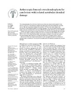
Background Gluteal muscle contracture (GMC), a debilitating disease is a clinical syndrome characterized by contracture of gluteal muscles, tensor fascia lata (TFL), iliotibial band (ITB), and related fascia, in severe cases it also involves hip external rotators and rarely the hip joint capsule [1–3]. GMC exists all across the globe but is more prevalent in China, with childhood incidence rate of 1–2.5% [4–8]. It is associated with intramuscular injections of antibiotics and antimalarial agents like quinine into buttocks [9–12]. Pathognomonic presentation of the disease is abduction and external rotation along with limited flexion and adduction of affected hip [1]. Other features include difficulty in crossing or overlapping the legs (cross sign) (Fig. 1a) and squatting (squatting test), positive Ober’s sign (Fig. 1b), frog leg sign, out-toeing gait, flattened and cone shaped buttock, apparent leg length discrepancy, pelvic obliquity, snapping sound, and a compensatory lumbar scoliosis [13].
Page 2 of 10
GMC usually starts in early childhood, if left untreated it worsens gradually and persists throughout the life [14]. Because the disease mostly affects adolescents and adults, there is always an aesthetic concern. For long, the conventional open release was regarded as the gold standard treatment method for GMC; however, the high rate of complications such as hypertrophic scar, postoperative adhesion and sciatic nerve injury tremendously decreased the patient’s satisfactions [15, 16]. Recently, arthroscopic release of GMC has been introduced as a minimally invasive technique and has dramatically gained popularity among orthopedic surgeons. It has been reported that it avoids extensive surgical trauma resulting in minimal complications in comparison to the conventional open surgery. It has high level of patient’s satisfactions and has excellent clinical outcome [4]. However, previous literatures regarding the comparison of surgical outcomes and complications of these two surgical procedures are still scarce. Currently, no standardized arthroscopic surgical
Fig. 1 Arthroscopic release of bilateral gluteal muscles contractures. a Pre-operatively, patient was unable to cross the legs completely, and b Ober’s sign was positive. c 3 days post-operative pictures, patient was able to cross the leg completely without any support, and d Ober’s sign was negative
Rai et al. BMC Musculoskeletal Disorders (2017) 18:113
technique for GMC release exists which can address the pathology in a systematic way. In this study, we performed arthroscopic release and conventional open release of GMC in adolescents and adult populations. The main purpose of this study was to introduce the arthroscopic F and C method of GMC release, and to compare its clinical efficiency with conventional open surgery in terms of subjective and objective clinical outcomes, patient’s satisfactions, complications and recurrence. Our hypothesis was that the arthroscopic release of GMC using F and C method would provide exceptionally precise and selective release of contractures which would improve the clinical outcomes, thus decreasing the complications associated with conventional open surgery.
Methods Patients
Between Jan 2013 and July 2015, 167 consecutive patients with GMC underwent surgical release using either arthroscopic technique or conventional open release technique. All the patients were carefully examined in the clinic and pre-operatively under anesthesia on the operating table by the senior surgeon in order to determine the severity of disease. Inclusion criteria involved primary GMC releases of 16 years or older patients who could complete the study and the strict rehabilitation protocol. Out of 167 patients, 146 patients provided written informed consent and were included in the study. Every patient was clearly informed about the disease conditions and the surgical procedures along with its benefits and risks. The types of procedures were selected according to the surgeon’s recommendations and patient’s choice, and were performed by or under direct supervision of senior surgeons HW and SHY. 75 patients (150 hips) (male = 25, and female = 50) with the mean age of 25.05 years (16 to 46 years) received the arthroscopic release and 71 patients (142 hips) (male = 33, and female = 38) with the mean age of 25.30 years (17 to 42 years) received the conventional open release. All the patients were classified according to Zhao et al. classification system [16]. In the arthroscopic surgery group, 25 patients were classified as mild, 40 as moderate, and 10 as severe diseases, whereas in the conventional open surgery group, 13 patients were classified as mild, 41 as moderate, and 17 as severe disease.
Surgical procedure Conventional open surgery
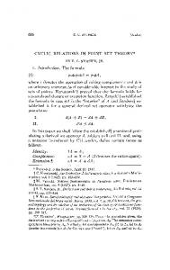
Variable lengths and shapes of skin incisions (5 cm −10 cm) were made in the lateral position over buttock and greater trochanter, followed by division of contracture bands (Fig. 2a). Contractile fibrotic bands were divided in a sequential order according to the muscle group involvement,
Page 3 of 10
(iliotibial bands, gluteus maximus, gluteus medius, gluteus minimus, piriformis and even hip joint capsule) starting from superficial to deeper structures until all the signs and symptoms completely disappeared intra-operatively. Any residual deformities were meticulously assessed, and complete release of contracture was confirmed by adduction, flexion, internal rotation, Ober’s sign, cross leg sign, and palpable click. Finally, appropriate haemostases were maintained, wounds were irrigated with normal saline, a drainage tube placed, and wounds were closed. Arthroscopic surgery (F and C method)
The procedure involved marking of all the anatomical landmarks, like greater trochanter (GT), anterior and posterior boarders of contracted glutei, and most importantly the course of the sciatic nerve in neutral lateral position of the hip. Two portals were usually made and sometimes three portals according to variations in the location and depths of GMC groups. First viewing portal (0.5 cm) was made just over the centre of GT (Fig. 2b). An artificial working space (6 cm × 8 cm) was created in the interval between the subcutaneous fascia and the contracture bands using curette. Silvery white contracture bands were visible when an arthroscope was introduced into the artificial space filled by continuous irrigation of normal saline. About 10 cm above the first portal in the longitudinal axis, second working portal was made under arthroscopy. Any fatty and fibrous tissues in the artificial space were meticulously removed by a shaver and a radio-frequency ablation device. There was always a chance of bleeding from muscles, which was prevented by the prophylactic use of adrenalin (1 mg in 3 l) in a continuous flow of saline, and any visible bleeders were coagulated instantly. Division of contracture bands using a radio-frequency ablation device was then performed using F and C method (Fig. 3a). Initially, division of the ITB was started from the centre of GT (approx. 4 cm below the superior pole of GT) and continued superiorly up to about 10 cm in the longitudinal axis (Fig. 3b). Then, the radiofrequency ablation device was faced anteriorly to divide contractures of tensor fascia latae (TFL), and continued up to the anterior superior iliac spine (ASIS) (Fig. 3c). Gluteus maximus contractures were then divided transversely from approximately 1 cm below the superior pole of GT until silvery white bands of contractures were visible, which completed the F shaped release of GMC (Fig. 3d). The arthroscopic instruments were then advanced further deep to visualize the contractures of gluteus medius, gluteus minimus and deeper structures, and were divided around the GT in the C shaped fashion (Fig. 3e). Finally, complete division of contracture bands were meticulously assessed using same technique as in the conventional open surgery.
Rai et al. BMC Musculoskeletal Disorders (2017) 18:113
Page 4 of 10
Fig. 2 Comparison of incision sizes of GMC release. a shows a big longitudinal incision, which was made for the conventional open surgery, and b shows 2 tiny incisions made for the arthroscopic 2 portal technique
Rehabilitation protocol
Drainage tube was placed routinely, and removed 24 to 48 h after surgery. Rehabilitation protocols were similar for both the groups, patients were instructed to do the functional exercises after elimination of post-surgical pain, or after the drainage tube was removed. At first, patients were placed in continuous passive motion (CPM) machine to allow passive hip and knee flexion exercises, followed by an active range of motion (ROM) exercises, then allowed to walk, and gradually to perform other exercises including crossing legs, straight walking, crouching with closed knees (Fig. 1c & d). Sutures were removed in 2 weeks for both the groups.
had trendelenburg gait consciously. Fourth, glide of fibrotic bands in the iliotibial tract: 2 points were given if the patient had no gliding of fibrotic band and no resistance, 1 point if the patient had gliding of fibrotic band and resistance could be felt, and 0 point if the patient had no fibrotic band, but resistance could be felt. Clinical grade was considered to be excellent if the points obtained was 9–10; good if 7–8; and poor if 0–6. A self administered questionnaire for patients’ satisfaction in terms of cosmetic and functional satisfaction was carried out and graded as satisfied or dissatisfied. Other parameters included incision lengths, duration of surgery, postoperative analgesia, off-bed activity time, complications, and recurrence.
Patient’s evaluation
All the patients were followed up for at least 18 months (mean, 22 months). Patients were clinically assessed by subjective and objective evaluations. Subjective evaluations were performed using hip outcome scores (HOS), which assesses activities of daily living (HOS-ADL) and sports activities (HOS-Sports), and a visual analogue scale (VAS) for pain [17]. Objective clinical evaluation was performed using evaluation criteria set by Ye et al. (2012) [14]. It includes 4 parameters. First, closing knees together while squatting and standing: 3 points were given if the patient could squat and stand freely, 2 points if the patient could squat and stand partly with help, 1 point if the patient could squat and stand wholly with help and 0 point if the patient was unable to stand or squat. Second, crossing and overlapping the legs with 90° of hip and knee flexion: 3 points were given if the patient could cross and overlap the legs freely, 2 points if the patient could cross or overlap the legs partly with help, 1 point if the patient could cross or overlap the legs wholly with help, and 0 point if the patient was unable to cross and overlap the legs. Third, ambulation: 2 points were given if the patient did not have trendelenburg gait involuntary, 1 point if the patient had no trendelenburg gait consciously, and 0 point if the patient
Statistical analysis
We used Statistical Package of Social Sciences (IBM SPSS Statistics 23) version 23 for statistical analysis. Categorical data were analyzed using Chi-square test and Fisher’s test, and independent t test (two tailed) was chosen for analysis of parametric continuous data, whereas the Mann-Whitney U test was used to compare non parametric continuous data. Results of categorical data were presented as frequencies and percentages, whereas results of continuous data were presented as mean ± standard deviation (SD). Statistical differences were considered significant for P values


![Romeo and Juliet Dire Straits Intro: [F] [C] [Bb] [C] [F] [C] [Bb] [C] [F ...](https://m.moam.info/img/260x300/romeo-and-juliet-dire-straits-intro-f-c-bb-c-f-c-b_5a0e82321723ddcfcd185c0e.jpg)