International Immunology, Vol. 14, No. 6, pp. 585±597
ã 2002 The Japanese Society for Immunology
CD6: expression during development, apoptosis and selection of human and mouse thymocytes Nora G. Singer1, David A. Fox2, Tariq M. Haqqi1, Laura Beretta2, Judith S. Endres2, Susan Prohaska3, Jane R. Parnes3, Jonathan Bromberg2,4 and R. Michael Sramkoski1 1Case Western Reserve University School of Medicine and Rainbow Babies and Children's Hospital/ University Hospitals of Cleveland, Cleveland, OH 44106, USA 2University of Michigan Medical Center, Ann Arbor, MI 48109, USA 3Stanford University Medical Center, Stanford, CA 94305, USA 4Present
address: Mt Sinai±NYU Health System, New York, NY 10029, USA
Keywords: apoptosis, comparative immunology, co-stimulation, T cells, thymus Abstract CD6, a 130-kDa surface glycoprotein, is expressed primarily on cells of T lineage. A co-stimulatory role for CD6 in mature T cells has been shown, but the function of CD6 during thymocyte development is unknown. Since CD6 ligands are expressed on thymic epithelium, their interactions with CD6 could be important in thymic selection. In this report we show that CD6 is developmentally regulated in human and mouse thymocytes, and further demonstrate that increase in the level of CD6 expression correlates with expression of the selection marker CD69. We also show that activation via CD2 induces CD6 expression on mature human thymocytes and on a subset of immature human thymocytes that are resistant to apoptosis. In human and mouse thymocytes that express heterogeneous TCR, CD6 increases occur as double-positive thymocytes are selected to a single-positive stage. In contrast, in thymocytes from TCR transgenic mice, CD6 is barely increased following selection, suggesting that as functional avidity increases, requirements for CD6-dependent co-stimulation decrease. Taken together, these results indicate that during thymic development CD6-dependent signals may contribute both to thymocyte survival, and to the overall functional avidity of selection in both man and mouse. Introduction Thymocyte selection is an ordered process during which T cells capable of mediating protective immunity are generated (1,2). To be selected, immature thymocytes must express TCR capable of interacting with antigen-presenting cell (APC)± MHC±antigen complexes that can then transduce signals favoring selection (2). This occurs in a manner appropriate for selection in a minority of thymocytes (3). A majority of thymocytes die due to insuf®cient functional avidity for APC± MHC±antigen complexes (`death by neglect') (2±4), while those thymocytes with excessive functional avidity suffer activation-induced cell death (AICD) (5,6). Therefore, successful thymocyte selection requires optimal functional avidity
so that the total signal transduced to immature thymocytes is suf®cient to avoid death by neglect, but not so great as to induce AICD (2±6). A variety of co-stimulatory molecules have been described that may alter the overall functional avidity of selection either by affecting TCR-dependent signals or by inducing signals independent of the TCR. CD5 is an example of a costimulatory protein that is important during thymocyte selection (7±10). Because CD5 knockout mice were initially reported to have normal T cell development, delineation of the importance of CD5 in thymic selection was delayed (10). However, studies of antigen-restricted thymocytes from TCR
Correspondence to: N. G. Singer, 504 Rainbow Babies and Children's Hospital, 11100 Euclid Avenue, Cleveland OH 44106, USA. E-mail:
[email protected] Transmitting editor: J. Allison Received 19 November 2001, accepted 28 February 2002
586 CD6 in thymocyte differentiation Table 1. Antigen Mouse anti-human
Anti-mouse
Second-step reagents
Other
Antibody
Source
Fluorochrome
CD2 CD2R CD3 CD4 CD6 CD8 CD69
T112 T113 OKT3 M-T441 UMCD6 14 HP-YB3
1 1 2 3 3 3 3
U U U F, PE F, B F, B F
CD3 CD4 CD6 CD8 CD16/ CD32 OVA-TCR CD69
17A2, 145-2C11 L3/T4 polyclonal 536.7 2.4G2 KJI-26 H1.2F3
4 4, 5 6 4 4 7 4
F, PE, B F, PE, allophycocyanin U, B F, PE, PerCP U U F
goat anti-mouse goat anti-rabbit streptavidin±Red 670 streptavidin±allophycocyanin
3 3 8 9
F B R670 allophycocyanin
Annexin V PI 7-AAD
4 10 11
F PI 7-AAD
The sources of the reagents used are as follows: (1) gift of E. Reinherz and S. Schlossman, (2) American Type Culture Collection (Rockville, MD), (3) Ancell (Bayport, MN), (4) PharMingen, (5) Biosource (Camarillo, CA), (6) produced in Parnes' laboratory (15), (7) gift of A. Levine (27), (8) Gibco/BRL (Rockville MD), (9) Caltag (Burlingame, CA), (10) Sigma and (11) Coulter Immunotech (Miami, FL). Fluorochromes (not de®ned in text): U = unconjugated, F = FITC, PE = R-phycoerythrin; B = biotin; R670 = covalent conjugated Cy5 and PE.
transgenic (Tg) mice that lack either CD5 or that lack a portion of the TCR required for signaling have highlighted the importance of CD5 in thymocyte development. These studies show that on mature single-positive (SP) thymocytes, CD5 surface expression directly parallels the avidity and/or signaling intensity of the positively selecting ligand (7). Furthermore, absence of CD5 increases response to TCR-dependent signals and CD5-dependent inhibition of selection appears to occur, in part, by decreasing ef®ciency of TCR-mediated signaling (7). Taken together, the data have led to identi®cation of CD5 as a negative regulator of selection (7±10) and it has been proposed that that proteins homologous to CD5, such as CD6, may also play a role in thymocyte selection (11). CD6 is located 5¢ to the CD5 gene and, like CD5, is a member of the macrophage scavenger receptor family of proteins (11±16). CD6 is a co-stimulatory protein that contains three extracellular cysteine-rich domains, which may differ from each other, both in ligand binding and signaling function(s) (17±23). The CD6 ligand, CD166 [activated leukocyte cell adhesion molecule (ALCAM)], is expressed on thymic epithelium and mediates the CD6-dependent binding of thymocytes to thymic epithelium via a membrane proximal extracellular scavenger receptor domain of CD6 (22±25). CD6 expression increases with thymocyte maturation and is up-regulated in vitro by anti-CD2 mAb. These anti-CD2 mAb activate both thymocytes and mature T cells, but alter the level of CD6 expression only in thymocytes (26). To delineate the function of CD6 in thymocyte development we characterized the expression of CD6 on developing human and mouse thymocyte subsets, determined to what degree human and
mouse thymocyte CD6 is similarly regulated, and obtained evidence regarding a possible role for CD6 in positive selection. We also analyzed whether CD6 expression correlated temporally with the appearance of other cell surface molecules that are reported to mark thymocyte selection, determined whether there is evidence for involvement of CD6 in cell survival and, ®nally, assessed whether CD6-dependent signals contribute to functional avidity during positive selection. Results of our studies suggested that expression of CD6 was an important contributor to immature thymocyte survival in both man and mouse. Furthermore, CD6-dependent signals may also be important in co-stimulation of thymocytes, allowing them to survive particularly when the overall functional avidity of thymocytes for APC±MHC±antigen complexes is low. This could have potential implications for autoimmunity.
Methods Human thymus Human thymic biopsy specimens derived from children CD8 SP > DP > DN thymocytes. Background staining is as indicated in (B) (dashed line). CD4 SP thymocytes are composed of a CD4intermediate and CD4high subset. The MCF for CD8 SP thymocytes is in between those of the CD4intermediate and CD4high SP subsets.
323±339 Tg mice are maintained by A. Levine (gift of K. Murphy) (27). OVA TCR 323±339 Rag-2±/± mice (H-2d), B10.D2 control mice (H-2d) and H-Y TCR Rag-2±/± mice (H-2b) (28) were purchased from Taconic (Germantown, NY). Isolation and tissue culture of thymocytes Thymocyte single-cell suspensions were generated by gently disrupting the tissue, followed by gradient centrifugation as previously described (26). Red cells were lysed using NH4Cl (15). Human thymocytes were cultured in RPMI supplemented with 10% FBS (Gibco/BRL, Gaithersburg, MD), 2 mm Lglutamine, and 100 U/ml penicillin and 100 mg streptomycin (complete media). For mouse thymocytes, 5.5 3 10±5 M 2mercaptoethanol was added to complete media (15). Thymocytes were cultured in six- or 24-well tissue culture dishes (Falcon, Lincoln Park, NJ) for 6±48 h at 37°C with 5% CO2. Antibodies, ¯uorochromes, chemicals and apoptosis detection reagents The antibodies used are shown in Table 1. Staining was performed by incubating cells with saturating concentrations of mAb for 30 min at 4°C. Mouse cells were pre-incubated with Fc block (CD16/CD32) (PharMingen, San Diego, CA) for 10 min at room temperature. Cells were washed twice with SMEM
supplemented with 2% FBS as previously described (17,18). When biotin conjugates were used for staining, staining was performed using PBS supplemented with 0.5% w/v biotin-free BSA (Sigma, St Louis, MO) instead of SMEM with 2% FBS. Cells were ®xed using 1% formaldehyde and stored at 4° C in the dark until analysis. For Annexin V, propidium iodide (PI) and 7-aminoactinomycin D (7-AAD) staining, cells were analyzed within 30±60 min of completion of the staining. For overnight culture, anti-CD2/CD2R mAb, anti-T112 and anti-T113 were used as ascites at 1:500 and 1:250 dilutions respectively, anti-CD3 mAb OKT-3 at 1:500 dilution, and puri®ed MsIgG1 at 2 mg/ml (29,30). Chemicals included phorbol myristate acetate used at 1 ng/ml (Sigma) and dexamethasone used at 1 3 10±5 M (American Regent, Shirley, NY) (26,31). To sort thymocytes, thymocytes stimulated overnight were labeled with UMCD6±FITC (19). mAb used for sorting were washed in Millipore ®lters that retain proteins >30,000 kDa (Millipore, Bedford, MA) to eliminate azide using PBS with Ca2+ and Mg2+. Flow cytometry and cell sorting Analysis of labeled cells was performed using either (i) an Epics XL-MCL (Beckman Coulter, Miami, FL) equipped with a 488 nm argon ion laser operating at 15 mW of power and System II (version 3.0) acquisition software for data collection
588 CD6 in thymocyte differentiation
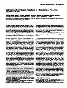
Fig. 2. Mouse CD6 expression is developmentally regulated. CD4, CD8 and CD6 expression was measured on thymocytes from a C57Bl/6 mouse. A representative experiment is shown. A double-color histogram for CD4 and CD8 (A) shows the thymocyte populations on which CD6 was measured. R2 (total thymocytes) is composed of the following subsets: CD8 SP (quadrant 2), CD4CD8 DP (quadrant 3), CD4CD8 DN (quadrant 4) and CD4 SP (quadrant 5). The percentage of total thymocytes within each of the quadrants is as indicated. Single-color histograms of CD6 expression (B±E, solid line) show that, as in human thymocytes, CD6 expression in mouse thymocytes is highest on CD4 SP > CD8 SP > DP > DN. Background staining is as indicated in (B) (dashed line).
or (ii) a BD LSR system (Becton Dickinson, San Jose, CA) equipped with a 633 nm helium neon laser operating at 17 mW of power, a 488 nm argon laser operating at 20 mW of power, a UV laser that was not used and CellQuest acquisition software for data collection. Fluorescence emission for FITC, phycoerythrin, Red 670, allophycocyanin and PerCP were collected with optical band pass ®lters of 525, 575, 675, 660 and 670 nm long pass respectively. Cell sorting experiments were done on an Elite ESP cell sorter (Beckman Coulter), equipped with an 15 mW argon ion laser (488 nm) and a 10 mW helium±neon (HeNe) laser (633 nm). List-mode data collected using Elite acquisition 4.01 software. The Coulter instruments were calibrated daily using Flow Check beads (Beckman Coulter) and pre-programmed computer settings with ®xed voltages to align the instruments, verify full CVs and assure equivalent mean channel ¯uorescence of controls run on different days. For the BD LSR Align Flow 2.25 mm beads were used instead (Molecular Probes, Eugene, OR). Live cells were gated on the basis of forward and side scatter characteristics. When staining was performed using Annexin V±FITC to tag apoptotic cells, dead cells were excluded from analysis with the viability dye (7-AAD) (Molecular Probes). 7-AAD ¯uorescence was collected with a 675 nm optical band pass ®lter. At least 10,000 gated events were acquired per sample in a list-mode ®le. Off-line data
analysis was done with WinList PC version 5.0 software (Verity Software House, Topsham ME). Apoptosis ELISA ELISAs were performed using a Cell Death Detection ELISA Plus kit (Boehringer Mannheim, Indianapolis, IN) according to the manufacturer's instructions. As breaks in the DNA strand occur early in apoptosis, histone proteins are exposed and are available to bind to the ELISA substrate (anti-histone antibody). Therefore, in this ELISA assay, the amount of histone proteins measured re¯ects the number of apoptotic cells. Cells (10,000) were lysed according to the protocol provided with the kit and an amount of lysate corresponding to 1000 cells plated in triplicate in 96-well ¯at-bottom plates. Results were read in an ELISA plate reader (MR 5000; Dynatech, Guernsey, UK) Results CD6 protein expression is developmentally regulated in human thymocytes Using ¯ow cytometry we measured CD6 expression on human and mouse thymocyte subsets, and characterized how CD6 expression varies with the stage of thymocyte development in both man and mouse (Figs 1 and 2). An increase in CD6
CD6 in thymocyte differentiation expression was observed as thymocytes progressed through development from being immature CD4 CD8 double-negative (DN) cells, to immature CD4 CD8 double-positive (DP) cells, and ®nally to mature CD4+ and CD8+ SP thymocytes (Figs 1 and 2). In man, the majority of DP thymocytes express CD6 at intermediate levels (Fig. 1C), between that of immature and mature thymocytes (Fig. 1B, D and E). Single-color histograms of CD6 expression revealed that in man, a minority of DP thymocytes express CD6 at levels comparable to those expressed by SP mature thymocytes (Fig. 1B±E). A small subset of mouse DP thymocytes expresses CD6 at levels clearly above background (Fig. 2C), whereas the majority of SP thymocytes from both mice and men express high levels of CD6 (Figs 1D and E and 2D and E). We also observed that CD4 SP mature human thymocytes contain a subpopulation of SP thymocytes with intermediate levels of CD6 (CD6intermediateCD4SP). These CD6intermediateCD4SP thymocytes are presumed, but not proven, to be more mature than CD6high SP thymocytes. Since CD6 surface expression is lower on resting peripheral blood T cells than on mature thymocytes, it seems logical that down-regulation of CD6 could begin prior to thymic emigration (26) (and our unpublished data). In mice, a CD6low CD4SP subset is present, but is less distinct then that seen in human thymus. Additionally, a CD8low SP subset was more evident among mouse thymocytes than in human thymocytes (Figs 1 and 2). Developmentally restricted regulation of thymocyte CD6 by CD2: CD2-dependent signals regulate expression of CD6 on thymocytes We have previously reported that human thymocyte CD6 expression was increased in vitro by cross-linking CD2 with combinations of anti-CD2 antibodies, whereas CD6 expression in mature lymphocytes remained unaffected by perturbation of CD2 (26). To understand the developmental stages at which thymocyte CD6 levels are tunable, we stimulated singlecell human thymocyte suspensions in vitro with mAb directed against CD3 or against both CD2 and CD2R. Cross-linking CD3 and CD2 using either anti-CD3 or anti-CD2 + anti-CD2R mAb can activate T cells (29). For comparison control conditions, we used MsIgG1 or media alone. In both the control group and the anti-CD3-stimulated group, staining with anti-CD4 and anti-CD8 mAb showed the predicted distributions of thymocytes into immature and mature subsets based on past studies (32). Notably, the group stimulated via CD2 demonstrated increases in absolute numbers of DP thymocytes despite the observation that culture with anti-CD2 mAb transiently modulates CD4 from the surface of DP thymocytes (total thymocyte number in CD2-treated group was increased 50% compared with control) (Fig. 3 and data not shown). Thus, a portion of those cells shown in Fig. 3(B, quadrant 1) are DP thymocytes in which CD4 has been modulated from the surface following perturbation of surface CD2 (32). Analysis of CD6 expression showed that the majority of CD4 CD8 DP thymocytes in the CD2-stimulated group expressed CD6 at higher levels than the CD3-stimulated or control thymocytes (Fig. 3E and F, and data not shown). A >1.5-fold increase in CD6 mean channel ¯uorescence (MCF) was observed. In contrast to increases in CD6 expression (Fig. 3G), we observed decreases in CD4 expression in a
589
subpopulation of DP thymocytes (Fig. 3B). In these studies, cross-linking of CD2 also increased CD6 on mature SP thymocytes [MCF 86.80 (anti-CD2/CD2R) versus 61.64 (medium) for CD4 SP, Fig. 3(G)], but not on peripheral blood lymphocytes (26). Taken together, the data showed that regulation of CD6 by CD2 in T cells is developmentally restricted. CD6 as a marker of positive selection Our observation of high levels of CD6 expression on a subset of both DP and SP thymocyte led us to hypothesize that increases in CD6 protein precede positive selection. Since it is already established that CD69 expression occurs on DP thymocytes near the time of selection, we reasoned that if CD6 marks selection, then CD6, like CD69, should also be increased on pre-selected DP thymocytes (33,34). CD69 expression was observed on ~15±23% of mature human thymocytes and up to 30% of mature mouse thymocytes (Fig. 4A and B). We observed that CD6 is expressed at high levels on the majority of CD69+ thymocytes in man and mouse (Fig. 4A and B). CD6 MCF is ~3-fold higher on human and up to 20fold higher on mouse CD69+ than on CD69± thymocytes (Fig. 4A and B). To explain the observation that some thymocytes were CD6high, but lacked CD69 surface expression, we hypothesized that the CD6highCD69± thymocytes had already undergone selection and had recently down-regulated CD69 (Fig. 4A and B). For human and mouse studies, we determined the percentage of CD69+ thymocytes in a given sample and then analyzed an identical percentage of thymocytes that expressed the highest levels of CD6: we discovered that the numbers of DP and SP thymocytes in both the CD69+ thymocyte subset and the CD6high subset were the same (data not shown). Since CD69 expression has been described on thymocytes undergoing both negative and positive selection (3), we tested whether the CD69+CD6+ thymocytes we detected were viable or apoptotic. We stained human thymocytes with either CD69 or CD6, Annexin V (binds to apoptotic cells) and either PI or 7-AAD (excludes dead cells). The data showed that the majority of CD69+ cells are viable, not apoptotic (Fig. 4C). These data are consistent both with studies showing that most CD69+ human thymocytes survive and differentiate in fetal organ cultures in vitro, and with studies showing human thymocytes in the thymic cortex undergo signi®cant apoptosis, whereas few mature CD3high thymocytes undergo apoptosis (34,35). Our data suggests that a CD6highCD69+ thymocyte subset is found both immediately prior to and following selection. CD6lowCD69± thymocytes are most likely DP thymocytes that have not yet acquired CD69. Triple staining of human thymocytes for CD4, CD8 and CD69 expression showed that CD69± thymocytes are comprised primarily of DP thymocytes, whereas CD69+ thymocytes contain more SP (presumed to be recently selected) than DP thymocytes (presumed to be undergoing positive selection) (Fig. 4D). We hypothesize, but have not directly shown, that the CD69+CD6± subpopulation (3±4%, Fig. 4A and B) of non-dead thymocytes is composed of CD69+AnnexinV+ apoptotic thymocytes (3.15%, Fig. 4C). CD6-dependent signals have been implicated in protecting B-CLL cells from antigen-mediated apoptosis, but not other
590 CD6 in thymocyte differentiation
Fig. 3. Human thymocyte CD6 expression is induced by a combination of anti-CD2 mAb in vitro. Human thymocytes were stimulated overnight with anti-CD2 mAb, anti-CD3 mAb, or control conditions using MsIgG1 or medium without antibody. A representative experiment in which thymocytes were stained for CD4, CD8 and CD6 expression is shown. (A±D). Expression of CD4 (x-axis) and CD8 (y-axis) is depicted for R1 (total thymocytes) as in Fig. 1. Distinct CD4highCD8+ versus CD4mediumCD8+ subpopulations can be distinguished within the DP subset (Fig. 2B) after stimulation via CD2, but not in control cultures with media alone (A), MsIgG1 (C) or anti-CD3 mAb (D), or control medium and MsIgG1 (data not shown). (E and F). CD6 staining on total thymocytes (R1, all quadrants) and DP thymocytes (R1, quadrant 2) are shown. CD6 expression is increased in thymocytes treated with anti-CD2 mAb to a greater extent than in those treated with anti-CD3 mAb (F). A dotted vertical line is drawn through the peak of the single-color CD6 histogram of thymocytes stimulated via CD3 (2F). A vertical line is placed at the same position in the single-color CD6 histogram from thymocytes stimulated via CD2/CD2R to more easily illustrate the relative increase in CD6 expression that occurs after stimulation with anti-CD2 mAb (Fig. 2E). (G). The MCF of CD6 expression for CD4 SP thymocytes is shown in table form along with the MCF for CD6 on DP thymocytes (R1, quadrant 2). (H) The percent increase in CD6 MCF due to cross-linking of CD2 or CD3 was compared to stimulation with phorbol myristate acetate, which is known to increase CD6 expression, relative to media control. The relative increase in total thymocyte CD6 MCF was calculated by dividing the absolute MCF (sample ± background) for each condition by the absolute MCF of thymocytes cultured in media alone and multiplying by 100. Not every condition was tested in each experiment. Results are expressed as the % increase in MCF 6 SEM.
CD6 in thymocyte differentiation
591
Fig. 4. CD69+ human and mouse thymocytes express high levels of CD6 and are not apoptotic. Freshly isolated human and mouse thymocytes were stained for CD6/CD69 expression (A and B) or CD4/CD8/CD6 and CD4/CD8/CD69 (D). In some experiments mouse thymocytes were stained for all four antigens simultaneously or for CD3/CD6/CD69 (data not shown). In representative experiments, CD69 was expressed on 20.3% of human thymocytes. The percent of thymocytes in each of the respective subsets is as indicated. High CD6 expression is demonstrated on mouse and human thymocytes that express CD69 (A and B). To verify that the CD69+ cells were viable and not apoptotic, human thymocytes cultured overnight in media alone were stained with Annexin V± FITC and CD69±phycoerythrin (C). Viable cells were gated based on absence of staining with 7-AAD. Results are expressed as double-color histograms of CD69 (x-axis) versus Annexin V binding (yaxis) (C). The percentages of immature DP and mature SP thymocytes were compared for total, CD69+ and CD69± thymocytes. A representative experiment shows the percent of immature DP versus mature SP thymocytes found within the CD69+ and CD69± subset (D).
types of apoptosis (36). Since resistance to apoptosis is fundamental to accomplishing positive selection and our data suggested that increases in CD6 mark selection, we next hypothesized that CD6 might be important for survival of developing thymocytes. Therefore, to determine if CD6 is involved in cell survival, we compared CD6 surface expression on viable and apoptotic thymocytes. Expression of CD6 was associated with thymocyte resistance to apoptosis To study the relationship between CD6 expression and apoptosis, an in vitro model in which CD6 levels could be manipulated was desirable. Since CD6 is regulated in thymocytes via CD2-dependent signals and cross-linking of CD2 may induce T cell apoptosis, we performed in vitro experiments in which thymocyte CD6 could be manipulated using anti-CD2 mAb to cross-link CD2 (32,37,38). We used a variety of conditions that have been previously demonstrated to induce thymocyte apoptosis, including serum withdrawal, culture with dexamethasone, and cross-linking of cell surface receptors such as CD3 and CD2 (31). We hypothesized that CD2-dependent signals could trigger either increases in CD6 expression or apoptosis in a given thymocyte, but that these events would be mutually exclusive.
To test our hypothesis, we cultured thymocytes for 16±24 h with anti-CD2 mAb, dexamethasone or in medium alone (Fig. 5). We then stained thymocytes with Annexin V±FITC, and compared CD6 expression on viable and apoptotic (but not dead) cells using ¯ow cytometry (Fig. 5). The data showed that when thymocytes were subjected to stimuli with the potential to induce apoptosis (especially activating stimuli transduced via CD2), CD6 was expressed at higher levels on the viable thymocytes compared with the apoptotic cells. Thymocytes cultured with dexamethasone showed signi®cantly higher rates of apoptosis than thymocytes cultured either with antiCD2 mAb or in medium alone. High CD6 expression correlated with resistance to apoptosis in sorted human thymocytes To verify that high CD6 expression on thymocytes marks a subset resistant to apoptosis, thymocytes were cultured overnight as described above and were sorted according to CD6 expression (Fig. 6). Cell lysates were used in an ELISA assay to measure the rate of apoptosis in both CD6high and CD6low thymocyte subsets. The data showed that CD6high thymocytes have a lower rate of apoptosis than CD6low thymocytes, especially following stimulation via CD2 (Fig. 6). Differences in apoptosis are not explained by varying proportions of mature and immature thymocytes in the samples
592 CD6 in thymocyte differentiation
Fig. 5. CD6 is expressed at high levels on viable thymocytes. Human thymocytes were stimulated as indicated in the middle column. Thymocytes were stained with Annexin V±FITC and CD6±phycoerythrin. Viable cells were gated based on absence of staining with 7-AAD. Results are expressed as (A) double-color histograms of CD6 (x-axis) versus Annexin V binding (y-axis) (A±C). Single-color histogram overlays of CD6 expression in non-apoptotic (Annexin V±FITC±) and apoptotic (Annexin V±FITC+) subpopulations are shown for stimulation with control (D), anti-CD2 mAb (E) or dexamethasone (F).
(calculations not shown). Taken together, the data suggest that even moderate increases in CD6 level correlate with decreases in the rate of apoptosis. Expression of a Tg TCR decreases requirement for CD6 expression: TCR-Tg thymocytes have minimal increase in CD6 during selection/maturation from DP to SP thymocyte
Fig. 6. Thymocytes that increased CD6 in response to CD2 stimulation were resistant to CD2-induced apoptosis. Human thymocytes were stimulated overnight with anti-CD2 mAb or cultured in control medium. Thymocytes were stained with anti-CD6 mAb and sorted based on CD6 expression. The 10% of thymocytes in each group that expressed the greatest and least amount of CD6 were collected using an Elite ESP cell sorter. CD6high (j) and CD6low (m) sorted thymocytes stimulated with anti-CD2 mAb or cultured in media alone were used in an ELISA designed to detect histone proteins as a measure of apoptosis. Absorbance was normalized to a blank and a positive control was off-scale for the assay but was in the middle of the range when used at a dilution of 1:5.
The observed pattern of CD6 expression also raises the possibility that CD6 may enhance the overall functional avidity between thymocytes and APC±MHC±antigen complexes, increasing the overall number of immature thymocytes selected. To pursue this issue we characterized the pattern of CD6 expression in thymocytes derived from two different antigen-speci®c TCR-Tg mice. We used OVA TCR mice to examine selection of CD4 SP thymocytes and H-Y TCR mice to examine selection of CD8 SP thymocytes (27,28). If CD6 is important for increasing the overall functional avidity of the T cell for MHC±antigen complexes, then the presence of a Tg TCR that boosts overall functional avidity during selection might decrease the need for co-stimulation or adhesion via CD6. Since the overall functional avidity of selection is increased by the presence of Tg TCR speci®c for OVA peptide 323±339, we hypothesized that OVA TCR thymocytes would require less co-stimulation via CD6 than thymocytes with heterogeneous TCR to achieve maturation to a CD4 SP status. We compared
CD6 in thymocyte differentiation
593
Fig. 7. OVA TCR thymocytes from TCR-Tg mice showed minimal increase in CD6 expression following selection. Thymocytes from OVA TCR DO 11.10 (H-2d) or control BALB/c mice were stained for CD4, CD8 and CD3 expression, and for OVA TCR, CD3 and CD6 expression. A representative experiment is shown. The percentage of total thymocytes within each of the quadrants is as indicated (A and B: R2, quadrants 2±5). OVA TCR mice preferentially select CD4 SP thymocytes (A: R2, quadrant 5) compared to wild-type mice with heterogeneous TCR (B: R2, quadrant 5). The CD3high subset (R6, middle) in both OVA TCR and wild-type mice is composed of SP thymocytes, whereas the CD3low subset (R7, middle) is composed of CD4 CD8 DP thymocytes (data not shown). In OVA TCR mice and wild-type mice there are subsets of DP thymocytes that express low levels and intermediate levels of CD6. The maximal amount of CD6 expressed by DP thymocytes is similar between the two mice, but control mice have more DP thymocytes with intermediate CD6 expression than do OVA TCR mice. As thymocytes mature, CD6 is increased on CD3high (R6) thymocytes from control mice (B, right lower) more than on CD3high (R6) thymocytes from OVA TCR mice (A, right upper). The antibodies and developing reagents used in these ®gures include CD3±FITC, CD4±FITC, CD8±phycoerythrin and CD6±biotin, and streptavidin±Red 670.
CD6 expression in OVA TCR-Tg mouse thymocytes (H-2d) to wild-type thymocytes (H-2d) (Fig. 7). We con®rmed that CD3high thymocytes contain almost exclusively mature SP thymocytes and that the CD3dull subset is comprised of immature DP thymocytes (data not shown). We then analyzed OVA TCR or BALB/c thymocytes for expression of OVA TCR, CD3 and CD6. The data show that a smaller subset of immature CD3dull thymocytes from OVA TCR mice and the major subset of immature CD3dull thymocytes in wild-type controls express similar levels of CD6 (R7; Fig. 7A, right upper and B right lower). A subset of DP and SP thymocytes from these OVA TCR mice expressed low levels of CD6 (R6, left peak; R7, left peak). Less extensive CD6 up-regulation was required for positive selection of OVA TCR thymocytes than was seen with wild-type thymocytes (Fig. 7). To determine whether diminished up-regulation of CD6 also accompanies selection of CD8 SP T cells in the presence of a Tg TCR, thymocytes from H-Y (male antigen) female mice (H2b) were used (28). These mice lacked Rag-2 and, thereby, expressed only H-Y TCR. The data show that both H-Y TCR DP
and CD8 SP thymocytes express less CD6 than do thymocytes from either syngeneic C57Bl/6 (H-2b) or allogeneic BALB/c (H-2d) mice at a similar maturational stage. Furthermore, in contrast to the increases in CD6 expression we described in thymocytes from wild-type mice and from humans, only a minimal increase in CD6 expression level was observed as H-Y TCR thymocytes progress from being DP to being CD8 SP (Fig. 8A). Since the two models of thymocyte selection (OVA TCR and H-Y TCR) we used differ in their potential to rearrange their TCR, we wanted to determine if increases in CD6 expression were attenuated in OVA TCR that lacked Rag-2. Therefore, we compared CD6 levels on OVA TCR Rag-2±/± mice (H-2d) to control B10.D2 (H-2d) mice (Fig. 9). In these thymocytes from OVA TCR Rag-2±/± mice, CD6 expression was similar on immature (CD3l°w) and mature (CD3high) thymocytes, whereas CD6 was increased on SP to DP thymocytes from control B10.D2 mice. The data from mouse thymocytes show that developmentally speci®c CD6 up-regulation was reduced in the presence
594 CD6 in thymocyte differentiation
Fig. 8. Minimal increase in CD6 expression occurs following selection of H-Y TCR thymocytes from female TCR-Tg mice. Thymocytes from female H-Y TCR Rag-2±/± (H-2b) or control C57Bl/6 mice were stained for CD4, CD8 and CD3 expression, CD4, CD8 and CD6 expression, and CD3 and CD6 expression. A representative experiment is shown. The percentage of total thymocytes within each of the quadrants is as indicated (A and B: R2, quadrants 2±5). Female H-Y TCR mice preferentially select CD8 SP thymocytes (A: R2, quadrant 2) compared to wildtype mice with heterogeneous TCR (B: R2, quadrant 2). The CD3high subset (R6) in both mice is composed of SP thymocytes, whereas the CD3low subset (R7) is composed of CD4 CD8 DP thymocytes (data not shown). Levels of CD6 are slightly lower on DP thymocytes (A: R7) from female H-Y TCR than from control female mice (A: F7). As thymocytes mature, CD6 is expressed at a higher level on CD3high thymocytes (R6) from control mice (B, right lower) than on CD3high thymocytes from H-Y TCR mice (A, right upper). The antibodies and developing reagents used in these ®gures include CD3±FITC, CD4±FITC, CD8±phycoerythrin and CD6±biotin, and streptavidin±Red 670.
of a Tg TCR for selection of either CD4 or CD8 SP thymocytes, consistent with a model in which CD6 is more important for costimulation when the overall functional avidity of selection is low. Discussion Human CD6 has been identi®ed and cloned, but its precise function(s) in both developing and mature T cells is unknown (15,16,39,40). Regulated CD6 expression accompanied by expression of CD6 ligands (CD6L) on thymic epithelium strongly suggest a speci®c role for CD6 in thymocyte development. Experiments performed in thymocytes from man and mouse and presented in this report suggest that physiologic CD6±CD6L interactions might act to: (i) send anti-apoptotic signals to immature thymocytes, and (ii) increase the functional avidity of binding between MHC±antigen complexes on thymic APC and thymocytes to levels suf®cient to enhance positive selection without inducing negative selection. Role of CD6-dependent signals in positive selection Evidence that CD6, like CD69, is also a marker for selection is strongly suggested by data showing that increases in CD6
expression are accompanied by expression of CD69 in human and mouse thymocytes. Data presented in this study, showing that stimulation via anti-CD2 increases CD6 expression as well as thymocyte numbers, together with previously published reports (41±47) provide strong support to our hypothesis that CD6, like CD2, is involved in thymocyte selection. The inverse correlation between thymocyte CD6 expression and rate of apoptosis favors a role for CD6-dependent effects on the balance between pro- and anti-apoptotic proteins in developing thymocytes. These observations are consistent with data showing that cross-linking of CD6 in B-CLL cells appears to protect against antigen-mediated apoptosis and induces the expression of anti-apoptotic proteins (36,42,48). We suggest that activation of DP thymocytes occurs in vivo due to antigen recognition accompanied by both CD2±CD2L interactions (e.g. LFA-3, CD58 in humans) and other co-stimulatory interactions while thymocytes are CD6low. Activated thymocytes ®rst up-regulate CD6 and then express CD69 or else undergo apoptosis. DP thymocytes with increased surface CD6 bind to CD6L on thymic epithelium at increased frequency and these interactions may help to trigger development from a DP to a SP stage. Selected SP thymocytes then down-regulate CD69 and eventually down-regulate CD6.
CD6 in thymocyte differentiation
595
Fig. 9. Thymocytes from OVA TCR Rag-2±/± Tg mice showed no increase in CD6 expression following selection. Thymocytes from OVA TCR DO 11.10 (H-2d) or control B10.D2 mice were stained for CD3 and CD6 expression. A representative experiment is shown. The CD3high subset (R6, middle) in both mice is composed of SP thymocytes, whereas the CD3low subset (R7, middle) is composed of CD4 CD8 DP thymocytes (data not shown). Levels of CD6 are comparable on DP (R7) from OVA TCR (A, right upper) and control mice (B, right lower). As thymocytes mature, CD6 is increased on CD3high (R6) thymocytes from control mice (B, right lower) more than on CD3high (R6) thymocytes from OVA TCR mice (A, right upper). The antibodies and developing reagents used in these ®gures include CD4±allophycocyanin, CD8±PerCP, CD3±FITC, CD6±unconjugated, goat anti-rabbit±biotin and streptavidin±phycoerythrin.
Role of CD6-dependent signals in functional avidity Results of our studies showed that CD6 was down-regulated in some SP thymocytes from humans and wild-type mice, and we hypothesize that this occurred at a time when CD6-dependent co-stimulation was no longer required because the selection process was complete. In H-Y TCR-Tg thymocytes, absolute levels of CD6 were lower in both DP and SP thymocytes than in wild-type mice. In OVA TCR mice the most obvious aberration in CD6 expression was that CD6 was only minimally increased during transition from DP to SP stage. This could be due to limited augmentation of CD6 in SP thymocytes from Tg mice because the Tg TCR acts to increase overall functional avidity of selection, thereby reducing relative requirements for CD6dependent signals. Differences between the various TCR-Tg mice (OVA versus H-Y TCR) may relate to the relative functional avidities of the respective TCR for APC±MHC± antigen complexes (7,44,45,49). However, further studies may be necessary to de®nitively con®rm or negate this assumption. In human thymocytes we observed a close association between CD4 and CD6 levels in CD4 SP mature thymocytes. It is conceivable that interactions between thymocyte CD6 and thymic epithelial CD6 ligands contribute to the positive selection of CD4 SP thymocytes in a manner similar to that of CD4±MHC class II interactions (49). Whether increases in CD6 occur in experimental systems that lack critical co-
stimulatory proteins (and thereby have less positive selection) is not yet known. We have previously demonstrated that CD6 is increased in mature T cell clones induced with drugs to become autoreactive and that anti-CD6 mAb inhibit reactivity with self-APC (18). Modest increases in CD6 were found to be functionally important in these studies (18). In mature T cells, excess functional avidity with self-antigen in APC±MHC± antigen complexes results in autoimmunity (50). In the periphery, excesses in CD6-dependent co-stimulation may augment functional avidity with self-antigen, predisposing to autoimmunity. Our new data suggest that human and mouse CD6 both function similarly during thymic selection, a process in which functional avidity is critical. Our new studies of CD6 in human and mouse thymocytes suggest that CD6-dependent signals tune selection in favor of thymocytes that have otherwise have insuf®cient functional avidity for MHC±antigen complexes to compete for selection. This model of CD6 requires additional proof using in vivo experimental systems. Nevertheless, our studies suggest that increases in CD6 expression facilitate co-stimulatory signaling, increase overall functional avidity and oppose apoptosis, in the end favoring thymocyte selection and maturation. Additionally, our data raise the possibility that supra-physiologic expression of CD6 might increase functional avidity, the consequences of which could either be increased positive
596 CD6 in thymocyte differentiation selection (with potential for autoreactivity) or even negative selection, depending on the avidity of a speci®c TCR for MHC± antigen. Demonstration that human and mouse CD6 have similar function(s) enhances the probability that use of models such as Tg mice that overexpress CD6 and CD6 `knockout' mice will be helpful in future evaluations of the physiologic function(s) of CD6.
12
13 14
Acknowledgements The authors thank Christine Anderson, Joseph Piktel for their technical assistance, Michelle Mor®n, Drs Michael Spector and Philip Smith and Edward Bove for help obtaining human thymic tissue, and Dr Melvin Berger for critical reading of this manuscript. This work was supported by NIH grants: AR-20618-20 to N. G. S., AR-44902 and AR-20618 to T. H., A107290 to S. P., CA68675 and AI38930 to J. P., R01 AI 41428 to J. B., and AR38477 to D.A.F.; and by a Lupus Foundation of America Award to N. G. S.; an Arthritis Foundation Biomedical Science Grant award to D. A. F.; and by the Northeastern Ohio and University of Michigan Multipurpose Arthritis Centers and the CWRU Flow Cytometry Core of the NCI-Comprehensive Cancer Center (CA44703).
Abbreviations AAD AICD APC CD6L DN DP MCF OVA PI SP Tg
7-aminoactinomycin D activation-induced cell death antigen-presenting cell CD6 ligand double negative double positive mean channel ¯uorescence ovalbumin propidium iodide single positive transgenic
15 16 17
18
19
20
21 22
References 1 Matzinger, P. 1993. Why positive selection? Immunol. Rev. 135:81. 2 Jameson, S., Hoquist, K. and Bevan, M. 1995. Positive selection of thymocytes. Annu. Rev. Immunol. 13:93. 3 Merkenschlager, M., Graf, D., Lovatt, M., Bommhardt, U., Zamoyska, R., and Fisher, A. 1997. How many thymocytes audition for selection? J. Exp. Med. 186:1149. 4 Fowlkes, B. and Schweighoffer, E. 1995. Positive selection of T cells. Curr. Opin. Immunol. 7:188. 5 Hogquist, K., Jameson, S., Heath, W., Howard, J., Bevan, M. and Carbone, F. 1994. T cell receptor antagonist peptides induce positive selection. Cell 76:17. 6 Jameson, S., Hogquist, K. and Bevan, M. 1994. Speci®city and ¯exibility in thymic selection. Nature 369:750. 7 Azzam, H., Grinberg, A., Lui, K., Shen, H., Shores, E. and Love, P. 1998. CD5 expression is developmentally regulated by T cell receptor (TCR). Signals and TCR Avidity. J. Exp. Med. 188:2301. 8 Chan, S., Waltzinger, C., Tarakhovsky, A., Benoist, C. and Mathis, D. 1999. An in¯uence of CD5 on the selection of CD4-lineage cells. Eur. J. Immunol. 29:2916. 9 Pena-Rossi, C. X., L., Strong, J., Kwan, J., Ferris, W., Chan, S., Tarakhovsky, A., Beyers, A. and Killeen N. 1999. Negative regulation of CD4 lineage development and responses by CD5. J. Immunol. 163:6494. 10 Tarakhovsky, A., Kanner, S., Hombach, J., Ledbetter, J., Muller, W., Killeen, N. and Rajewsky, K. 1995. A role for CD5 in TCRmediated signal transduction and thymocyte selection. Science 269:535. 11 Aruffo, A., Melnick, M., Linsley, P. and Seed, B. 1991. The lymphocyte glycoprotein CD6 contains a repeated domain
23
24
25
26 27 28 29
30
structure characteristic of a new family of cell surface and secreted proteins. J. Exp. Med. 174:949. Bowen, M., Whitney, G., Neubauer, M., Starling, G., Palmer, D., Zhang, J., Nowak, N., Shows, T. and Aruffo, A. 1997. Structure and chromosomal location of the human CD6 gene. J. Immunol. 158:1149. Lecomte, O., Bock, J., Birren, B., Vollrath, D. and Parnes, J. 1996. Molecular linkage of the mouse CD5 and CD6 genes. Immunogenetics 44:385. Robinson, W., Prohaska, S., Santoro, J., Robinson, H. and Parnes, J. 1995. Identi®cation of a mouse protein homologous to the human CD6 T cell surface protein and sequence of the corresponding cDNA. Immunology 155:4739. Pal, A., Romain, P., Singer, N., Fox, D. and Stavnezer, J. 1996. Mouse CD6: sequence of cDNA and expression of mRNA. Immunol. Lett. 49:133. Whitney, G., Bowen, M., Neubauer, M. and Aruffo, A. 1995. Cloning and characterization of murine CD6. Mol. Immunol. 32:89. Singer, N., Mitra, R., Lialios, F., Richardson, B., Marks, R., Pesando, J., Fox, D. and Nickoloff, B. 1997. CD6 dependent interactions of T cells and keratinocytes: functional evidence for a second CD6 ligand on gamma-interferon activated keratinocytes. Immunol. Lett. 58:9. Singer, N., Richardson, B., Powers, D., Hooper, F., Lialios, F., Endres, J., Bott, C. and Fox, D. 1996. Role of the CD6 glycoprotein in antigen-speci®c and autoreactive responses of cloned human T lymphocytes. Immunology 88:537. Bott, C., Doshi, J., Morimoto, C., Romain, P. and Fox, D. 1993. Activation of human T cells through CD6: functional effects of a novel anti-CD6 monoclonal antibody and de®nition of four epitopes of the CD6 glycoprotein. Int. Immunol. 5:783. Osorio, L., Garcia, C., Jondal, M. and Chow, S. 1994. The antiCD6 mAb, IOR-T1, de®ned a new epitope on the human CD6 molecule that induces greater responsiveness in T cell receptor/ CD3-mediated T cell proliferation. Cell. Immunol. 154:123. Osorio, L., Ordonez, C., Garcia, C., Jondal, M. and Chow, S. 1995. Evidence for protein tyrosine kinase involvement in CD6-induced T cell proliferation. Cell. Immunol. 166:44. Bowen, M., Bajorath, J., Siadak, A., Modrell, B., Malacko, A., Marquardt, H., Nadler, S. and Aruffo, A. 1996. The amino-terminal immunoglobulin-like domain of activated leukocyte cell adhesion molecule binds speci®cally to the membrane-proximal scavenger receptor cysteine-rich domain of CD6 with a 1:1 stoichiometry. J. Biol. Chem. 271:17390. Bowen, M., Patel, D., Li, X., Modrell, B., Malacko, A., Wang, W., Marquardt, H., Neubauer, M., Pesando, J., Francke, U., et al. 1995. Cloning, mapping. and characterization of activated leukocyte-cell adhesion molecule (ALCAM), a CD6 ligand. J. Exp. Med. 181:2213. Patel, D., Wee, S., Whichard, L., Aruffo, A. and Haynes, B. 1995. Identi®cation of a ligand for CD6 on human thymic epithelial cells. In Schlossman, S., Boumsell, L., Gilks, W. et al. eds, Leukocyte Typing V: White Cell Differentiation Antigens, p. 1684. Oxford University Press, Oxford. Patel, D., Wee, S., Whichard, L., Bowen, M., Pesando, J., Aruffo, A. and Haynes, B. 1995. Identi®cation and characterization of a 100-kD ligand for CD6 on human thymic epithelial cells. J. Exp. Med. 181:1563. Bott, C., Doshi, J., Li, L., Mcmurtry, S., Sanders, J. and Fox, D. 1994. Transcriptional regulation of CD6 expression on human T lymphocytes by phorbol ester. J. Immunol. 153:1. Murphy, K., Heimberger, A. and Loh, D. 1990. Induction by antigen of intrathymic apoptosis of CD4+CD8+TCRlo thymocytes in vivo. Science 250:1720. Kisielow, P., Bluthmann, H., Staerz, U., Steinmetz, M. and Vonboehmer, H. 1988. tolerance in T cell receptor mice involves deletion of nonmature CD4+CD8+ thymocytes. Nature 333:742. Meuer, S., Hussey, R., Fabbi, M., Fox, D., Acuto, O., Fitzgerald, K., Hodgdon, J., Protenetis, J., Schlossman, S. and Reinherz, E. 1984. An alternative pathway of T cell activation: a functional role for the 50kd T11 sheep erythrocyte protein. Cell 36:897. Reinherz, E., Kung, P., Goldstein, G. and Schlossman, R. 1980. Discrete stages of human intrathymic differentiation: analysis of
CD6 in thymocyte differentiation 31
32
33 34
35
36
37
38
39 40
normal thymocytes and leukemic lymphoblasts of T cell lineage. Proc. Natl Acad. Sci. USA 77:1588. Kirsch, A., Mahmood, A., Endres, J., Bohra, L., Bonish, B., Weber, K. and Fox, D. 1999. Apoptosis of human T cells: induction by glucocorticoids or surface receptor ligation in vitro and ex vivo (published erratum appears in J. Biol. Regul. Homeost. Agents 1999; 134: following 267). J. Biol. Regul. Homeost. Agents 13:80. Blue, M., Daley, J., Levine, H., Branton, K., Jr and Schlossman, S. 1989. Regulation of CD4 and CD8 surface expression on human thymocyte sub-populations by triggering through CD2 and CD3 T cell receptor. J. Immunol. 142:374. Kersh, G. and Hedrick, S. 1995. Role of TCR specify in CD4 versus CD8 lineage commitment. J. Immunol. 154:1057. Vanhecke, D., Verhasselt, B., De Smedt, M., Leclercq, G., Plum, J. and Vandekerckhove, B. 1997. Human thymocytes become lineage committed at an early postselection CD69+ stage, before the onset of functional maturation. J. Immunol. 159:5973. Varas, A., Jimenez, E., Sacedon, R., Rodriquez-Mahou, M., Maroto, E., Zapata, A. and Vincente, A. 2000. Analysis of human neonatal thymus: evidence for a transient thymic evolution. J. Immunol. 164:6260. Osorio, L., De Santiago, A., Aguilar-Santelises, M., Mellstedt, H. and Jondal, M. 1997. CD6 ligation modulates the Bcl-2/Bax ratio and protects chronic lymphocytic leukemia B cells from apoptosis induced by anti-IgM. Blood 89:2833. Rouleau, M., Bernard, A., Lantz, O., Vernant, J., Charpentier, B. and Senik, A. 1993. Apoptosis of activated CD8+/CD57+ T cells is induced by some combinations of anti-CD2 mAb. J. Immunol. 151:3547. Rouleau, M., Mollereau, B., Bernard, A., Metivier, D., RosenthalAllieri, M., Charpentier, B. and Senik, A. 1996. Mitogenic CD2 monoclonal antibody pairs predispose peripheral T cells to undergo apoptosis on interaction with a third CD2 monoclonal antibody. J. Immunol. 152:4861. Aruffo, A., Bowen, M., Patel, D., Haynes, B., Starling, G., Gebe, J. and Bajorath, J. 1997. CD6±ligand interactions: a paradigm for SRCR domain function? Immunol. Today 18:498. Robinson, W., Prohaska, S., Santoro, J., Robinson, H. and Parnes,
41
42 43 44
45 46
47
48 49 50
597
J. 1995. Identi®cation of a mouse protein homologous to the human CD6 T cell surface protein and sequence of the corresponding cDNA. J. Immunol. 155:4739. Vollger, L., Tuck, D., Springer, T., Haynes, B. and Singer, K. 1987. Thymocyte binding to human thymic epithelial cells is inhibited by monoclonal antibodies to CD2 and LFA-3 antigens. J. Immunol. 138:358. Le, P., Vollger, L., Haynes, B. and Singer, K. 1990. Ligand binding to the LFA-3 cell adhesion molecule induces IL-1 production by human thymic epithelial cells. J. Immunol. 144:4541. Killeen, N., Stuart, S. and Littman, D. 1992. Development and function of T cells in mice with a disrupted CD2 gene. EMBO J. 11:4329. Melton, E., Sarner, N., Torkar, M., Van Der Merwe, P., Russel, J., Budd, R., Mamlaki, C., Tolaini, M., Kioussis, D. and Zamoyska, R. 1996. Transgene-encoded human CD2 acts in a dominant negative fashion to modify thymic selection signals in mice. Eur. J. Immunol. 26:2952. Teh, S., Killeen, N., Tarakhovsky, A., Littman, D. and Teh, H. 1997. CD2 Regulates the positive selection and function of antigenspeci®c CD4±CD8+ T cells. Blood 89:1308. Van Der Merwe, P., Barclay, A., Mason, D., Davies, E., Morgan, B., Krishnam, A., Ianelli, C. and Davis, S. 1994. Human cell-adhesion molecule CD2 binds CD58 LFA-3. with a low af®nity and an extremely fast dissociation rate but does not bind CD48 or CD59. Biochemistry 33:10149. Cibotti, R., Punt, J., Dash, K., Sharrow, S. and Singer, A. 1997. Surface molecules that drive T cell development in vitro in the absence of thymic epithelium and in the absence of lineagespeci®c signals. Immunity 6:245. Osorio, L., Jondal, M. and Aguilar-Santelises, M. 1998. Regulation of B-CLL apoptosis through membrane receptors and Bcl-2 family proteins. Leuk. Lymphoma 30:247. Frank, GD. and Parnes, JR. 1998. The level of CD4 surface protein in¯uences T cell selection in the thymus. J. Immunol. 160:634. Lehman, P., Tarqari, U. and Forsthubr, T. 1998. Shifting T cell activation thresholds in autoimmunity and determent spreading. Immunol. Rev. 164:53.