(ES2), and (3) changes in headache locus of control and self-efficacy. Forty-four young adults with chronic tension-type headaches were assigned either to six ...
Applied Psychophysiology and Biofeedback, Vol. 22, No. 1, 1997
Change Mechanisms Associated with Combined Relaxation/EMG Biofeedback Training for Chronic Tension Headache Lori A. Rokicki,13 Kenneth A. Holroyd,1 Christopher R. France,1 Gay L. Lipchik,1 Janis L. France,1 and Steven A. Kvaal1,2
Therapeutic mechanisms hypothesized to underlie improvements in tension headache activity achieved with combined relaxation and eleclromyographic (EMG) biofeedback therapy were examined. These therapeutic mechanisms included (I) changes in EMG activity in frontal and trapezii muscles, (2) changes in central pain modulation as indexed by the duration of the second exteroceptive silent period (ES2), and (3) changes in headache locus of control and self-efficacy. Forty-four young adults with chronic tension-type headaches were assigned either to six sessions of relaxation and EMG biofeedback training (N = 30) or to an assessment only control group (N = 14) that required three assessment sessions. Measures of self-efficacy and locus of control were collected at pre- and posttreatment, and ES2 was evaluated at the beginning and end of the first, third, and lost session. EMG was monitored before, during, and following training trials. Relaxation/EMC biofeedback training effectively reduced headache activity: 51.7% of subjects who received relaxation/biofeedback therapy recorded at least a 50% reduction in headache activity following treatment, while controls failed to improve on any measure. Improvements in headache activity in treated subjects were correlated with increases in self-efficacy induced by biofeedback training but not with changes in EMG activity or in ES2 durations. These results provide additional support for the hypothesis that cognitive changes underlie the effectiveness of relaxation and biofeedback therapies, at least in young adult tension-type headache sufferers. KEY WORDS: biofeedback; tension type headache; change mechanisms; self-efficacy; exteroceptive suppression.
1Department of Psychology and Institute of Health and Behavioral Sciences. Ohio University, Athens, Ohio. 2Currently affiliated with the Department of Psychology. Roosevelt University. Chicago, Illinois. 3Address all correspondence to Lori A. Rokicki, who is now at the Illinois Institute of Technology, Institute of Psychology, 3101 South Dearborn, Chicago, Illinois 60616-3793. 21 lOW-OSSimWKMXKISIZ-SQOe 1997 Plenum Publishing Corporation
22
Rokicki, Holroyd, France, Lipchik, France, and Kvaal
INTRODUCTION
In the 25 years since Budzynski, Stoyva, and Adler (1970) introduced the use of electromyographic (EMG) biofeedback in the treatment of tension headache, biofeedback training has been intensively evaluated. Meta-analytic reviews of this literature reveal that EMG biofeedback training administered either alone or combined with relaxation training typically reduces tension headache activity by 40% to 60% (Blanchard, Andrasik, Ahles, Teders, & O'Keefe, 1980; Bogaards & ter Kuile, 1994; Holroyd & Penzien, 1986). In spite of the fact (hat the efficacy of EMG biofeedback training has been well established for some time, the therapeutic mechanism whereby this treatment produces improvements remains unknown. If this therapeutic mechanism could be specified, it might not only facilitate the development of more effective biofeedback training procedures, but also shed light on the ways biological and psychological variables interact in producing chronic pain disorders. The initial hypothesis was that EMG biofeedback training reduced tension headache activity by reducing the sustained contraction of pericranial muscles (Budzynski, Stoyva, Adler, & Mullaney, 1973). However, this physiological learning hypothesis has failed to receive empirical support: Changes in EMG activity during and following biofeedback tiaining are typically unrelated to improvements in tension headache activity. This has been true irrespective of whether frontal EMG (Andrasik & Holroyd, 1980; Cox, Freundlich, & Meyer, 1975; Holroyd, Andrasik, & Noble, 1980; Holroyd et al., 1984; Kroener-Herwig & Weich, 1989; Lacroix, Clarke, Bock, & Doxey, 1986), cervical EMG (Hart & Cichanski, 1981), or trapezii EMG feedback is administered (Arena, Bruno, Hannah, & Meador, 1995). To date, no attempt has been made to examine relationships between changes in EMG activity and improvements in headache activity when both frontal and trapezii or cervical EMG feedback are provided as they often are in clinical practice. It is possible that when F.MG feedback is provided to multiple muscle groups changes in EMG activity in at least one of these muscle groups would be associated with observed improvements in headache activity. An alternate hypothesis postulates that cognitive changes induced by biofeedback training, rather than changes in EMG activity, are responsible for improvement in headache activity (Blanchard, Kirn, Hermann, & Steffek, 1993; Holroyd, 1979; Holroyd & Penzien, 1983; Holroyd et al., 1984). Specifically, tension headache sufferers who perceive EMG biofeedback training as a credible treatment and perceive themselves as able to control EMG activity are postulated to begin viewing their headaches as being under their own control (i.e., increased internal or decreased external locus of control) and to begin believing that they are able to carry out the necessary, behaviors to manage headaches (i.e., increased self-efficacy), These cognitive changes are hypothesized to lead tension headache sufferers to initiate and persist in efforts to cope with headache-related stressors that lead to reductions in headache activity. A study by Holroyd and colleagues (1984) provides some support for this cognitive hypothesis. In that study, actual changes in EMG activity and subjects' perceptions that they were successfully controlling EMG activity were manipulated independently. Subjects' perceptions (but not actual
EMG Biofeedback Change Mechanisms
23
changes in EMG activity) predicted both changes in cognitive variables during biofeedback training and subsequent improvements in headache activity. One limitation of the cognitive hypothesis, at least as articulated in the Holroyd et al. (1984) report, is that while cognitive change mechanisms arc clearly specified, little is said about psychophysiological change mechanisms. Recently it has been proposed that improvements in tension headache activity following biofeedback training result from biofeedback training's ability to enhance central pain modulation, not from its direct effects on peripheral EMG. Noninvasive measures of activity in supraspinal pain modulation systems that might allow this intriguing possibility to be investigated have only recently become available. Of particular relevance is the second exteroceptive suppression of masseter and temporalis muscle activity, which is claimed to provide an index of the functional integrity of central pain modulation systems involved in the pathogenesis of chronic tension-type headaches. Normally, perioral electrical stimulation elicits two successive suppression periods in jaw-ciosing muscles; however, the second of these exteroceptive suppression periods (ES2) has been shown to be shortened and sometimes absent in patients with chronic tension-type headaches (e.g., Gobel & Weigle, 1991; Mathew, Leis, Ali, & Dimitrijevic, 1992; Nakashima & Takahashi, 1991; Paulus, Raubuchi, Straube, & Schoenen, 1992; Schoenen, Jamart, Gerard, Lenarduzzi, & Delwaide, 1987; Wallash, Reinecke, & Langhor, 1991). The brainstem region that mediates ES2 receives afferents from structures implicated in endogenous pain control (e.g., the nucleus raphe magnus and the periaqueductal grey), and in animal models, ES2 is shortened or eliminated by electrically disrupting these antinociceptive structures (e.g., Dostrovsky, Hu, Sessle, & Sumino, 1982; Sessle & Hu, 1981). Based on these animal models, investigators have interpreted the abnormal ES2 responses observed in chronic tension-type headache as reflecting a dysfunction in the functional integrity of supraspinal pain modulation associated with this disorder. Thus, chronic tension headaches may develop in the absence of input from peripheral nociceptors in pericranial muscles as a result of a dysfunction in supraspinal pain modulation systems. Jean Schoenen has recently summarized the anatomical, pharmacologic, and clinical evidence supporting this formulation in recent reviews (Schoenen, 1993a, b). In a study of particular relevance to understanding therapeutic mechanisms in biofeedback training, Schoenen (1989) found that combined relaxation and frontal EMG biofeedback training normalized abnormal ES2 responses in chronic tension headache sufferers. Moreover, changes in the duration of ES2 induced by biofeedback training were correlated with reductions in tension headache activity (r = .52). Unfortunately, control groups were not included in this study, so these results must be interpreted cautiously. Nonetheless, this study raises the possibility that relaxation/EMG biofeedback treatments control chronic tension headaches primarily through their ability to enhance supraspinal pain modulation. These supraspinal effects might be associated with the cognitive changes that have been shown to be produced by biofeedback training and are emphasized in the cognitive model described above, or they might be independent of these cognitive changes.
24
Rokicki, Holroyd, France, Lipchik, France, and Kvaal
In the present study we examined the three types of change mechanisms that have been hypothesized to underlie the effectiveness of combined relaxation/EMG biofeedback training. We also attempted to address some of the limitations of previous studies that have examined change mechanisms in EMG biofeedback training. Thus, more than one muscle group was monitored during biofeedback training, and feedback was provided from more than one location; changes in both central and peripheral physiological responses (ES2 responses and EMG activity) as well as cognitive changes induced by training were assessed; and relationships between each hypothesized change mechanisms and improvements in headache activity were examined.
METHOD Participants Over 2,500 undergraduate college students completed a screening questionnaire designed to identify individuals with recurrent headaches (Holm, 1983). Individuals who indicated that they experienced more than 2 headaches per week and endorsed several items characteristic of tension headache (e.g., bilateral onset, pressing or tightening pain quality, short duration, and no nausea, vomiting, photophobia, or phonophobia) were invited to participate in a detailed diagnostic interview designed to identify individuals who met the diagnostic criteria for chronic tension headache established by the Headache Classification Committee of the International Headache Society (1988). Informed consent was obtained prior to the interview, and the interview was conducted by graduate students trained in the IHS diagnostic system. Individuals who had experienced a recent change in headache activity were referred to their family physician. Individuals who met IHS diagnostic criteria for chronic tension-type headache and indicated that they were not currently taking any antidepressant medications completed a second informed consent form prior to further participation and were asked to record their headache pain four times daily for a 2-week baseline period. Following 2 weeks of baseline headache recording, 45 individuals (aged 18 to 22) met the IHS diagnostic criteria for chronic tension-type headache and recorded at least three headache episodes per week. These individuals recorded taking less than 7 analgesic medication tablets per week. Of this sample, 52% described a pressing or tightening pain quality, 86% reported a mild to moderate pain intensity, 74% reported bilateral onset, 79% stated that their headaches were not aggravated by routine physical activity, 34% reported experiencing photophobia, and 14% stated that phonophobia accompanies a typical headache. Fifty-one percent had previously sought treatment for their headaches. It is noteworthy that of the 2,500 students screened, 2% were eligible for the study; this proportion of chronic tension headache sufferers is considerably smaller than identified in previous surveys (Andrasik, Holroyd, & Abell, 1979; Linet, Stewart, Celentano, Ziegler, & Sprecher, 1989; Rasmussen, Jensen, & Olesen, 1991). Thus, the extensive screening procedure that we used may have increased the likelihood that our sample is similar to a clinical sample.
25
EMG Biofeedback Change Mechanisms
Apparatus
Experimental Setting All sessions were conducted in a sound-attenuated suite of rooms. For treatment sessions, participants were seated in a comfortable reclining chair in a room that contained the biofeedback equipment. For ES2 assessments, participants were seated in a heavily padded chair in a room adjacent to the recording and stimulation equipment.
Physiological Measures ES2 Assessment. Masseter electromyographic activity (EMG; in uV) was recorded using a pair of 10-mm Beckman silver/silver-chloride electrodes (Sensor Medics, Yorba Linda, CA) and a Sensor Medics silver earclip reference electrode. EMG activity was amplified using a World Precision Instruments (Sarasota, FL) DAM-50 differential amplifier with a 10-3000 Hz bandwidth and a 1000 AC gain. Electrical stimulation was applied using a Digitimer DS7 electrical stimulator and an Electrode Store DDY-15 reusable stainless steel pediatric bar electrode with a 15-mm spacing (center to center) of 6-mm contacts. EMG activity was sampled at 2 kHz using RC Electronics (Goleta, CA) Computerscope data acquisition and analysis software and a Dell (Austin, TX) 486 personal computer. All electrode placements were palpated, cleansed with 70% isopropyl alcohol, and gently abraded to ensure a maximum resistance of 10 kii. Beckman electrolyte gel was used as a conductive medium. The right masseter muscle was palpated during voluntary jaw occlusion. The first electrode was placed 2 cm from the most lateral point on the mandibular angle. The second electrode was placed 1 cm (measured center to center) superior and slightly medial to the first electrode along the length of the masseter in a bipolar placement. The earclip reference electrode was clipped to the right earlobe. The stimulating electrode was taped 5-mm posterior to the right labial commissure with the anode superior to cathode. In order to ensure maximal clenching during each stimulation, two additional Beckman electrodes were attached to the left masseter and connected to an EMG J33 electromyogram biofeedback unit (Cyborg, Boston, MA) to provide audio feedback to the participant. Individuals inserted a Lancer Orthodontics (Carlsbad, CA) Therabite rubber dental wafer between their upper and lower teeth, and a threshold level was preset for each individual by asking him or her to clench as hard as possible on the dental wafer in order to elicit a tone. The electrical stimulation to the labial commissure was applied gradually, starting at 0 mA; the intensity was increased in 2-mA steps up to 30 mA. Eleven trials of electrical stimulation were then applied to the ipsilateral labial commissure during jaw occlusion at an intensity of 30 mA, a duration of 0.1 ms, and a stimulation rate of 0.1 Hz; there was a 10-second interstimulation interval between each trial. A 250-ms sweep was recorded for each stimulation trial (50-ms prestimulation, and 200-ms poststimulation). Each of the 11 raw EMG sweeps were rectified and then averaged on-line to create a single waveform for scoring ES responses.
26
Rokicki, Holroyd, France, Lipchik, France, and Kvaal
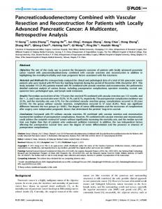
Fig. 1. Example of ES response and scoring method used. The shaded areas represent the durations of the first and second exteroceptive silent periods.
£5 Scoring. Figure 1 illustrates a sample waveform after 11 individual 250-ms sweeps of EMG activity have been rectified, averaged, and plotted in relation to prestimulus baseline levels. A mean baseline was calculated by averaging the 50 ms of EMG data prior to stimulation. The onset of ES2 was determined at the point of crossing the calculated EMG baseline; a return to the baseline for greater than 1-ms was considered the end of ES2 (Nakashima, Takahashi, Azumi, & Ishida, 1990). EMG Activity. Participants receiving relaxation/EMG biofeedback training had frontalis, right trapezius, and left trapezius muscle activity monitored using the Biolab system, which was connected to an IBM PS/2 computer, and was run using Biotext software (Version 1.61). This system has a bandpass of 100-250 kHz. All EMG recordings were integrated and averaged for each trial. The muscle sites were cleaned with a 70% isopropyl alcohol solution and then gently abraded to ensure a resistance of less than 10 kQ. All recording electrodes (two per muscle site) were 10-mm Beckman silver/silver-chloride electrodes and were directed to three EMG modules (module M130) of the Biolab system. Beckman electrode paste served as the electrolyte. For the frontalis, an electrode was placed approximately 2.5 cm above each eyebrow and centered over each eye (Andrasik, 1979). For each of the trapezius muscles, electrodes were placed in a small oval area, approximately 4 cm long, such that the long horizontal axis of the oval was halfway between the angle of the acromion and vertebra C7 (Basmajian, 1989). A common ground SensorMedics earclip was coated with electrolyte and clipped to the right earlobe.
27
EMG Biofeedback Change Mechanisms
Headache Variables Participants were asked to record their headache pain four times daily using an 11-point rating scale. The following anchors were used: 0 = No pain; 2 = Slightly Painful — 1 only notice my headaches when I focus my attention on them; 4 = Mildly Painful — I can ignore my headaches most of the time; 6 = Painful — My headache is painful, but I can continue what I am doing; 8 = Very Painful — Concentration is difficult, but I can perform tasks of an undemanding nature; and 10 = Extremely Painful — I can't do anything when I have a headache (Haynes, Griffin, Mooney, & Parise, 1975). We defined a headache episode as a peak intensity rating greater than 2. We calculated a Headache Activity score by summing the ratings from the week (typically, 4 ratings per day for 7 days) and dividing this sum by the number of ratings for that week (Blanchard & Andrasik, 1985). The number of headache-free days per week was calculated and also examined.
Medication Intake Participants recorded the names of any medication taken and the number of tablets consumed. Only over-the-counter analgesic medications were taken; thus, the number of tablets consumed weekly was used.
Psychological Measures All participants completed two measures that were designed to assess cognitions hypothesized to mediate improvements in headache activity. The HeadacheSpecific Locus of Control Scale (HSLC; Martin, Holroyd, & Penzien, 1990) is a 33-item scale consisting of three subscales. The Internal subscale indicates the extent to which the individual believes headaches are influenced by personal actions. The Health Care Professionals subscale indicates the extent to which the individual believes the source and relief of headache problems lies with health care professionals. The Chance subscale measures the extent to which the individual believes that headache activity is due to pure chance. The scores on the Health Care Professionals subscale and the Chance subscale were combined to create an External Locus of Control Score. The Headache Self-Efficacy Scale (HSES; Martin, Holroyd, & Rokicki, 1993) is a Si-item questionnaire designed to assess individuals' beliefs that they can manage their headache pain. Items reflect an individual's confidence in his or her ability to prevent headaches across a number of situations. An average self-efficacy score was calculated by dividing the total score by the number of items endorsed; lower HSES scores indicate higher self-efficacy.
28
Rokicki, Holroyd, France, Lipchik, France, and Kvaal
Procedure
At the end of the 2-week baseline assessment, participants were randomized to one of the two groups by a within-sample matching technique on the basis of their mean Headache Activity scores. Namely, three individuals with similar Headache Activity scores were grouped together and one was randomly chosen to be assigned to the control group in an attempt to reduce the possibility that baseline Headache Activity scores would differ between groups. Participants assigned to the EMG biofeedback group received six sessions of combined relaxation/EMG biofeedback training (two sessions per week). ES2 was evaluated prior to and at the end of the first, third, and sixth treatment session. Individuals assigned to the record-only control group attended a weekly session similar in duration to the treatment group. ES2 was evaluated at the beginning and end of each session. Individuals in both groups were asked to record their headache activity and medication intake throughout their participation in the study and at least two weeks following their last session. Participants were asked to bring in their headache recording sheets during each session, and 80% replied with this request. The majority of those who did not comply stated that they forgot to bring in their form and brought it in the next session. Pretreatment questionnaires were readministered at the last session to individuals in both groups. Participants were compensated for their considerable time commitment to the study by receiving $15 and an entry into a $100 lottery drawing. Relaxation/Biofeedback
Croup
Therapists were trained using a biofeedback manual designed for this study to ensure similar treatment between subjects. The beginning of each treatment session consisted of the following sequence: (1) review of headache recording sheets and attachment of electrodes (approximately 10 minutes), (2) an adaptation period (5 minutes), and (3) a baseline EMG recording in which the participant was asked to sit upright with his or her eyes closed and to remain as still as possible (5 minutes). During Session 1, electrodes were removed after the baseline EMG recording and the therapist instructed the individual in progressive muscle relaxation training according to the study manual (PMRT; see Blanchard & Andrasik, 1985). Participants were asked to engage in home practice of PMRT at least once a day and were given an audiotape as an aid; practice times were recorded on the headache recording sheets. During Sessions 2 through 6, the following sequence occurred after the baseline EMG recording: First, two 10-minute trials of auditory feedback on the specified muscle group using the Biolab system (Session 2: frontalis; Session 3: frontalis; Session 4: right trapezius; Session 5: left trapezius; Session 6: frontalis) during which participants were provided auditory feedback directly proportional to EMG activity of the specified muscle site. Participants were asked to use the relaxation skills that they had learned to try to "make the beeping slow down." In addition, participants
29
EMG Biofeedback Change Mechanisms
were given some additional techniques that they could use to try to "make the beeping slow down"; therapists gave the same suggestions to all participants, as the suggestions were included in a treatment manual designed for the study. Second, a self-control trial in which participants were asked to continue reducing muscle rension without feedback was provided (5 minutes). Individuals were not pushed to reach a particular criterion level, as there was concern that frustration and anxiety might be experienced if he or she did not reach the criterion level. Beginning at Session 3, participants were encouraged to deter the build-up of tension throughout the day by choosing everyday situations that would serve as cues to monitor tension levels and to begin using brief forms of relaxation. Individuals were asked to practice these skills daily to prevent headaches in their everyday life.
Record-Only Control Group The record-only control group attended three one-hour sessions. At the beginning of each session, headache recording sheets were reviewed, electrodes were attached, and ES2 was evaluated. Participants then read a short story from "The Secret Sharer" and Other Great Stories (Lass & Tasman, 1969) and were asked to answer questions about the story. Participants had at least 30 minutes to read and answer the questions to ensure that the time between within-session ES2 assessments was relatively similar between the treatment and control group (if an individual finished early, he or she was asked to review the answers). Upon completion of the reading period, ES2 was evaluated again. Appointments were scheduled approximately a week-and-a-half apart to ensure that the time between ES2 assessments was similar between the treatment and control group. The control group did not have EMG activity measured as we believed that this would place too much demand on the participants, and between-group comparisons would be difficult because the control group would only have weekly EMG readings.
RESULTS We used an alpha levels of .05 for all statistical tests.
Participant Characteristics The mean age of the treated participants was 19.00 years (n = 29), and the mean age of the individuals in the record-only control group was 18.64 years (n - 14). Females were predominant in both groups (86% in both the treatment and control group). Medical treatment had been sought previously by 59% of the subjects in the treatment group and 36% of the participants in the control group (X2 [1] - 1-98, p > .1). The chronicity of frequent headaches was 4.13 years for the treatment group and 3.96 years for the record-only control group.
JO
Rokicki, Holroyd, France, Lipchik, France, and Kvaal
EMG Activity Across Treatment Pre-Post Changes The mean pretreatment baseline EMG activity of each muscle was compared to the mean EMG activity of the self-control trial of Session 6. Subjects exhibited decreased levels of muscle activity in all monitored muscles at posttraining compared to baseline (mean frontalis decrease = 6.2 uV, mean right trapezius decrease = 5.7 uV, and mean left trapezius decrease = 6.5 uV). A 3 (muscle group) x 2 (pre-post) repeated-measures MANOVA confirmed this observation (F [3,27] = 7.93, p < .01). Univariate F-tests revealed that participants showed significant reduction in all three muscle groups from the pre- to posttreatment assessment (all p's < .01).
Changes across and Within Sessions Figure 2 and Table I present pretraining, training (average of the two 10minute feedback trials), and posttraining EMG activity for each treatment session. The first session (progressive muscle relaxation training) includes only the pretreatment EMG baseline, as EMG activity was not monitored during tensing and relaxing of muscles. A 5 (session) x 3 (trial) repeated-measures MANOVA for all three muscle groups was performed. The multivariate F-tests were significant for the Session x Trial interaction (F [24,4] = 6.06, p < .05), and for the main effects of session (F [12,16] = 2.60, p < .05) and trial (F [6,22] = 9.57, p < .001). To interpret the interaction, each muscle group was analyzed separately. Significant trial effects were observed for all three muscles. Polynomial decomposition of the trial effect in each case revealed significant linear decreases in EMG activity across trials. For the frontalis, a significant Session x Trial interaction also revealed that larger reductions in EMG activity were observed on the second, third, and fifth treatment sessions. A significant Session x Trial interaction for the left trapezius further revealed that there were significant reductions in EMG activity for each session; however, reductions in some trials were greater than in others.
Outcome Measures4 Headache Variables Posttreatment data were unavailable for one subject in the treatment group and one subject in the control group. No pretreatment differences were observed between the treatment and record-only control group on headache activity, headache-free days, and medication intake. A 2 (pre-post) x 2 (group) repeated measures MANOVA was performed using the two headache measures and medication 4Table II lists the means and standard deviations for the outcome measures.
EMG Biofeedback Change Mechanisms
31
Fig. 2. Mean EMG levels for each muscle group during pretraining, training, and self-control trials of each biofeedback training session.
intake. The Group x Pre-Post interaction was significant (F [2,39] = 4.35, p < .05). Univariate f-tests indicated that the treated participants showed greater improvements in headache activity (F [1,40] = 4.91, p < .05), reported more headache-free days (F [1,40] = 4.11, p < .05), and tended to show larger reduction in analgesic consumption (F [1,40] = 3.9, p = .055) than participants in the record-only control group.
32
Rokicki, Holroyd, France, Lipchik, France, and Kvaal Table I. Means and Standard Deviations (or the Frontalis, Right Trapezius, and Left Trapezius from Pre- to Posttreatment Session
Mean
Standard deviation
Baseline
Frontalis EMG 10.27
10.22
Session 2 Baseline Feedback No-feedback
10.27 4.84 4.80
9.95 4.63 4.07
Session 3
Baseline
12.34 5.23 4.28
12.00 4.04 2.62
6.52 5-39 4.29
5.59 5.86 3.19
No-feedback
6.81 4.22 4.22
5.98 4.07 4.34
Session 6 Baseline Feedback No-feedback
4.53 3.97 4.09
2.44 3.17 3.57
Right trapezius EMG 17.26
17.21
Session 2 Baseline Feedback No-feedback
19.85 8.43 5.43
20.05 8.10 3.62
Session 3 Baseline Feedback No-feedback
15.42 8.25 7.92
13.27 9.32 1073
Session 4 Baseline Feedback No-feedback
12.22 6.24 5.80
13.84 6.00 6.21
Feedback No-feedback
7.98 6.35 5.87
8.25 9.94 10.24
Session 6 Baseline Feedback No-feedback
9.31 7.08 5.53
8.51 8.53 6.44
Feedback No-feedback Session 4
Baseline Feedback No-feedback Session 5 Baseline
Feedback
Baseline
Session 5
Baseline
(Continued)
EMG Biofeedback Change Mechanisms
33
Table I, Continued
Session
Mean
Standard deviation
Left trapezius EMG Baseline
9.54
9.02
Session 2 Baseline Feedback No- feedback
8.5: 4.54 3.14
10.96 6.00
Baseline Feedback No-feedback
10.54 7.51 5.49
11.94 9.70 6.22
Session 4 Baseline Feedback No-feedback
6.67 4.33 3.12
8.59 5.80 4.87
3.80 2.28
3.17 3.55 3.14
3.57 3.56 3.07
3.16 5.00 3.95
232
Session 3
Session 5 Baseline Feedback No-feedback Session 6 Baseline Feedback No-feedback
207
Note: n = 28.
Following popular convention (Blanchard & Andrasik, 1985; Hugdahl & Ost, 1981), a 50% reduction in headache activity was considered to be clinically significant improvement. Examination of the cell frequencies indicated that 51.7% of the treated subjects were classified as clinically improved, while only 15% of the individuals in the record-only control group were classified as unproved (X2[1] = 4.92, p < .05).
ES2 and Cognitive Variables5
£52 Valid ES2 responses were obtained at all six assessments from 23 treatment and 10 control group participants. Overall, ES2 responses were stable. A 3 (day) x 2 (pre-post) x 2 (group) repeated-measures ANOVA was performed on the durations of ES2. The Group x Day interaction was significant (F [2,30] = 3.34, p < .05). Because the pre- and postsession ES2 responses did not differ, the two 5Table III lists the means, standard deviations, and within group t-test values of these variables.
Rokicki, Holroyd, France, Lipchik, France, and Kvaal
34
Table II. Means, Standard Deviations, and t-Test Values for the Outcome Variables from Pre- to Posttreatment
Mean
Groups/session
Standard deviation
Within-group changes: t-test
Headache activity scores (range 0-40)a Treatment group pretreatmcnt Posttreatment
2.1 1.4
.8 1.2
4.6c
Control group Pre treatment Posttreatment
2.4 2.5
1-0 1.5
-.1
Headache-free days (range 0-7)a Treatment group Pretreatment Posttreatment
2.2 4.1
1.2 2.3
Control group Pretreatment Posttreatment
1.7 2.4
2.1
-5.0c
1.5 -2.0
Medication intakea Treatment group Pretreatment Posttreatment
4.5 1.2
4.4
Control group Pretreatment Posttreatment
4.4 3.4
3.0
1.8
2.9
4.6'
2.0
aTreatment n = 29, control n = 13. bTreatment n = 30, control n = 14. cp < .001.
assessments for each day were averaged. However, Tukey posttests indicated that differences in the ES2 responses of treatment and control subjects did not differ on any day. The interaction might have been significant because there was a trend for control subjects to have slightly shorter ES2 responses on the third assessment day. Analgesic use did not appear to affect ES2 durations, as the correlation between baseline medication consumption and baseline ES2 duration using all participants was not significant (r = -.07).
Cognitive Changes No differences were observed between groups on the pretreatment internal locus of control scores and self-efficacy scores; however, pretreatment differences were found on the external locus of control scores. Individuals assigned to the treatment group reported slightly higher external locus of control scores than control
35
EMG Biofeedback Change Mechanisms Table III. Means, Standard Deviations, and t-Test Values for the ES2 and the Cognitive Variables from Pre- to Posttreatment Groups/session
Mean
Standard deviation
Within-group changes: t-test
ES2 durationsa Treatment group Session 1 Session 3 Session 6
32.2 32.9 33J
1Z7
Control group Session l Session 3 Session 6
32.4 33.6 29.1
11.3
10.1 10.0
8.9 9.2
-.1
1.3
Internal locus of controlb Treatment group Pretreatment Posttreatment Control group Pretreatment Posttreatment
42.3
5.3 5.7
-1.8
40.8 41.4
5.4 6.1
.4
40.4
External locus of controlb
Treatment group Pretreatmcnt Posttreatment
55.3 46.3
12.0 13.9
Control group Pretreatment Posttreatment
46.8 51.4
10.4
7.6
4.9c
-2.1
Self-efficacy*
Treatment group Pretrcatment Positreaiment
3.3 2.8
.7 .7
4.5c
Control group Prctrcatmcnl Postireatment
3.2 2.9
.6 .8
1.8
aTreatment n = 23, control n = 10. bTreatment n = 30. control n = 14. cp < .001.
subjects prior to treatment (F [1,42) = 5.15, p < .05). Analysis of covariance with pretreatment scores serving as the covariate was thus used to examine group differences. The F-test from this one-way ANCOVA was significant (F [2,41] = 19.41, p < .001). A significant decrease in external locus of control scores from pre- to posttreatment was observed for treated participants, but not for individuals in the control group.
Kokicki, Holroyd, France, Lipchik, France, and Kvaal
36
Table IV. Correlations Between the Changes in the Variables of Interest from Pre- to Posttreatment and Headache Improvement at the End of Treatment and Posttreatment Assessments Headache improvement at end of treatment
Variable
Headache improvement at posttreatment
Treatment group
ES2
.34
Frontal!: Right trapezius Left trapezius
.20 .25
.30 .18 .27 .01 .20 .24
.36a
.43a
-.22 -.04 -.28
Internal locus of control External locus of control Self-efficacy
Control group ES2 Internal locus of control
-.53
.03
-.31 -.06
External locus of control Self-efficacy
-.15 -.50
.16 .38
ap < .05.
A 2 (pre-post) x 2 (group) repeated-measures ANOVA on the internal locus of control scores revealed no main effect or interaction. It is noteworthy that both groups reported rather high internal locus of control scores at pretreatment. Hence, it is possible that change on this measure may have been limited by ceiling effects. A 2 (pre-post) x 2 (group) repeated-measures ANOVA conducted on the average self-efficacy scores revealed only a significant Pre-Post main effect (F [1,42] = 15.99, p < .001), indicating that both the treatment and control participants as a combined group reported a greater sense of self-efficacy (a lower HSES score) at posttreatment. However, repeated-measures t-tests revealed that only the treatment group reported a significant increase in self-efficacy from pre- to posttreatment (see Table III).
Correlates of Improvement Pre- to posttreatment change scores on the internal locus of control scale, external locus of control scale, the HSES, EMG activity for each muscle group, and ES2 durations were correlated with the headache improvement scores at the end of treatment and at posttreatment. The correlation coefficients are listed in Table IV. A significant positive relationship was found between self-efficacy assessed at the end of treatment and headache activity improvement scores at both the end of treatment and at posttreatment for treated participants. Moreover, increases in self-efficacy (decrease in self-efficacy score) during treatment were related to subsequent improvements in headache activity during posttreatment weeks one (r = -.41, p < .05) and two (r = -.37, p < .05), but were unrelated to concurrent improvement, suggesting that changes in self-efficacy preceded change in headache activity.
EMG Biofeedback Change Mechanisms
37
No significant correlations were obtained between improvements in headache activity and changes in EMG activity for any muscle group, suggesting that decreases in muscle tension were unrelated to headache improvement. Likewise, changes in ES2 were not significantly related to headache improvement.
DISCUSSION Relaxation/EMG biofeedback training produced statistically and clinically significant improvements in headache activity and analgesic use, while untreated subjects failed to improve on any of these measures. Fifty-one percent of treated subjects showed what have come to be termed clinically significant improvements (