enzyme has a high turnover rate and can be inhib- ited by specific .... cations for this assumption were found by Burtin ... Further studies will therefore include a.
Agents and Actions,vol. 27, 1/2 (1989)
0065-4299/89/020227-05$1.50 + 0.20/0 9 1989 Birkhauser Veflag, Basel
Changes in histamine synthesis, tissue content and catabolism in human breast cancer s M. Garcia-Caballero 1, E. Neugebauer and C, Vara Thorbeck I
2 F.
Rodriguez 1, I, Nufiez de Castro 3, A. Heredia 3, E. Oosting 4
1 Department of Surgery, University of Malaga, 29080 Malaga, Spain; 2 Institute for Theoretical Surgery, Centre Operative Medicine I, Phillips-UniversityMarburg, BaldingerstraBe, D-3550 Marburg, FRG; 3 BiochemistryDepartment, University of Malaga, 29080 Malaga, Spain and 4 Bergschot Centre for Research, 69, Bergschot,4817 PA Breda, The Netherlands
Abstract
The present study in 10 breast cancer patients supports the concept that newly synthetized, nascent histamine is involved in tumour growth. Histidine decarboxylase (HDC) activity is increased in m a m m a ry tumour tissue compared to healthy m a m m a r y gland-, skin- and muscle tissue in all but one patient studied. The newly formed histamine is probably not stored in the turnout tissue. Significantly decreased histamine concentrations were measured in parallel samples in the turnout tissue. Moreover, the preliminary results from urinary analysis of histamine and N~-methylhistamine in 3 of the 10 patients studied showed a significant decline after tumour extirpation compared to preoperative values.
Introduction
There is growing evidence suggesting that histamine is of major importance in tumour development. Two lines of evidence exist. Firstly, it has been demonstrated that histamine is newly synthesized by many tumours via activation of an inducible form of histidine decarboxylase (HDC). This enzyme has a high turnover rate and can be inhibited by specific HDC-blockers, such as monofluoromethylhistamine (MFMH), resulting in a marked inhibition of tumour growth in different animal models [1]. Second, it has also been demonstrated that the administration of histamine leads to decreased tumour growth and increased survival time [2]. This transient beneficial effect is more pronounced by combination treatment with Correspondence: Dr. Garcia-CaballeroManuel, I Department of Surgery, Facultad de Medicina, 29080 Malaga, Spain. s This study is part of a joint project between the University of Malaga and the Phillips-University of Marburg. The project is supported with a grant from the CAICYT(Spain) (PA-85-0371).
H2-receptor antagonists, such as cimetidine [3]. In contrast, treatment with antagonists of Ha-receptors favours growth of some tumours and abolishes the antitumoural effects of Hz-receptor antagonists in some animal models [1]. In line with these findings, activation of Hi-receptors with selective agonists produce antitumoural effects [3]. To integrate these mutually opposing findings, the following current working hypothesis has been developed [4, 5]. During tumour growth, host suppressor Tlymphocytes bearing H2-receptors on their cell surface become activated by newly synthesized histamine released by the tumours. H2-receptor antagonists may block this activation and facilitate the natural immune response. In parallel, hista. mine may also activate T-contrasuppressor lymphocytes or effector cells bearing Hi-receptors or their surface. Deleterious effects of Hl-receptol antagonists could be explained via blockade of th~ potential immunostimulatory effect of histamim on these effector cells. Evidence supporting thi:
228
Agents and Actions, vol. 27, I/2 (1989)
Table 1 Clinical characteristics of the ten breast cancer patients under study.
Case 1 2 3 4 5 6 7 8 9 10
Patient (Age)
Clinical Stage
Pathological Diagnosis
Hormonal Treatment
Time of Evolution
RBH (65) EPR (41) IBZ (47) JGJ (50) Clum (76) MCC (58) DGR (48) OBG (47) MDGM (40) CRR (44)
T4 N 1 M o
Lobular Ca. Infiltrating Ductal Ca. Infiltrating Lobular Ca. Infiltrating Medular Ca. Infiltrating Ductal Ca. Infiltrating Lobular Ca. Infiltrating Lobular Ca. Infiltrating Ductal Ca. Infiltrating Medular Ca. atypical Lobular Ca. Infiltrating
0
2 Weeks
Anovulatory for 15 yrs 0
1 Month 1 Month
0
7 Months
0
1 Month
0
3 Months
Anovulatory for 6 Months Anovulatory for 7 Years Anovulatory
2 Weeks
Anovulatory for 8 Months
?
T 2 NI
Mo
T 2 N 1M o T 3 N 1M o
T 3N o M o T 2 N oMo
T2 N o Mo T 2 No Mo
T2 N o M o T 2 N OM o
h y p o t h e s i s a n d the whote c o n c e p t o f h i s t a m i n e in t u m o u r g r o w t h is, however, m a i n l y based o n a n i m a l studies. Studies in h u m a n subjects are rare [5, 6]. We therefore aimed to study changes in H D C - activity, tissue histamine c o n t e n t a n d cat a b o l i s m in breast cancer patients. T h e results o f the first ten patients o f an o n g o i n g study are rep o r t e d here. Materials and methods
Patients T h e clinical characteristics o f the ten patients w i t h breast c a n c e r e x a m i n e d are s u m m a r i z e d in Table 1. T h e age o f the patients varied between 41 a n d 76 years. T h e clinical staging o f the t u m o u r was perf o r m e d by the T N M classification; in 4 cases the axillary l y m p h n o d e s were involved. H a l f o f the patients received p r i o r a n o v u l a t o r y treatment. T h e time b e t w e e n the detection o f the n o d u l e a n d surgical i n t e r v e n t i o n r a n g e d f r o m I w e e k to 7 m o n t h s .
6 Months 1 Week
and healthy m a m m a r y tissue was collected after p a t h o l o g i c a l e x a m i n a t i o n d u r i n g surgical intervention. H e a l t h y tissue o f the m a m m a r y gland was t a k e n f r o m the u p p e r i n n e r q u a d r a n t . I n addition, small parts o f tissue were t a k e n f r o m the underlying pectoralis m a j o r muscle a n d the skin o f the breast. The tissues were w a s h e d in ice-cold physiological R i n g e r s o l u t i o n i m m e d i a t e l y after r e m o v a l , dried quickly on a filter p a p e r and frozen in liquid nitrogen. T h e y were k e p t frozen at - 80 ~ until analysis for H D C - a c t i v i t y a n d histamine c o n tent. In three o f the patients (Patients 2, 4, 5) urine samples were t a k e n f r o m the 24 h urine p r e o p e r a tively and at days 1, 3 a n d 7 after the operation. T h e urine samples were m i x e d with 2 ml chlorhexidine solution ( 2 % ) to ensure sterility a n d stored at - 3 0 ~ until analysis for h i s t a m i n e and its metabolites.
Sample collection
Determination of histamine decarboxylase ( HDC) activity
Different kinds o f breast tissue were t a k e n d u r i n g the c o u r s e o f the t u r n o u t operation. T u m o u r o u s
T h e e n z y m i c activity was m e a s u r e d by the isotope d i l u t i o n m e t h o d described p r e v i o u s l y [6, 7].
229
Agents and Actions, vol. 27, 1/2 (1989)
a) Histidine decarboxylase activity
b} Histamine content
A 2 #"
500
/
,'/
~ ',
25
A ', /" ', \
, ',
~00
20
,r
"7 c
9
;
"7c_
,
~';
o Q.
300
,,~
z. 9
'~,
'b 2
g 15 /_-_ g
~tp-. iL
e
E
E 200
10
100
Skin Tumorous Heaflhy Muscle mammary
Skin Tumorous Healthy mammary
Musc)e
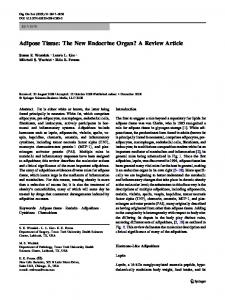
Figure 1
Histidine decarboxylase (HDC) activity (a) and histamine content (b) in cancerous and healthy tissues of the breast. The columns indicated t h e median values of the 10 patients under study. *p < 0.01 (Wilcoxon test): healthy mammary tissue compared to tulnour tissue. The numbers in the figure refers to the patients numbers indicated in Table 1. For further information see "Materials and methods" section.
Determination of tissue histamine content
Statistical methods
The histamine concentration of each tissue sample was measured in the same crude enzyme extract used to determine the H D C - activity with the fluorometric - fluoroenzymatic assay described by Hesterberg et al. [8].
The results are expressed individually. Comparisons between healthy mammary and cancer tissue results were performed with the Wilcoxon test for independant data. Results
Determination of urinary histamine and N'-methylhistamine Both, histamine and its metabolite were determined by the mass-fragmentographic technique described by Oosting et al. [9].
I. Histidine decarboxylase (HDC) activity and histamine content in cancerous and healthy tissues of the breast Fig. 1 represents the results obtained for (a) H D C activity and (b) histamine content in the four dif-
230 ferent tissues examined. The H D C activity in all except 1 patient (Pat. 1), is higher in the t u m o u r tissue than in the healthy m a m m a r y gland tissue. The median value differs by 22%, the greatest difference is 45% (Pat. 4). This difference, however, is not significant on the basis o f p < 0.05. The cancer tissue also shows higher HDC-activity than skin and muscle (significant ( p < 0 . 0 5 ) c o m p a r e d to muscle). In contrast, the histamine content is significantly lower ( p < 0 . 0 1 ) in the cancer tissue c o m p a r e d to healthy m a m m a r y gland tissue. The median value differs by 53%. All o f the patients had lower concentrations in the cancer tissue. C o m p a r i s o n o f the histamine contents o f healthy m a m m a r y tissue and skin showed similar values in each o f the patients; the values in muscle, however, were approximately 10 times higher. 2. U r i n a r y levels o f h i s t a m i n e a n d N~-methylhbtamine
The urinary excretion pattern o f histamine and N~-methylhistamine was similar in the 3 patients studied (not shown). The value showed a permanent decline from the preoperative values to day 7 after the operation. The difference was statistically significant (p