Epilepsia, 50(6):1385–1395, 2009 doi: 10.1111/j.1528-1167.2008.01730.x
FULL-LENGTH ORIGINAL RESEARCH
Changes in individual and group spatial and verbal learning characteristics after anterior temporal lobectomy ∗
yMario F. Dulay, ∗ yzHarvey S. Levin, yxMichele K. York, zXiaoqi Li, yx{#Eli M. Mizrahi, yx{Ian Goldsmith, ∗∗ Amit Verma, yx{Alica Goldman, yyRobert G. Grossman, and ∗ yDaniel Yoshor Departments of ∗ Neurosurgery, yBaylor Comprehensive Epilepsy Center, zPhysical Medicine and Rehabilitation, xNeurology, {Peter Kellaway Section of Neurophysiology in Neurology, and #Pediatric Neurology, Baylor College of Medicine, Houston, Texas, U.S.A.; and Departments of ∗∗ Neurology and yyNeurosurgery, The Methodist Hospital Neurological Institute, Houston, Texas, U.S.A.
SUMMARY Purpose: To evaluate the effects of anterior temporal lobectomy (ATL) on individual and group spatial and verbal learning and memory abilities as a function of side of surgery and seizure control outcome. Methods: We evaluated pre- and postsurgical learning and memory abilities of 75 left-hemisphere language dominant individuals who underwent ATL (33 left, 42 right) using the 8-trial Nonverbal Selective Reminding test and the 12-trial Verbal Selective Reminding test. Results: Reliable change index methods indicated that 40.5% of individuals who underwent rightATL had a clinically significant decline in spatial memory, and 62.5% of individuals who underwent left-ATL had a significant reduction in verbal memory. Growth curve analyses indicated that both side of surgery and poor seizure outcome
Modern theories of medial temporal lobe functioning agree that the hippocampus and parahippocampal gyrus are important for learning and memory (Squire et al., 2004; Moscovitch et al., 2006). While verbal memory impairment is a robust finding after individuals undergo left-anterior temporal lobectomy (ATL) (Milner, 1968; Novelly et al.,
Accepted May 29, 2008; Early View publication July 24, 2008. Address correspondence to Mario F. Dulay, Department of Neurosurgery, Methodist Neurological Institute, 6560 Fannin St., Suite 944, Houston, TX 77030. E-mail:
[email protected] Wiley Periodicals, Inc. ª 2008 International League Against Epilepsy
independently affected the learning slope in the best fitting models. Left-ATL reduced the slope, but did not affect the overall shape, of verbal learning across trials. On the other hand, poor seizure control outcome affected the slope of spatial learning regardless of the side of surgery. Discussion: Results demonstrate both individual and group declines in spatial memory and learning after ATL. Results suggest that individuals who undergo right-ATL should be counseled regarding the likelihood of a decline in spatial memory and learning abilities after ATL. Results also suggest that individuals with poor seizure control after ATL should be referred for rehabilitation services given the significant declines in spatial and verbal memory that occurred in our sample regardless of side of surgery. KEY WORDS: Nonverbal memory, Spatial learning, Verbal learning, Surgery outcome, Anterior temporal lobectomy.
1984; Hermann et al., 1992a, 1992b; Naugle et al., 1993; Sass et al., 1994), results are equivocal regarding decline in nonverbal/visual memory after right-ATL (see Lee et al., 2002 and Vaz, 2004 for reviews). Several studies report significant declines in nonverbal memory (Chiaravalloti & Glosser, 2004; Helmstaedter et al., 2004), others report no significant change (Martin et al., 1998; Rausch et al., 2003), and still others report improvements (Saykin et al., 1989; Graydon et al., 2001) after right-ATL. The variability among studies has been attributed to use of single trial nonverbal memory tests, use of test materials and procedures that allow for verbal processing of the information, and task complexity that necessitates involvement of other
1385
1386 M. F. Dulay et al. cognitive functions in addition to visual memory (Barr, 1997; Jones-Gotman et al., 1997). One aim of the present study was to evaluate the effects of ATL on visual learning and memory as a function of side of surgery. Whereas many studies have evaluated change in free-hand drawing or recognition memory of geometric designs from before to after ATL (Novelly et al., 1984; Ivnik et al., 1987; Saykin et al., 1989; Goldstein & Polkey, 1993; Seidenberg et al., 1998; Graydon et al., 2001; Rausch et al., 2003; Jansky et al., 2005; Morino et al., 2006), few studies have evaluated change in spatial learning (Trenerry et al., 1993; Chiaravalloti & Glosser, 2004; Kneebone et al., 2007). Memory for spatial locations, such as route finding of a location on a map, may be a sensitive indicator of right temporal lobe dysfunction according to neuroimaging studies (Crane et al., 1997; Burgess et al., 2001; Duzel et al., 2003). Many studies in patients with intractable epilepsy have demonstrated impaired spatial memory for individuals who underwent right-ATL compared to left-ATL (left side of Table 1), but it is unclear from these studies whether or not the impairment existed before surgery. Further, other studies reported no lateralized differences (right side of Table 1). We evaluated both individual and group changes in spatial memory and learning using the Nonverbal Selective Reminding test (NVSRT) (Fletcher, 1985). Poor performance on visual selective reminding tests occurs in individuals with right temporal lobe dysfunction (Giovagnolli et al., 1995; Plenger et al., 1996; Bell et al., 2005; Giovagnolli et al., 2005) although no data exist regarding post-ATL performance. Another goal of the present study was to evaluate change in learning characteristics (rate of acquisition, shape of the learning function) on tests of spatial and verbal learning and memory after ATL. Several studies reported on the slope of verbal learning before and after left-ATL (Hermann et al., 1992a, 1992b; Hermann et al., 1994). However, these slope functions were calculated based on a simple linear model, which may be misleading given that research in other populations indicate that learning occurs in a curvilinear fashion (Jones et al., 2005; Warschausky et al., 2005). Importantly, change in the slope of a spatial learning function has not been previously characterized, because examiners routinely conduct only a single learning trial or have tested spatial learning only after ATL. Change in learning as a function of seizure frequency outcome was also considered given the finding that poor seizure control after ATL is associated with impaired cognitive functioning (Novelly et al., 1984; Sanyal et al., 2005). In summary, the present study compared performance of patients who underwent left-ATL with patients who underwent right-ATL on an 8-trial NVSRT and a 12-trial Verbal Selective Reminding test (VSRT). The goals were to evaluate the following: (1) Individual and group changes in spatial and verbal learning and memory scores after ATL; (2) change in learning characteristics (rate of acquisition, Epilepsia, 50(6):1385–1395, 2009 doi: 10.1111/j.1528-1167.2008.01730.x
Table 1. Studies comparing postsurgical performance between right- and left-ATL patients on measures of spatial learning and memory Significantly poorer performance for right-ATL versus left-ATL
No significant difference between right-ATL and left-ATL
Abrahams et al. (1997) Corkin (1965) Feigenbaum & Morris (2004)a Goldstein et al. (1989)b,c Milner (1965) Nunn et al. (1998, 1999) Parslow et al. (2005) Petrides (1985) Pigott & Milner (1993) Pillon et al. (1999) Smith & Milner (1981) Spiers et al. (2001) Worsley et al. (2001)
Chiaravalloti & Glosser (2004)wnl Feigenbaum & Morris (2004)a,wnl Goldstein et al. (1989)b,c Kessels et al. (2004) Kneebone et al. (2007) Maguire et al. (1996)c Owen et al. (1995)c Trenerry et al. (1993)
wnl, indicates that spatial memory scores were within normal limits compared to normative data or a normal control group. a There was no significant difference between left/right or controls when using an egocentric spatial memory task, but the right-ATL group performed significantly poorer using an allocentric spatial memory task. b Goldstein et al. (1989) found no significant difference among left/right or controls when using an egocentric spatial memory task, but the right-ATL and left-ATL groups performed significantly poorer than controls using an allocentric spatial memory task. c Indicates memory test performance was impaired compared to normative data, but not significantly different between left-ATL and right-ATL patients.
shape of the learning function) across repeated trials before and after ATL; and (3) taking into account the effects of side of surgery and seizure control outcome on learning and memory.
Methods Sample The Baylor College of Medicine Institutional Review Board approved the study. Seventy-five individuals who sought treatment for intractable epileptic seizures at the Baylor College Comprehensive Epilepsy Center in Houston, TX, between 1986 and 2003 were included in the study. Forty-two individuals underwent right-ATL and 33 underwent left-ATL. Patients in the study had complete pre- and postsurgical neuropsychological data and a presurgical full-scale intelligence quotient (FSIQ) greater than 75 (Wechsler, 1981, 1997). Only patients who were lefthemisphere language-dominant, based on the intracarotid amobarbital procedure testing (Loring et al., 1994), were included in the study. Sixty-seven percent of the sample
1387 Learning Before and After ATL Table 2. Demographic, seizure-related, IQ, and pathology information divided by side of surgery, and then divided by good or poor seizure outcome Side of surgery
Seizure control outcome
Right-ATL (n ¼ 42) M Age (years) Education (years) Age at onset (years) Duration of illness (years) Pre-ATL seizure frequency (month) Post-ATL seizure frequency (month) Presurgical FSIQ Neuropathology Classic AHS Atypical MTS Ganglioglioma Other
31.5 12.3 14.4 17.2 28.5 2.2 92.6
Left-ATL (n ¼ 33) SD
10.4 1.9 8.9 8.9 24.5 6.5 13.5
64.3% 14.3% 14.3% 7.1%
M 29.5 13.1 13.2 16.3 18.0 2.4 93.1 62.5% 11.8% 15.6% 10.1%
Good (n ¼ 60) SD
7.8 2.1 8.7 9.1 23.9 5.9 13.4
M 30.7 12.6 13.6 17.1 19.3 0.6 92.3 70.0% 05.0% 13.3% 11.7%
Poor (n ¼ 15) SD
M
SD
9.8 2.1 9.1 9.2 30.0 1.7 13.5
30.6 12.9 14.8 15.8 38.1a 9.0 b 94.8
6.7 1.9 7.8 7.7 39.9 9.2 14.6
20.0% 46.7%b 13.3% 20.0%
Preoperative assessment was performed an average of 6.2 months (SD ¼ 1.7) prior to surgery and the postoperative findings were obtained an average of 11.8 months (SD ¼ 6.7) after surgery. There were no significant side of surgery differences for any of the variables. a p ¼ 0.07 compared to the good outcome group. b p < 0.001 compared to the good outcome group. AHS, Ammon’s horn sclerosis; MTS, mesial temporal sclerosis; ATL, anterior temporal lobectomy; M, mean; SD, standard deviation; FSIQ, full-scale intelligence quotient.
was female. Ninety-two percent of the sample was right-handed, 6% left-handed, and 2% ambidextrous (Oldfield, 1971). Other demographic, disease-related, IQ, and pathology variables separated by side of surgery and seizure control outcome are presented in Table 2. Seizure localization Seizure localization and lateralization were confirmed upon concordance of 24-hour video electroencephalography (EEG) monitoring and neuroimaging data (York et al., 2003). Seizure monitoring involved scalp electrodes, often with follow-up unilateral or bilateral sphenoidal, depth, or subdural electrodes. Seizures were typically recorded over a period lasting about a week (Binne & Mizrahi, 1997). Neuropathology Resected tissue was analyzed based on criteria established at the 1987 International Conference on the Surgical Treatment of Epilepsy (Armstrong, 1993). The four pathology groups were classic Ammon’s horn sclerosis, atypical mesial temporal lobe sclerosis (Armstrong, 2005), low-grade tumoral tissue (primarily ganglioglioma, with several dysembroplastic neuroepithelial tumor), or other (inconclusive, infarct, arteriovenous malformation, microdysgenesis). Surgical procedure A detailed description of the standard surgical procedure used with our patients has been published (Yoshor et al.,
2006). Briefly, the anterior 3.5–4.0 cm of lateral temporal neocortex was resected en bloc from the superior or middle temporal gyrus to the collateral fissure. Then, the medial temporal lobe, including the anterior 2.5–3.0 cm of the hippocampal formation, the subiculum, the parahippocampal gyrus, and the inferior amygadala were removed using an operating microscope. The extent of resection of mesial temporal lobe structures and lateral temporal neocortex did not vary significantly according to the side of surgery. No instances of wound or intracerebral infection, hemiparesis, or death occurred following ATL. Surgeries were performed by one surgeon (R.G.G.) between 1986 and 1999 and by another neurosurgeon (trained by R.G.G.) between 2000 and 2003 (D.Y.). Surgical outcome for seizure control was based on Engel’s classification scheme (Engel et al., 1993). Engel class I was defined as a good outcome of surgery, and Engel classes II to IV were defined as poor outcome of surgery. Retrospective chart review indicated that only one patient from the good seizure outcome group was not taking antiepileptic medication, because he had taken himself off of the medicine, at the postsurgical neuropsychological evaluation session. Memory testing procedures Learning and memory were tested an average of 6.2 months (sd ¼ 1.7) prior to surgery and an average of 11.8 months (sd ¼ 6.7) after surgery. Outcome measures included the consistent long-term retrieval and delayed free recall indices of the NVSRT (Fletcher, 1985) and the VSRT Epilepsia, 50(6):1385–1395, 2009 doi: 10.1111/j.1528-1167.2008.01730.x
1388 M. F. Dulay et al. (Buscke & Fuld, 1974), as well as a percent retention index calculated for this study. Testing followed standardized procedures. Detailed description of the procedures is provided in Supplementary Text 1. Statistical analyses First, possible between-group (side of surgery, seizure outcome) differences in demographic, seizure related, IQ, and pathology variables were assessed using analysis of variance (ANOVA) and chi-square (v2). Second, the reliable change index (RCI) method was used to determine the rate of clinically significant reductions, no change or clinically significant improvements in individual patient memory and learning scores from before to after surgery using a 95% RCI criterion (i.e., €1.64 z-score change) (Jacobson & Truax, 1991). Supplementary Text 2 provides details about the parameters used when calculating the RCIs. Third, a doubly multivariate analysis with repeated measures was carried out with side of surgery (right, left) and seizure outcome (good, poor) as between-group factors and time (presurgery, postsurgery) as the within-subject factor with consistent long-term retrieval (CLTR) nonverbal and verbal memory included simultaneously. The same analysis was conducted separately substituting delayed recall, and then percent retention, for a total of three multivariate repeated measures analyses. Effect sizes (gp2) were reported using guidelines recommended by Green and Salkind (2003) for interpretation: 0.01 represents a small effect size, above 0.06 a medium effect size, and above 0.14 a large effect size. Raw scores were used in all analyses calculated with SPSS 15.0 (SPSS Inc., Chicago, IL, U.S.A.). Finally, four growth curve models were calculated using PROC MIXED in SAS (Littell et al., 1996) to evaluate the changes in learning characteristics over trials. The goals of evaluating these models were to (1) describe the trajectories of spatial and verbal learning functions before surgery and after surgery and to (2) evaluate the effects of side of surgery and seizure outcome on the intercept, slope, and change in slope of the postsurgical learning functions. Evaluating the growth curve models involved the following: Level 1, tests four random coefficients models of individual changes over trials (linear, quadratic, cubic) based on goodness of model fit (compared the -2 log likelihood statistics and computed the likelihood ratio v2 statistics and p-values). Level 2, enter seizure outcome and side of surgery as trial invariant variables into the models examined in level 1 analyses to test their effect on the intercept, slope, and change in slope on postsurgery spatial and verbal learning. For interpretation purposes, ‘‘trial’’ was centered at the fourth trial for spatial learning and was centered at the sixth trial for verbal learning. The intercept tells us the level of a learning score at that corresponding trial, the slope tells the rate of change in the score and the curvature tells us the degree to which the slope is changing. Epilepsia, 50(6):1385–1395, 2009 doi: 10.1111/j.1528-1167.2008.01730.x
Results Descriptive statistics No missing data or out of range values were encountered. Studentized residuals were calculated to evaluate the presence of outliers. An outlier was found for number of monthly seizures in the left-ATL group (120 events a month, or an average of four a day), and two outliers were found in the right ATL group (116 and 123 events a month). A series of one-way ANOVAs and v2 analyses were computed to examine potential differences by side of surgery. The left-ATL and right-ATL groups did not differ significantly in age, education, age at onset of seizures, duration of illness, pre- or postsurgical seizure frequency, presurgical FSIQ, rate of underlying neuropathology, or time since surgery when neuropsychological testing occurred (left side of Table 2). The rate of seizure cessation was similar after left-ATL (78.6%) or right-ATL (83.3%) (Fisher’s exact test p-value ¼ 0.77). One-way ANOVA indicated that good seizure and poor seizure outcome groups did not differ significantly in age, education, age at onset of seizures, duration of illness or FSIQ (right side of Table 2). Chi square analyses indicated that patients with atypical mesial temporal sclerosis were more likely to have poor seizure outcome compared to other groups (v2 ¼ 18.6, p < 0.001), and patients with classic Ammon’s horn sclerosis were more likely to have good seizure outcome. Individual changes: RCI RCIs were calculated for each dependent variable to determine whether change in learning and memory scores from before to after surgery were clinically meaningful and significant. Table 3 shows the rates of learning and memory changes after ATL as a function of side of surgery. Post hoc v2 analysis comparing individual cells indicated that significantly more right-ATL patients declined on spatial memory and learning indices than left-ATL patients. For example, 40.5% right-ATL patients demonstrated a clinically significant decline on spatial delayed recall as compared to 12.5% left-ATL patients (v2 ¼ 7.1, p < 0.01). On the other hand, 62.5% left-ATL patients showed clinically significant decline on verbal delayed recall as compared 28.6% right-ATL patients (v2 ¼ 8.4, p < 0.01). Doubly multivariate analysis with repeated measures A doubly multivariate analysis with repeated measures was carried out with side of surgery (right, left) and seizure outcome (good, poor) as between-group factors and time (presurgery, postsurgery) as the within-subject factor with the CLTR memory type (spatial, verbal) dependent variables tested simultaneously. There was no significant fourway interaction. Results indicated a significant three-way memory type · side of surgery · time interaction (Hotelling’s Trace F[1,71] ¼ 14.6, p < 0.0001, gp2 ¼ 0.17). Review of the CLTR sum across trials scores ( Table 4) shows the
1389 Learning Before and After ATL Table 3. Reliable change index percentages for memory and learning scores for the right-ATL and left-ATL groups using a 95% RCI criterion Right-ATL Domain/test Spatial learning and memory CLTR sum across trials Delayed recall Verbal learning and memory CLTR sum across trials Delayed recall
Left-ATL
Decline
No change
Improve
Decline
No change
Improve
35.7% 40.5%
40.5% 47.6%
23.8% 11.9%
25.0% 12.5%
56.2% 68.7%
18.8% 18.8%
21.4% 28.6%
38.1% 28.6%
40.5% 42.8%
50.0% 62.5%
37.5% 9.4%
12.5% 28.1%
ATL, anterior temporal lobectomy; CLTR, consistent long-term retrieval.
pattern of the interaction revealing that spatial learning declined from before to after right-ATL and improved from before to after left-ATL. On the other hand, verbal learning improved from before to after right-ATL and declined from before to after left-ATL. Two-way side of surgery interactions were not interpreted because of the signification three-way interaction. Results also indicated significant seizure outcome by time interactions for spatial CLTR (Hotelling’s Trace F[1,71] ¼ 3.8, p ¼ 0.047, gp2 ¼ 0.06) and verbal CLTR (Hotelling’s Trace F[1,71] ¼ 9.4, p ¼ 0.003, gp2 ¼ 0.12) revealing that the poor seizure outcome group had significantly greater drops in learning scores from before to after surgery, and the good seizure outcome group maintained their level of learning from before to after surgery. Another doubly multivariate repeated measures analysis was carried out with spatial and verbal delayed recall tested simultaneously. There was no four-way interaction. A significant three-way memory type · side of surgery · time interaction (Hotelling’s Trace F[1,71] ¼ 14.4, p < 0.0001, gp2 ¼ 0.17) was found revealing that spatial delayed recall (Table 4) declined from before to after rightATL and remained relatively the same from before to after left-ATL. On the other hand, verbal delayed recall
improved from before to after right-ATL and remained the same from before to after left-ATL. Results also indicated a significant seizure outcome by time interaction for verbal delayed recall (Hotelling’s Trace F[1,71] ¼ 11.9, p ¼ 0.001, gp2 ¼ 0.15), revealing that the poor seizure outcome group had a significantly greater drop in recall scores from before to after surgery, but that the good seizure outcome group maintained their level of performance from before to after surgery. A final doubly multivariate repeated measures analysis was carried out with spatial and verbal percent retention tested simultaneously. There was no four-way interaction. A significant three-way memory type · side of surgery · time interaction (Hotelling’s Trace F[1,71] ¼ 4.9, p ¼ 0.030, gp2 ¼ 0.07) was found. Interpretation of the pattern of results was exactly the same as those found for delayed recall reported above. Results indicated significant seizure outcome by time interactions for spatial percent retention (Hotelling’s Trace F[1,71] ¼ 3.7, p ¼ 0.045, gp2 ¼ 0.06) and verbal percent retention (Hotelling’s Trace F[1,71] ¼ 4.9, p ¼ 0.029, gp2 ¼ 0.07) revealing that the poor seizure outcome group had a significantly greater drop in percent retention scores from before to after surgery, but that the
Table 4. Learning and memory data divided by side of surgery Right-ATL (n ¼ 42)
Spatial learning and memory CLTR sum across trials Delayed recall Percent retention Verbal learning and memory CLTR sum across trials Delayed recall Percent retention
Left-ATL (n ¼ 33)
pre mean (SD)
post mean (SD)
pre mean (SD)
post mean (SD)
32.0 (16.1) 5.1 (1.5) 79% (3.6)
28.9 (17.0) 4.2 (2.4)a 65% (4.1)
27.9 (15.7) 5.6 (1.5) 82% (2.9)
29.2 (17.3) 5.7 (1.6) 78% (3.8)
73.2 (32.8) 9.6 (2.3) 84% (3.1)
84.4 (35.2)b 9.6 (2.8)b 83% (4.2)b
67.2 (30.4) 8.1 (2.8) 53% (3.2)
45.6 (34.0) 6.0 (3.4) 40% (4.1)
ATL, anterior temporal lobectomy; SD, standard deviation; CLTR, consistent long-term retrieval. a p < 0.05 showing poorer performance for right-ATL compared to left-ATL for post-ATL delayed spatial recall. b Significantly better performance for right-ATL compared to left-ATL for post-ATL verbal learning and memory scores. Epilepsia, 50(6):1385–1395, 2009 doi: 10.1111/j.1528-1167.2008.01730.x
1390 M. F. Dulay et al. good seizure outcome group maintained their level of memory from before to after surgery. Growth curve analyses Curve fit (level 1) Four random coefficient models were examined with the intercept, slope, quadratic, and the cubic terms used as outcome measures for pre-ATL and post-ATL spatial and verbal learning. The likelihood ratio tests indicated that a quadratic model fit the data significantly better than a cubic or linear model for all four models. For presurgery spatial learning, variance estimates were significantly greater than zero indicating that learning scores were increasing with each trial with a deceleration of learning as trials progressed indicative of a quadratic function (intercept, Z ¼ 6.05, p < 0.0001; slope, Z ¼ 5.48, p < 0.0001; slope estimate ¼ 0.58; quadratic term, Z ¼ 4.05, p < 0.0001). A similar pattern was found for postsurgery spatial learning (intercept, Z ¼ 6.11, p < 0.0001; slope, Z ¼ 5.56, p < 0.0001; slope estimate ¼ 0.52; quadratic term, Z ¼ 4.64, p < 0.0001). Verbal learning scores showed a similar pattern with increased scores by trial and a deceleration of learning as trials progressed for both presurgery (intercept, Z ¼ 6.25, p < 0.0001; slope, Z ¼ 6.12, p < 0.0001; slope estimate ¼ 0.67; quadratic term, Z ¼ 5.54, p < 0.0001) and postsurgery (intercept, Z ¼ 6.15, p < 0.0001; slope, Z ¼ 6.11, p < 0.0001; slope estimate ¼ 0.63; quadratic term, Z ¼ 5.76, p < 0.0001). Effects of moderators (side of surgery, seizure outcome) on learning (level 2) The next step was to model the effects of side of surgery, seizure outcome, and the interaction of side of surgery/ seizure outcome on the shape of the postsurgical learning functions. Since the variances of intercept, slope, and curvature were significantly random in the level 1 analyses, these terms were set as random in the level 2 models. For spatial learning, the final model showed that seizure outcome had a significant effect on learning. Specifically, patients with a good seizure outcome had a larger intercept (average of 2.21 points higher at trial 4, t[390] ¼ 3.46, p ¼ 0.009) and had a larger slope (patients with good seizure outcome had a 0.21 points larger slope than those with poor outcome, t[390] ¼ 2.29, p ¼ .019) compared to patients with a poor seizure outcome. There was no significant effect of side of surgery or an interaction between side of surgery and seizure outcome for overall spatial learning across trials. For verbal learning, the final model showed that the good outcome group had a larger slope compared to the poor outcome group (average of 0.18 points higher, t[702] ¼ 2.22, p ¼ 0.021). Further, right-ATL patients had a larger intercept (average of 3.62 points higher at trial 6, t[702] ¼ 4.50, p < 0.0001), larger slope (average of 0.29 points higher, t[702] ¼ 3.72, p ¼ 0.0007), but quicker Epilepsia, 50(6):1385–1395, 2009 doi: 10.1111/j.1528-1167.2008.01730.x
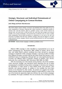
Figure 1. Shows the trial-by-trial learning rate of verbal learning (Verbal Selective Reminding test consistent long-term retrieval scores) before and after surgery divided by side of surgery. Left-ATL patients show a significant drop in verbal learning within and across trials from before to after surgery while right-ATL patients showed improvements. Epilepsia ILAE
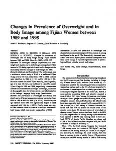
decreasing rate in the slope at later trials (rate ¼ )0.039, t[702] ¼ -2.42, p ¼ 0.023) compared to left-ATL patients. Fig. 1 shows the trial-by-trial verbal CLTR scores by side of surgery and pre- and postsurgery occasions. As can be seen pictorially, the slope of verbal learning, as reflected by the number of words consistently retrieved across trials, was significantly reduced following left-ATL and improved after right-ATL. The top panel in Fig. 2 shows the trial-by-trial rate of postsurgical spatial learning divided by side of surgery and seizure outcome. As the growth curve analyses demonstrated, seizure cessation partly moderated spatial learning ability such that patients who were seizure-free after surgery remembered the most spatial locations across trials regardless of side of surgery. The bottom panel in Fig. 2 shows the trial-by-trial rate of postsurgical verbal learning divided by side of surgery and seizure outcome. Side of surgery and seizure outcome (but not the interaction) influenced verbal learning across trials.
Discussion The purpose of this investigation was to characterize changes in spatial and verbal learning and memory abilities in left-hemisphere language dominant individuals who underwent unilateral ATL. This study is unique in that few studies have evaluated change in spatial learning and memory from before to after ATL (and no study has evaluated changes using a selective reminding procedure), no study has reported on the individual rates of clinically meaningful change in spatial learning after ATL, and no study has
1391 Learning Before and After ATL reduced the slope, but did not affect the overall shape, of verbal learning across trials. On the other hand, poor seizure control outcome affected the slope of spatial learning regardless of the side of surgery.
Figure 2. Shows the trial-by-trial rate of postsurgical spatial learning (top panel) and verbal learning (bottom panel) divided by side of surgery and seizure outcome. (Top panel) Seizure freedom partly moderated spatial learning ability such that patients who were seizurefree after surgery remembered the most spatial locations across trials regardless of side of surgery. (Bottom panel) Side of surgery and seizure outcome (but not the interaction) influenced verbal learning test performance across. Error bars represent standard error of the mean. Epilepsia ILAE
documented the effects of ATL on spatial learning characteristics. RCI methods indicated that 40.5% of individuals who underwent right-ATL had a clinically significant decline in spatial memory, and 62.5% of individuals who underwent left-ATL had a significant reduction in verbal memory. Growth curve analyses indicated that both side of surgery and poor seizure outcome independently affected the learning slope in the best fitting models. Left-ATL
Individual rates of change in spatial learning after ATL Our results demonstrate that more than a third of individuals who underwent right-ATL (36% to 41%) had clinically meaningful change in spatial learning and memory (Table 3), which was statistically greater than the declines that occurred after left-ATL (13% to 25%). However, approximately 48% of the sample showed no change after right-ATL and 12% showed improvements in spatial delayed recall. Results further demonstrate the problem that arises when using group means as the individual changes in performance cancel each other out when impairments are subtracted from improvements in performance. Table 1 documents the conflicting data when using group means for spatial memory data. A review of the verbal memory scores shows, as has been previously documented, that verbal learning and memory declines were large (between 50% to 63%) after left-ATL with unexpected but smaller declines after right-ATL (between 21% to 29%). Our review of the literature found eight studies that reported individual nonverbal memory changes after a unilateral temporal lobectomy (Hermann & Wyler, 1988; Chelune et al., 1993; Phillips & McGlone, 1995; Martin et al., 1998; Walton et al., 1999; Gleissner et al., 2002; Henke et al., 2003; Lineweaver et al., 2006). Each study used different criteria to define significant change after ATL and used different measures of nonverbal memory, thus direct comparisons are difficult. The most commonly used measure was the Wechsler Memory Scale-R (WMS-R) or III (e.g., figural reproduction) with rates of decline after right-ATL ranging from 0%–28% (Martin et al., 1998; Walton et al., 1999; Chelune et al., 1993; Gleissner et al., 2002; Lineweaver et al., 2006). Walton et al. (1999) used multiple visual memory measures and found a 20% reduction in Warrington’s Recognition Memory test performance and 11% reduction in Rey-Complex Figure test performance after right-ATL. Henke et al. (2003) found that 76% of individuals declined on the WMS-R nonverbal index, although the definition of decline was any raw score reduction after surgery. We found that decline in spatial memory after nonlanguage dominant ATL was more frequent than the common view that there are few adverse effects of this surgery. Group change after ATL Even though group change can mask individual differences, analyses of these effects are informative since the bulk of literature has reported group data. Our results indicated significant declines in performance after right-ATL for delayed spatial recall. Thus, our results partially Epilepsia, 50(6):1385–1395, 2009 doi: 10.1111/j.1528-1167.2008.01730.x
1392 M. F. Dulay et al. support the material specificity hypothesis of memory functioning that proposes that human left and right mesial temporal lobes are differentially involved in the delayed retrieval of verbal or nonverbal (typically visual) memories (Milner, 1974; Milner et al., 1998). The major problem with reaching a more definitive conclusion based on our data is that a proportion of individuals (Table 3) actually show improvements in spatial learning after right-ATL, and there are unexplained improvements in verbal learning after left-ATL (see Supplementary Text 3 for a more thorough discussion regarding the validity of nonverbal memory as a construct). One reason that visual memory impairments are not consistently found across studies may be because a larger distributed neuroanatomical network is involved besides those structures removed during a right temporal lobe resection (Elger et al., 2004; and see Supplementary Text 3 for an extensive discussion regarding the neural networks involved in the processing of different types of visual memories). We found a significant three-way interaction, such that spatial learning declined after right-ATL and improved after left-ATL and verbal learning improved after rightATL and declined after left-ATL. However, decline in spatial learning was small (3 points or 10%) after right-ATL and not significantly different postoperatively compared to the left-ATL patients ( Table 4). We infer that patients who undergo right-ATL have a similar ability to encode locations of information compared to left-ATL given the similar performance across learning trials (Fig. 2, top), but that right-ATL leads to rapid forgetting of spatial locations after a delay for some patients (Smith & Milner, 1989; see Burgess, 2002 for a review). Results are concordant with some studies, but not with others. Jones-Gotman (1986) showed slow learning for the first five trials of design learning for right-ATL, but by trials 6–10, the learning was equivalent between left-ATL and right-ATL patients. Others have shown impaired nonverbal learning with intact delayed recall in individuals with right-sided temporal lobe epilepsy (Giovagnoli et al., 1995) or post–right-ATL (Majdan et al., 1996; Jones-Gotman et al., 1997) compared to left-sided patients using design memory across trials. Jones-Gotman attributed this pattern to ‘‘the novelty of the designs which results in a more wide-spread cerebral representation of those stimuli, making them more resistant to forgetting’’ (Jones-Gotman et al., 1997, p. 971). There are also studies that show no impairments for both nonverbal learning trials or delayed recall (Barr et al., 2004; Bell et al., 2005) or impaired performance on both indices (Helmstaedter et al., 1991; Glosser et al., 2002; Crane & Milner, 2005). Importantly, all but one of these studies pertained to design memory. The one study that tested objectlocation memory across trials (Crane & Milner, 2005) found impairments for both left-ATL and right-ATL patients for learning and delayed recall. Our results are inconsistent with other studies that reported no change in Epilepsia, 50(6):1385–1395, 2009 doi: 10.1111/j.1528-1167.2008.01730.x
group means from before to after ATL using different spatial memory methods including an adapted Corsi-block test called the Spatial Sequential Learning Task (Chiaravalloti & Glosser, 2004), the Rey-Complex Figure test modified to evaluate spatial memory (Kneebone et al., 2007), and the Visual Spatial Learning test where abstract designs in specific locations are learned (Trennery et al., 1993). Slope and shape learning characteristics Growth curve analyses indicated that the best-fitting model for the shape of the learning function was curvilinear (quadratic or decelerating) both before and after ATL for spatial and verbal learning. Left-ATL affected the slope, but not the shape, of verbal learning (Fig. 1). On the other hand, poor seizure control was the dominant factor moderating change in slope (but not shape) after surgery for spatial learning regardless of side of surgery (Fig. 2). Results are consistent with other studies that demonstrate verbal learning changes from before to after left-ATL (Hermann et al., 1992a, 1992b; Hermann et al., 1994). However, results call into question the validity of using an index such as a learning slope calculated based on a simple linear function (e.g., as commonly used by the California Verbal Learning test and other list-learning tests) given that the learning slope is a decelerating function. The learning slope is a measure of average gains in performance across learning trials. Slope calculations based on a simple linear regression model assumes continued improvement across trials. The linear slope statistic would be misleading because it would average early learning trials (where learning occurs at a greater rate) and later learning trials (where there is a leveling off) leading to an underestimate in performance, especially for those individuals who initially perform well. This would be especially problematic when group comparisons are made because the incremental changes in slope would be misleading as they would suggest no difference in rate of learning across trials when learning has either reached a ceiling or optimal performance. As recommended by others (e.g., Warschausky et al., 2005), when studying learning in a different patient population, calculation of learning functions should either be based on curvilinear computations or should be reported as a limited slope (e.g., exclusion of the trials where learning has leveled off or flattened near the end of trials) though these methods have not fully been validated. Application We suggest presurgical counseling is necessary regarding the likelihood of a decline in spatial memory and learning abilities after right-ATL, which could help to minimize frustration associated with new onset problems. Episodic spatial recall difficulties found in a clinical setting may signify spatial memory impairment in every day life, such as getting lost when navigating in a new environment or misplacing ones keys and other belongings. The decline after
1393 Learning Before and After ATL surgery may be noticeable if the person is in a spatialrelated job (e.g., architect, machine operator), especially for patients with better presurgical nonverbal memory ability who are more likely to have a noticeable drop (Gleissner et al., 2002). Referral for rehabilitation services would be important to help mitigate the effects of surgery. Recent research suggests that rehabilitation interventions that combine memory-encoding strategies that actively involve patient participation with the use of external cues (e.g., reminder notes) are effective at mitigating memory difficulties in seizure disorder patients (Schefft et al., 2008). Rehabilitation may be particularly important for individuals with poor memory and poor seizure outcome given that self-report quality of life is good for individuals with postsurgical memory impairment and no seizures, but poor for individuals with both memory impairment and continued seizures (Langfitt et al., 2007).
Conclusion In summary, our results demonstrate significant individual declines in spatial memory after ATL. Group means were somewhat misleading in that there were both declines and improvements in spatial learning after right-ATL that canceled each other out to some degree based on individual data. Both side of surgery and poor seizure outcome independently influenced the slopes of the learning functions in the best fitting growth curve models. Results suggest that individuals who will undergo right-ATL should be counseled regarding the likelihood of a decline in spatial memory abilities after ATL. Results also suggest that individuals with poor seizure outcome should be referred for rehabilitation services given the significant impairments that exist after ATL regardless of side of surgery. Further study of learning and memory characteristics of persons with epileptic seizures could lead to effective rehabilitation programs tailored to specific learning difficulties.
Acknowledgments Conflict of interest: We confirm that we have read the Epilepsia position on issues involved in ethical publication and affirm that this report is consistent with those guidelines. The authors have no conflicts of interest to disclose.
References Abrahams S, Pickering A, Polkey CE, Morris RG. (1997) Spatial memory deficits in patients with unilateral damage to the right hippocampal formation. Neuropsychologia 35:11–24. Armstrong DD. (1993) The neuropathology of temporal lobe epilepsy. J Neuro Pathol Exp Neurol 52:541–552. Armstrong DD. (2005) Epilepsy-induced microarchitectural changes in the brain. Pediatr Dev Pathol 8:607–614. Barr WB. (1997) Examining the right temporal lobe’s role in nonverbal memory. Brain Cogn 35:26–41. Barr W, Morrison C, Zaroff C, Devinsky O. (2004) Use of the Brief Visuospatial Memory Test-Revised (BVMT-R) in neuropsychologi-
cal evaluation of epilepsy surgery candidates. Epilepsy Behav 5: 175–179. Bell BD, Fine J, Dow C, Seidenberg M, Hermann BP. (2005) Temporal lobe epilepsy and the selective reminding test: the conventional 30-minute delay suffices. Psychol Assess 17:103–109. Binne CD, Mizrahi EM. (1997) The epilepsy-monitoring unit. In Engel J, Pedley TA (Eds) Epilepsy: a comprehensive textbook. LippincottRaven Publishers, Philadelphia, pp. 1011–1019. Burgess N. (2002) The hippocampus, space, and viewpoints in episodic memory. Q J Exp Psychol 55:1057–1080. Burgess N, Maguire EA, Spiers H, O’Keefe J. (2001) A temporoparietal and prefrontal network for retrieving the spatial context of lifelike events. Neuroimage 14:439–453. Buschke H, Fuld PA. (1974) Evaluation of storage, retention, and retrieval in disordered memory and learning. Neurol 11:1019–1025. Chelune GJ, Naugle RI, Luders H, Sedlak J, Awad IA. (1993) Individual change after epilepsy surgery: practice effects and base-rate information. Neuropsychology 7:41–52. Chiaravalloti ND, Glosser G. (2004) Memory for faces dissociates from memory for location following anterior temporal lobectomy. Brain Cogn 54:35–42. Corkin S. (1965) Tactually-guided maze learning in man: effects of unilateral cortical excisions and bilateral hippocampal lesions. Neuropsychologia 3:339–351. Crane J, Milner B. (2005) What went where? Impaired object-location learning in patients with right hippocampal lesions. Hippocampus 15:216–231. Crane J, Johnsrude IS, Milner BA, Owen AM, Evans AC. (1997) Right hippocampal activation during recognition tests of object location memory: a PET study. NeuroImage 5:S625. Elger CE, Helmstaedter C, Kurthen M. (2004) Chronic epilepsy and cognition. Lancet Neurol 3:663–672. Engel J Jr, Van Ness PC, Rasmussen TB, Ojemann LM. (1993) Outcome with respect to epileptic seizures. In Engel J Jr (Ed) Surgical treatment of the epilepsies. Raven Press, New York, pp. 609–621. Feigenbaum JD, Morris RG. (2004) Allocentric versus egocentric spatial memory after unilateral temporal lobectomy in humans. Neuropsychology 18:462–472. Fletcher JM. (1985) Memory for verbal and nonverbal stimuli in learning disability subgroups: analysis by selective reminding. J Exp Child Psychol 40:244–259. Giovagnoli AR, Casazza M, Avanzini G. (1995) Visual learning on a selective reminding procedure and delayed recall in patients with temporal lobe epilepsy. Epilepsia 36:704–711. Giovagnoli AR, Erbetta A, Villani F, Avanzini G. (2005) Semantic memory in partial epilepsy: verbal and non-verbal deficits and neuroanatomical relationships. Neuropsychologia 43:1482–1492. Gleissner U, Helmstaedter C, Schramm J, Elger CE. (2002) Memory outcome after selective amygdalohippocampectomy: a study in 140 patients with temporal lobe epilepsy. Epilepsia 43:87–95. Glosser G, Cole L, Khatri U, DellaPietra L, Kaplan E. (2002) Assessing nonverbal memory with the Biber Figure Learning Test-Extended in temporal lobe epilepsy patients. Arch Clin Neuropsychol 17:25–35. Goldstein LH, Polkey CE. (1993) Short-term cognitive changes after unilateral temporal lobectomy or unilateral amygdalo-hippocampectomy for the relief of temporal lobe epilepsy. J Neurol Neurosurg Psychiatry 56:135–140. Goldstein LH, Canavan AG, Polkey CE. (1989) Cognitive mapping after unilateral temporal lobectomy. Neuropsychologia 27:167–177. Graydon FJ, Nunn JA, Polkey CE, Morris RG. (2001) Neuropsychological outcome and the extent of resection in the unilateral temporal lobectomy. Epilepsy Behav 2:140–151. Green SB, Salkind NJ. (2003) Using SPSS for Windows and Macintosh: analyzing and understanding data. 3rd ed. Prentice Hall, Upper Saddle River, NJ. Helmstaedter C, Pohl C, Hufnagel A, Elger CE. (1991) Visual learning deficits in nonresected patients with right temporal lobe epilepsy. Cortex 27:547–555. Helmstaedter C, Van Roost D, Clusmann H, Urbach H, Elger CE, Schramm J. (2004) Collateral brain damage, a potential source of cognitive impairment after selective surgery for control of mesial temporal lobe epilepsy. J Neurol Neurosurg Psychiatry 75:323–326. Epilepsia, 50(6):1385–1395, 2009 doi: 10.1111/j.1528-1167.2008.01730.x
1394 M. F. Dulay et al. Henke K, Treyer V, Weber B, Nitsch RM, Hock C, Wieser HG, Buck A. (2003) Functional neuroimaging predicts individual memory outcome after amygdalohippocampectomy. Neuroreport 14:1197–1202. Hermann BP, Wyler AR. (1988) Neuropsychological outcome of anterior temporal lobectomy. J Epilepsy 1:35–45. Hermann BP, Wyler AR, Bush AJ, Tabatabai FR. (1992a) Differential effects of left and right anterior temporal lobectomy on verbal learning and memory performance. Epilepsia 33:289–297. Hermann BP, Wyler AR, Somes G, Berry AD III, Dohan FC. (1992b) Pathological status of the mesial temporal lobe predicts memory outcome from left anterior temporal lobectomy. Neurosurgery 31: 652–657. Hermann BP, Wyler AR, Somes G, Dohan FC Jr, Berry AD 3rd, Clement L. (1994) Declarative memory following anterior temporal lobectomy in humans. Behav Neurosci 108:3–10. Ivnik RJ, Sharbrough FW, Laws ER Jr. (1987) Effects of anterior temporal lobectomy on cognitive function. J Clin Psychol 43:128–137. Janszky J, Jokeit H, Kontopoulou K, Mertens M, Ebner A, Pohlmann-Eden B, Woermann FG. (2005) Functional MRI predicts memory performance after right mesiotemporal epilepsy surgery. Epilepsia 46:244–250. Jacobson NS, Truax P. (1991) Clinical significance: a statistical approach to defining meaningful change in psychotherapy research. J Consult Clin Psychol 59:12–19. Jones RN, Rosenberg AL, Morris JN, Allaire JC, McCoy KJ, Marsiske M, Kleinman KP, Rebok GW, Malloy PF. (2005) A growth curve model of learning acquisition among cognitively normal older adults. Exp Aging Res 31:291–312. Jones-Gotman M. (1986) Right hippocampal excision impairs learning and recall of a list of abstract designs. Neuropsychologia 24: 659–670. Jones-Gotman M, Zatorre RJ, Olivier A, Andermann F, Cendes F, Staunton H, McMackin D, Siegel AM, Wieser HG. (1997) Learning and retention of words and designs following excision from medial or lateral temporal-lobe structures. Neuropsychologia 35:963–973. Kessels RP, Hendriks M, Schouten J, Van Asselen M, Postma A. (2004) Spatial memory deficits in patients after unilateral selective amygdalohippocampectomy. J Int Neuropsychol Soc 10:907–912. Kneebone AC, Lee GP, Wade LT, Loring DW. (2007) Rey Complex Figure: figural and spatial memory before and after temporal lobectomy for intractable epilepsy. J Int Neuropsychol Soc 13:664–671. Langfitt JT, Westerveld M, Hamberger MJ, Walczak TS, Cicchetti DV, Berg AT, Vickrey BG, Barr WB, Sperling MR, Masur D, Spencer SS. (2007) Worsening of quality of life after epilepsy surgery: effect of seizures and memory decline. Neurology 68:1988–1994. Lee TM, Yip JT, Jones-Gotman M. (2002) Memory deficits after resection from left or right anterior temporal lobe in humans: a metaanalytic review. Epilepsia 43:283–291. Lineweaver TT, Morris HH, Naugle RI, Najm IM, Diehl B, Bingaman W. (2006) Evaluating the contributions of state-of-the-art assessment techniques to predicting memory outcome after unilateral anterior temporal lobectomy. Epilepsia 47:1895–1903. Littell RC, Milliken GA, Stroup WW, Wolfinger RD. (1996) SAS system for mixed models. SAS Institute, Cary, NC. Loring DW, Lee GP, Meador KJ. (1994) Intracarotid amobarbital (Wada) assessment. In Wyler AR, Hermann BP (Eds) The surgical management of epilepsy. Butterworth Heinemann, Boston, pp. 97–110. Maguire EA, Burke T, Phillips J, Staunton H. (1996) Topographical disorientation following unilateral temporal lobe lesions in humans. Neuropsychologia 34:993–1001. Majdan A, Sziklas V, Jones-Gotman M. (1996) Performance of healthy subjects and patients with resection from the anterior temporal lobe on matched tests of verbal and visuoperceptual learning. J Clin Exp Neuropsychol 18:416–430. Martin RC, Sawrie SM, Roth DL, Gilliam FG, Faught E, Morawetz RB, Kuzniecky R. (1998) Individual memory change after anterior temporal lobectomy: a base rate analysis using regression-based outcome methodology. Epilepsia 39:1075–1082. Milner B. (1965) Visually-guided maze learning in man: effects of bilateral hippocampal, bilateral frontal, and unilateral cerebral lesions. Neuropsychologia 3:317–313. Milner B. (1968) Disorders of memory after brain lesions in man: preface: material-specfic and generalized memory loss. Neuropsychologia 6:175–179. Epilepsia, 50(6):1385–1395, 2009 doi: 10.1111/j.1528-1167.2008.01730.x
Milner B. (1974) Hemispheric specialization: scope and limits. In Milner B (Ed.) Hemispheric specialization and interaction. MIT Press, Cambridge. Milner B, Johnsrude I, Crane J. (1998) Right medial temporal-lobe contribution to object-location memory. In O’Keefe JM, Burgess N (Eds) Parietal and hippocampal contributions to spatial cognition. Oxford University Press, Oxford, pp. 247–258. Morino M, Uda T, Naito K, Yoshimura M, Ishibashi K, Goto T, Ohata K, Hara M. (2006) Comparison of neuropsychological outcomes after selective amygdalohippocampectomy versus anterior temporal lobectomy. Epilepsy Behav 9:95–100. Moscovitch M, Nadel L, Winocur G, Gilboa A, Rosenbaum RS. (2006) The cognitive neuroscience of remote episodic, semantic and spatial memory. Curr Opin Neurobiol 16:179–190. Naugle RI, Chelune GJ, Cheek R, Luders H, Awad IA. (1993) Detection of changes in material-specific memory following temporal lobectomy using the Wechsler memory scale-revised. Arch Clin Neuropsychol 8:381–395. Novelly RA, Augustine EA, Mattson RH, Glaser GH, Williamson PD, Spencer DD, Spencer SS. (1984) Selective memory improvement and impairment in temporal lobectomy for epilepsy. Ann Neurol 15:64–67. Nunn JA, Polkey CE, Morris RG. (1998) Selective spatial memory impairment after right unilateral temporal lobectomy. Neuropsychologia 36:837–848. Nunn JA, Graydon FJ, Polkey CE, Morris RG. (1999) Differential spatial memory impairment after right temporal lobectomy demonstrated using temporal titration. Brain 122:47–59. Oldfield RC. (1971) The assessment and analysis of handedness: the Edinburgh inventory. Neuropsychologia 9:97–113. Owen AM, Sahakian BJ, Semple J, Polkey CE, Robbins TW. (1995) Visuo-spatial short-term recognition memory and learning after temporal lobe excisions, frontal lobe excisions or amygdalohippocampectomy in man. Neuropsychologia 33:1–24. Parslow DM, Morris RG, Fleminger S, Rahman Q, Abrahams S, Recce M. (2005) Allocentric spatial memory in humans with hippocampal lesions. Acta Psychol (Amst) 118:123–147. Petrides M. (1985) Deficits on conditional associative-learning tasks after frontal- and temporal-lobe lesions in man. Neuropsychologia 23:601–614. Phillips NA, McGlone J. (1995) Grouped data do not tell the whole story: individual analysis of cognitive change after temporal lobectomy. J Clin Exp Neuropsychol 17:713–724. Pigott S, Milner B. (1993) Memory for different aspects of complex visual scenes after unilateral temporal- or frontal-lobe resection. Neuropsychologia 31:1–15. Pillon B, Bazin B, Deweer B, Ehrle N, Baulac M, Dubois B. (1999) Specificity of memory deficits after right or left temporal lobectomy. Cortex 35:561–571. Plenger PM, Breier JI, Wheless JW, Papanicolaou AC. (1996) Nonverbal selective reminding test: Efficacy in the assessment of adults with temporal lobe epilepsy. J Epilepsy 9:65–69. Rausch R, Kraemer S, Pietras CJ, Le M, Vickrey BG, Passaro EA. (2003) Early and late cognitive changes following temporal lobe surgery for epilepsy. Neurology 60:951–959. Sanyal SK, Chandra PS, Gupta S, Tripathi M, Singh VP, Jain S, Padma MV, Mehta VS. (2005) Memory and intelligence outcome following surgery for intractable temporal lobe epilepsy: relationship to seizure outcome and evaluation using a customized neuropsychological battery. Epilepsy Behav 6:147–155. Sass KJ, Westerveld M, Buchanan CP, Spencer SS, Kim JH, Spencer DD. (1994) Degree of hippocampal neuron loss determines severity of verbal memory decrease after left anteromesiotemporal lobectomy. Epilepsia 35:1179–1186. Saykin AJ, Gur RC, Sussman NM, O’Connor MJ, Gur RE. (1989) Memory deficits before and after temporal lobectomy: effect of laterality and age of onset. Brain Cogn 9:191–200. Seidenberg M, Hermann B, Wyler AR, Davies K, Dohan FC Jr, Leveroni C. (1998). Neuropsychological outcome following anterior temporal lobectomy in patients with and without the syndrome of mesial temporal lobe epilepsy. Neuropsychology 12:303–316. Schefft BK, Dulay MF, Fargo JD, Szaflarski JP, Yeh HS, Privitera M. (2008). The use of self-generation procedures facilitates verbal memory in individuals with seizure disorders. Epilepsy Behav 13:162–168.
1395 Learning Before and After ATL Smith ML, Milner B. (1981) The role of the right hippocampus in the recall of spatial location. Neuropsychologia 19:781–793. Smith ML, Milner B. (1989) Right hippocampal impairment in the recall of spatial location: encoding deficit or rapid forgetting? Neuropsychologia 27:71–81. Spiers HJ, Burgess N, Maguire EA, Baxendale SA, Hartley T, Thompson PJ, O’Keefe J. (2001) Unilateral temporal lobectomy patients show lateralized topographical and episodic memory deficits in a virtual town. Brain 124:2476–2489. Squire LR, Stark CE, Clark RE. (2004) The medial temporal lobe. Annu Rev Neurosci 27:279–306. Trenerry MR, Jack CR Jr, Ivnik RJ, Sharbrough FW, Cascino GD, Hirschorn KA, Marsh WR, Kelly PJ, Meyer FB. (1993) MRI hippocampal volumes and memory function before and after temporal lobectomy. Neurology 43:1800–1805. Vaz SA. (2004) Nonverbal memory functioning following right anterior temporal lobectomy: a meta-analytic review. Seizure 13:446–452. Walton NH, Goodsman C, McCarter R, Sandeman DR, Bird JM. (1999) An analysis of neuropsychological change scores following selective temporal resection of the non-dominant temporal lobe. Seizure 8:241–245. Warschausky S, Kay JB, Chi P, Donders J. (2005) Hierarchical linear modeling of California verbal learning test—children’s version learning curve characteristics following childhood traumatic head injury. Neuropsychology 19:193–198. Wechsler D. (1981) Wechsler adult intelligence scale revised. The Psychological Corporation, San Antonio, TX. Wechsler D. (1997) Administration and scoring manual. The Psychological Corporation, San Antonio, TX. Worsley CL, Recce M, Spiers HJ, Marley J, Polkey CE, Morris RG. (2001) Path integration following temporal lobectomy in humans. Neuropsychologia 39:452–464.
York MK, Rettig G, Levin HS, Armstrong DD, Hamilton W, Mizrahi E, Grossman RG. (2003) Seizure control and cognitive outcome after temporal lobectomy: a comparison of classic Ammon’s horn sclerosis, atypical mesial temporal sclerosis, and tumoral pathologies. Epilepsia 44:387–398. Yoshor D, Hamilton WJ, Grossman RG. (2006) Temporal lobe operations for drug-resistant epilepsy. In Schmidek HH, Sweet WH (Eds) Operative neurosurgical techniques: indications, methods, and results. Saunders Co Ltd, Philadelphia, pp. 1383–1392.
Supporting Information Additional Supporting Information may be found in the online version of this article: Supplementary Text 1. Detailed description of selective reminding procedures. Supplementary Text 2. Parameters used when calculating the RCIs. Supplementary Text 3. Discussion regarding the validity of visual memory as a construct, and discussion regarding the neural networks involved in the processing of different types of visual memories. Please note: Wiley-Blackwell is not responsible for the content or functionality of any supporting information supplied by the authors. Any queries (other than missing material) should be directed to the corresponding author for the article.
Epilepsia, 50(6):1385–1395, 2009 doi: 10.1111/j.1528-1167.2008.01730.x