Genes & Genomics (2010) 32: 487-497 DOI 10.1007/s13258-010-0072-z
RESEARCH ARTICLE
Characterization and expression pattern of IbPRP1 and IbPRP2 stress‐related genes from sweetpotato Sang‐Gyu Seo · Seo Bum Jeon · Ji‐Seoung Kim · Ji‐Min Shin · Jong‐Bo Kim · Seung‐Won Kang · Gung‐Pyo Lee · Sun‐Hyung Kim 1)
Received: 13 June 2010 / Accepted: 29 July 2010 / Published online: 31 October 2010 © The Genetics Society of Korea and Springer 2010
Abstract Two putative stress‐related genes were isolated from sweetpotato and were designated as IbPRP1 and IbPRP2 (Ipomoea batatas proline‐rich proteins 1 and 2). The deduced amino acid aligment of IbPRP1 and IbPRP2 shows that these two genes belong to the AAI_LTSS superfamily. Proteins in this family are known to play primary roles including defending plants from pathogens and insects, lipid transport between intracellular membranes, and nutrient storage. The mRNA expression of IbPRP1 and IbPRP2 were investigated and the results demonstrate that IbPRP2 has tissue‐specific expression. Moreover, IbPRP1 and IbPRP2 may be involved in response to various stresses including drought, pathogen, and oxidative stress. In addition, when leaf disc test was performed, the IbPRP1 transgenic tobacco plants showed increase in tolerance to salt (100 mM, 200 mM, and 300 mM). Moreover, IbPRP1 and IbPRP2 may have some roles of transmembrane protein in sweetpotato when checked through GFP fusion cell localization and transmembrane analysis. All of these results indicate that IbPRP1 and IbPRP2 might play an important role in plant stress responses. S.‐G. Seo · S.B. Jeon · J.‐M. Shin · J.‐S. Kim · S.‐H. Kim( ) Department of Environmental Horticulture, The University of Seoul, Jeonnong‐dong 90, Dongdaemun‐gu, Seoul 130‐734, Korea e-mail:
[email protected] J.‐B. Kim Division of Life Resources & Environmental Science, College of Natural Science, Konkuk University, Chungju‐si, Chungcheongbuk‐do, 380‐701, Korea S.‐W. Kang · G.‐P. Lee Department of Applied Plant Science, College of Industrial Science, Chung‐Ang University, Anseong‐Si, Gyeonggi‐Do, 456‐756, Korea The first two authors, S.-G. Seo and S.B. Jeon, contributed equally to this work.
Keywords Proline‐rich Protein (PRP); Sweetpotato; Cell wall protein
Introduction Biotic and abiotic stresses such as drought stress, salt stress, and pathogen attack cause serious problems to plant growth and crop yield (Boyer, 1982; Bartels and Nelson, 1994). In order to overcome many biotic and abiotic stresses, genetic improvement of stress tolerance is necessary for agriculture in the future (Bartels and Nelson, 1994). Although some of the gene functions related to biotic and abiotic stress have been well studied, discovering various kinds of genes related to stress is necessary to overcome different stress conditions. Sweetpotato (‘Yulmi’) is an important crop in developing countries because it can be used as food like other crops (Food and Agriculture Organization, 2002). In spite of this importance, only few studies about stress‐related genes using sweetpotato have been conducted. Therefore, finding new stress‐related genes and studying their functions in sweetpotato are necessary to overcome diverse stress conditions. The present study focuses on the mRNA expression patterns, cell localization, and promoter analysis on putative stress‐related genes named IbPRP1 and IbPRP2 (Ipomoea batatas proline‐rich protein gene 1 and 2). Proline‐rich proteins (PRPs) are a group of structural cell wall proteins which have been studied in higher plants and have been found to have crucial functions in plant development (Showalter, 1993). PRPs had been expressed when plants exposed to physical damages such as pathogen infection wounding and drought (Chen and Varner, 1985; Tierney et al., 1988). Moreover, PRPs rapidly insolubilized into the cell wall after pathogen infection (Bradley et al., 1992; Brisson et al., 1994;
488
Fowler et al., 1999). The expression patterns of PRPs have been well studied in many plant species. In legumes, there are different patterns of PRPs expressions depending on the organs such as leaf, stem seed, and root (Hong et al., 1989; Kleis‐San Francisco and Tierney, 1990; Wyatt et al., 1992). In this study, putative stress‐related genes IbPRP1 and IbPRP2 were isolated from sweetpotato and analyzed their gene structures. In addition, mRNA expression patterns were examined under various stress situations. Moreover, cell localization of IbPRP1 and IbPRP2 via GFP fusion vector and stress‐related cis‐elements in the promoter region were carried out. Finally, transgenic tobacco plants that overexpressed these genes were examined for salt stress response.
Genes & Genomics (2010) 32:481-491
To obtain the IbPRP1 and IbPRP2 genes, total genomic DNA was extracted from sweetpotato using the modified CTAB method (Kim and Hamada, 2005) and PCR was carried out using IbPRP1 and IbPRP2 ORF‐specific primers. (IbPRP1 : 5'‐ ATG GCT TCC AAG AAA ACT TCA GT 3' and 5' TCA AGG GCA TTG GAA CCC AGA A ‐3', IbPRP2 : 5'‐ ATG GAT TCC AAG AGC ACC AGG G‐ 3' and 5'‐ GGA TCC CTA GGG GCA GGT GAA GCC CTT ‐3'). PCR was run for 35 cycles of 30 sec at 94℃, 30 sec at 60℃, and for 1 min at 72℃, with a 5 min final extension at 72℃. The amplified PCR products were visualized by electrophoresis on 1% agarose gel. The PCR fragments were inserted into the T&A vector (Real Biotech, Taiwan) and sequenced. Sequence alignment was performed using the CLC sequence viewer program (CLC Bio, Denmark).
Materials and methods Southern hybridization analysis Plant materials Sweetpotato (Ipomeoa batatas L. ‘Yulmi’) and tobacco plants (Nicotians tabacum ‘Xanti’) were used in this study. Plants were cultured in a plant culture room at 24–26℃ within a 16 h photoperiod. Cloning of full‐length IbPRP1 and IbPRP2 cDNA To obtain the cDNA of IbPRP1 and IbPRP2, total RNA was extracted from sweetpotato using the modified CTAB method (Kim and Hamada, 2005). cDNA was synthesized by 3'RACE (Rapid Amplification of cDNA Ends) system (Clontech Laboboratories, CA, USA). In order to acquire the sweetpotato IbPRP1 and IbPRP2 genes, RACE‐PCR was conducted with the two primers (Forward: 5'‐ACTTGCCCTAGCCCTAAACC‐3'; Reverse: 5'‐GCTGAGGGAAAGTGGAACAT‐3') that were designed on the basis of PRP gene of Sweetpotato ‘White star’ (accession no. ABP35529) found our previous study (Kim et al. 2009). RACE‐PCR was conducted in accordance with the manufacturer’s instructions for the SMART RACE cDNA Amplification Kit (Clontech Laboboratories, CA, USA). The bands were subcloned and clones encoding the part of PRP were selected and isolated by sequencing. After the fulllength cDNAs were reconstructed by perfect overlapping, IbPRP1 and IbPRP2 genes were isolated. The CLC sequence viewer program (CLC Bio, Denmark), protein BLAST program in the NCBI database, and the Clustal W program (www.ebi. ac.uk/clustalw) were used to investigate the detail sequence alignment and comparison. Structure analysis of IbPRP1 and IbPRP2 genes
Total genomic DNA was isolated from leaves of sweetpotato plants using the modified CTAB method (Kim and Hamada, 2005). Isolated DNA equivalent to 20 µg was digested for 18 h with the enzymes, and BamHI, EcoRI, and XbaI. The digested DNA were purified and resolved by electrophoresis in 0.8% agarose gel at 20V/cm. After electrophoresis, the agarose gel was soaked in 0.25N HCl solution for 15 min with brief shaking and rinsing with dH2O afterward. The gel was soaked and shaken in a tray containing 0.4N NaOH for 30 min. Thereafter, the DNAs were blotted on positively charged nylon membrane (11209272001, Roche Molecular Biochemicals, Germany) by the capillary transfer method (Sambrook and Russel, 2001). At the end of the transfer period, the membrane was rinsed in 2X SSC for 5 min and then dried. The 399bp IbPRP1 and 402bp IbPRP2 fragments were each labeled as a probe by two primers (IbPRP1 5'‐TCC AAT GCC CTT GAA CAA TAA T‐3' and 5'‐ACA CAG CAA ATG AAA GCA GAG A‐3' IbPRP2 5'‐CAC CTG CCC CTA GAT ACA TCA T‐3' and 5'‐TTA CAT GCT CTC CAA ACA ATG G‐3') using a PCR DIG probe synthesis kit (Roche Molecular Biochemicals, Germany). The genomic DNAs of IbPRP1 and IbPRP2 T0 tobacco plants were isolated from different transgenic plant lines using the modified CTAB method (Kim and Hamada, 2005). The genomic DNAs (20μg/each line) were digested with XbaI and separated by 0.8% agarose gel. The following methods are identical with Southern hybridization from sweetpotato plants. Reverse Transcription (RT)‐PCR analysis To check the abiotic stresses on transcript expression, sweetpotatoes were used. For dehydration treatment, sweetpotato
Genes & Genomics (2010) 32:481-491
489
plants were gently taken out from the pots and transferred onto the filter paper to dry. The leaves and roots of sweetpotatoes were harvested at 0 (control), 1, 2, 4, and 8 h after drought stress treatments. Sweetpotato plants were treated with 0.1 mM abscisic acid (ABA), ethephone (ET), benzol thiadiazole (BTH), salicylic acid (SA), methyl jasmonate (MeJA) for 48 h. To test the effects of oxidative stress, sweetpotato plants were exposed to 0.05mM methyl viologen (MV) and 440mM H2O2 for 24 hr. To study their salt composition, sweetpotato plants were soaked in 200mM NaCl for 48 hr. After various stress treatments, the roots and leaves of sweetpotato were directly soaked in liquid nitrogen and stored at ‐80℃ for RNA extraction. Total RNA was extracted from stress‐treated plant and untreated control plant using the modified CTAB method (Kim and Hamada, 2005). First‐strand cDNA was synthesized from 1μg of total RNA using MMLV Reverse Transcriptase (Clontech, USA) and oligo dTprimer. After reverse transcription reaction (RT) was performed for 2 hr at 42℃, the 1μl RT reaction mixture was used as template in 50μℓ PCR‐ amplification reaction.
326 GFP vector using the XbaI and BamHI sites. The GFP::IbPRP1 and GFP::IbPRP2 vectors were introduced into onion epidermal cells via particle bombardment using a PDS‐ 1000/He Particle Delivery System (Bio Rad, USA). The onion epidermal cells were incubated at 25℃ for 20 hr and transient expression was observed with a confocal laser scanning microscope (Carl Zeiss, LSM510) at 488nm. Fluorescence was detected in green (BP 505‐530) channels.
Generation of transgenic tobacco plant
The IbPRP1 and IbPRP2 promoter regions were amplified from sweetpotato DNA fragments using the genome walking method (Genome Walker Universal Kit PR742239, Clontech Laboratories, USA). For the PCR amplification, gene specific primers were designed from IbPRP1 and IbPRP2 sequences; IbPRP1 gene specific primer (GSP)‐1: 5'‐AAC CAC AAG GCC AAG CAA GT‐3', IbPRP1‐GSP‐2: 5'‐TGG CAC AAA CAC CCA ACT TTA G‐3', IbPRP2‐GSP‐1: 5'‐ TAA TGT TGA GCA ACC CAT TCA G‐3', IbPRP2‐GSP‐2: 5'‐ CTTAGGGCTAGGGCAAGTGTTA‐3'. Acquired sequence of promoter regions were analyzed using the PLACE program (http://www.dna.affre.go.jp/PLACE) and CLC Free workbench program (CLC Bio, Denmark).
For plant transformation, IbPRP1 and IbPRP2 cDNA was amplified by PCR using forward (IbPRP1 : 5' TCT AGA ATG GCT TCC AAG AAA ACT TCA GT 3', IbPRP2 : 5' TCT AGA ATG GAT TCC AAG AGC ACC AGG G 3') and reverse primers (IbPRP1 : 5' GGA TCC TCA AGG GCA TTG GAA CCC AGA A 3', IbPRP2 : 5' GGA TCC CTA GGG GCA GGT GAA GCC CTT 3') containing XbaI and BamHI enzyme site, respectively. The digested fragment was inserted into the pCAMLA (pCAMBIA 1300 + P35‐Tnos cassette) vector (Lee et al., 2005). The pCAMLA vector containing IbPRP1 or IbPRP2 genes was introduced into the Agrobacterium tumefaciens strain EHA 105 using the freeze‐thaw method (An, 1987) and transformed in tobacco (Nicotians tabacum ‘Xanti’).
Leaf disc test and estimation of chlorophyll Wild‐type and transgenic tobacco leaf discs (1.3 cm in diameter) were used. The discs were floated on distilled water (control) and various concentrations of NaCl (0, 100, 200, 300mM) for five days. Ten discs (0.4g each) were used at each treatment. The leaf discs were then immersed in 80% (v/v) acetone for seven days at 4℃. The OD of supernatant was taken at 663 and 645 nm. The chlorophyll was calculated per gram fresh weight of tissue using Arnon’s equation (1949). Sequence analysis of IbPRP1 and IbPRP2 promoter regions
Results Subcellular localization analysis Cloning and sequence analysis of IbPRP1 and IbPRP2 cDNA The entire coding region of IbPRP1 and IbPRP2 cDNA was amplified with PCR using forward primer (IbPRP1 : 5' TCT AGA ATG GCT TCC AAG AAA ACT TCA GT 3', IbPRP2 : 5' TCT AGA ATG GAT TCC AAG AGC ACC AGG G 3') and reverse primers (IbPRP1 : 5' GGA TCC AAG GGC ATT GGA ACC CAG AA 3', IbPRP2 : 5' GGA TCC AGG GGC AGG TGA AGC CCT T 3'), containing XbaI and BamHI enzyme sites, respectively. The PCR fragment was ligated to the T&A vector (Real Biotech, Taiwan). The coding region of IbPRP1 and IbPRP2 was restricted and inserted into the
A cDNA was obtained from drought‐treated sweetpotato roots in an attempt to clone drought stress‐related genes. The sequence analysis revealed that it encoded a protein homologous to a series of proline‐rich proteins (PRPs). The genes corresponding to these cDNAs were designated as IbPRP 1 and IbPRP2. The full‐length clone of IbPRP1 cDNA was 726 bp comprising a 72 bp 5' untranslated region, 255 bp 3' untranslated region, and 399 bp open reading frame (ORF) encoding 132 amino acids (Fig. 1A). In contrast, the full length
490
Genes & Genomics (2010) 32:481-491
Figure 1. DNA and predicted amino acid sequence. IbPRP1 cDNA (A) and IbPRP2 cDNA (B). The start codon is indicated by a box and the termination codon is indicated by an asterisk. The underlined nucleotide sequences are 5' ‐UTR and 3' ‐UTR, respectively.
of IbPRP2 cDNA was 670 bp and consisted of 47 bp 5' untranslated region, 221 bp 3' untranslated region, and 402 bp open reading frame (ORF) encoding 133 amino acids (Fig. 1B). Interestingly, the two genes were composed of only exon (Fig. 1A and 1B). The IbPRP1 and IbPRP2 are composed of 132 and 133 amino acids and their calculated molecular weights are 13630 and 14021 Da, respectively. IbPRP1 and IbPRP2 share 81.2 % sequence identity and both of them contain 15 (11.4%) proline residues, which is the second most abundant amino acid next to leucine. The C‐terminal region of PRP contains a plant lipid transfer/hydrophobic domain that are known to form a four helical bundle in a right‐handed superhelix with an internal cavity stabilized by disulphide bonds. The N‐terminus contains a signal sequence that directs the protein to the secretary pathway. The deduced amino acid aligments of IbPRP1 and IbPRP2 show that these two genes belong to the AAI_LTSS superfamily [Alpha‐Amylase Inhibitors (AAI), Lipid Transfer (LT), and Seed Storage (SS) Protein family]. Proteins in this family are known to play primary roles such as defending plants from pathogens and insects, lipid transport between intracellular membranes, and nutrient storage (Molina et al., 1993; Terras et al., 1992; Kader, 1996). Therefore, these results suggested that IbPRP1 and IbPRP2 are involved in defense mechanism against variety stresses and are also involved in membrane interaction. The amino acid sequence of IbPRP1 indicates that it is similar to other plant proline‐rich proteins such as Medicago sativa
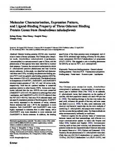
(accession no. L22305) (49.7%), soybean (59%), Phaseolus vulgaris (accession no. U34333) (54.7%), Arabidopsis thaliana (accession no. X67421) (61%), Catharanthus roseus (accession no. X85206) (71.7%), Zea mays (accession no. AB018587) (55.8%) and Asparagus officinalis (X82413) (30.2%) but IbPRP2 has similarity with these proteins 45.3%, 53.3%, 52.9%, 54.5%, 69.6%, 52.2%, and 30%, respectively (Figs. 2A and 2B). Prediction analysis of the hydrophobicity of the deduced amino acid sequences indicated that IbPRP1 and IbPRP2 proteins most likely contain one specific transmembrane‐spanning domain (Fig. 2C) similar to the other SbPRP from soybean (He et al., 2002). We used the TMHMM program (Sonnhammer et al., 1998) to estimate the length of the transmembrane domain. IbPRP1 and IbPRP2 clearly harbor 25 hydrophobic amino acids, all of them having the highest chance to be integral to a membrane. IbPRP1 and IbPRP2 may be located on plasma membrane PRPs are known as structural cell wall proteins that have been identified in higher plants (Cassab, 1998; Showalter, 1993). The question arises as to how the IbPRP1 and IbPRP2 function in regulating the signaling pathway? A plausible answer is that like other plant PRPs, IbPRP1 and IbPRP2 might be targeted to the plasma membrane (PM) in order to establish an interaction with a PM‐localized receptor for transmitting extracellular signals to its downstream signaling pathway(s). The
Genes & Genomics (2010) 32:481-491
491
Figure 2. (A) Comparison of the amino acid sequences of IbPRP1, IbPRP2, and other PRP proteins. The sequences were aligned using the ClustalW program. Identical amino acid residues in all sequences are indicated by asterisks. (B) Phylogenic tree of the genes with other PRP proteins. Analysis was performed using the CLC workbench program. (C) The putative transmembrane domains position of the IbPRP1(A) and IbPRP2(B). The transmembrane regions were estimated using the TMHMM program (http://www.cbs.dtu.dk/servieces/TMHMM).
Figure 3. Subcellular localization (introduced into onion epidermal cells) of the GFP::IbPRP1 and GFP::IbPRP2. The onion epidermal cells were incubated for 24 h at 24℃ and transient expression was observed using a Confocal Laser Scanning Microscope (Carl Zeiss, LSM510) at 488nm. Fluorescence was detected in green (BP 505‐530) channels.
492
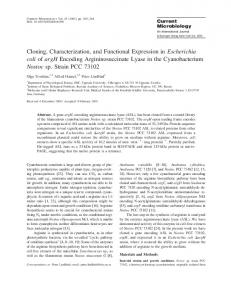
first 25 residues represented a putative signal peptide (Fig. 2A) which has been shown to be involved in the localization of IbPRP1 and IbPRP2. To examine whether IbPRP1 and IbPRP2 are localized at the PM, we employed three different approaches using the same CaMV35::GFP: IbPRP1 and IbPRP2 chimeric construct. The entire periphery of GFP::IbPRP1 and IbPRP2‐transformed cells fluoresced distinctively, whereas the GFP‐only control cells fluoresced primarily in the cytoplasm and nucleus (Fig. 3). Microscopy imaging under UV light confirmed that the GFP::IbPRP1 and IbPRP2 fluorescence was almost exclusively at the cell periphery, and thus the fusion was associated with either cell wall or plasma membrane. Proline Extension‐like Receptor Kinase 1, MsPRP2, RhoGTPase::GFP fusion indicated similar localization patterns that compare with IbPRP1 and IbPRP2 (Nancy and Daphne, 2002; Bischoff et al., 2000). These observations, together with the prediction analysis of the hydrophobicity, indicate that IbPRP1 and IbPRP2 may have the role of transmembrane protein in sweetpotato. Southern hybridization from sweetpotato and differential expression patterns of IbPRP1 and IbPRP2 in RT‐PCR analysis To confirm the complexity of the IbPRP1 and IbPRP2 genes, Southern blot analysis was performed under highly stringent condition. IbPRP1 and IbPRP2 genes existed more than two copies in sweetpotato genome when digested with diverse enzymes (BamHI, EcoRI, and XbaI), it suggests that IbPRP1 and IbPRP2 seem to exist as members of a multigene family in the sweetpotato genome (Fig. 4A).
Figure 4. Southern blot and RT‐PCR analyses of the IbPRP1 and IbPRP2 gene activity in sweetpotato. (A) Southern blot analysis of sweetpotato genomic DNA. Genomic DNA was digested with BamHI (B), EcoRI (E), and XbaI (X). The genomic DNAs were separated on 0.8% agarose gel, blotted, and probed with a DIG. (B) Expression patterns of the IbPRP1 and IbPRP2 mRNA in different tissues of sweetpotato. Total RNA was isolated from leaf (L), petiole (P), and stem (S), tuberous root (T), thick pigmented root (TPR), and white fibrous root (WFR). Tubulin was used as a control.
Genes & Genomics (2010) 32:481-491
To confirm the mRNA expression of IbPRP1 and IbPRP2 genes depending on various tissues, RT‐PCR analysis was conducted using leaf, petiole, stem, tuberous root, thick pigmented root, and white fibrous root cDNAs from sweetpotato. IbPRP1 was expressed in the whole tissues (leaf, petiole, stem, tuberous root, thick pigmented root, and white fibrous root) and the expressed levels were not different among tissues. In contrast, IbPRP2 was only expressed in the tuberous root, thick pigmented root, and white fibrous root, and highly expressed in the tuberous root (Fig. 4B). The IbPRP2 mRNA expression levels in the thick pigmented root and white fibrous root were not different from those expressed in IbPRP1 (Fig. 4B). These results indicate that IbPRP2 seems to have a tissue‐specific expression. Also, even if two genes have a similar gene structure, the tissue expression can be changed. Tissue‐specific expression was already reported in several PRP genes. PVR5 from bean was only expressed in the root (Choi et al., 1996), AtPRP1,3 were only detected in arabidopsis root (Fowler et al., 1999) and SbPRP only accumulated in epicotyls of soybean seedling (He et al., 2002). To examine the mRNA expression of IbPRP1 and IbPRP2 in more detail, RT‐PCR analysis was performed using cDNAs from sweetpotato leaf and root under various stresses. When treated with plant growth regulators and oxidative stress, the expression of IbPRP1 gene in the leaf increased by H2O2 (Fig. 5). Also, there was significant IbPRP1 mRNA accumulation in the leaf after drought stress treatment, especially 1 h after treatment (Fig. 5). However, IbPRP2 mRNA was not expressed in the leaf after various stress treatments (Fig. 5). H2O2 is well known as a major reactive oxygen species (ROS) involved in the defense and developmental processes in plants (Slesak et al., 2007). This result on the higher mRNA expression of IbPRP1 by H2O2 in the leaf indicate that IbPRP1 may be involved in oxidative stress, and that IbPRP1 may also be related to ROS and plant developmental mechanism. The mRNA expression of IbPRP1 and IbPRP2 in the root was different from that in the leaf. The expression level of IbPRP2 was increased by ET, BTH, SA, and MeJA, and was
Figure 5. Expression patterns of the IbPRP1 and IbPRP2 mRNA in root and leaf of sweetpotato after various stress treatments with 0.1 mM abscisic acid (ABA), ethephone (ET), benzol thiadiazole (BTH), salicylic acid (SA), methyl jasmonate (MeJA) for 48 h, 0.05mM methyl viologen (MV), 440mM H2O2 and drought, salt stress (0, 1, 2, 4, and 8 h drought stress and 200mM NaCl for 48 h salt stress).
Genes & Genomics (2010) 32:481-491
particularly increased by SA (Fig. 5). However, the level of expression was decreased by ABA (Fig. 5). The significant increase of mRNA expression in IbPRP1 was not detected under various stresses in the root (Fig. 5). He et al. (2002) reported that SbPRP gene was induced by SA treatment but was inhibited by ABA treatment, a similar result when compared to IbPRP2. Based on the previous research, SA related to plant disease resistance, Durner et al., 1997; Martinez et al., 2000; and He et al. (2002) reported that SbPRP gene might play a role in the soybean disease response. According to the structural analysis on IbPRP1 and IbPRP2, these genes were related with the AAI_LTSS superfamily. Also, this superfamily was involved in pathogen attack (Molina et al., 1993; Terras et al., 1992; Kader, 1996). These results indicate that IbPRP1 and IbPRP2 are involved in the response to various stresses
Figure 6. The conformation of gene integration in transgenic plants. The conformation of IbPRP1(A) and IbPRP2(B) gene integration in transgenic plants using PCR and Southern blot. PCR analysis of genomic DNA isolated from transgenic IbPRP1 and IbPRP2 T1 tobacco plants. M, marker; PC, Positive control; NC, Negative Control (lines 1, 2, and 5 represent individual lines of transgenic tobacco plants in IbPRP1 and lines 2, 3, 4 and 5 represent individual lines of transgenic tobacco plants in IbPRP2). Southern hybridization analysis of genomic DNA from transgenic T1 tobacco plants. Genomic DNA was digested with XbaI. M, DIG‐labeled DNA molecular weight marker; C, control (Wild‐type plant); Lines 1, 2, and 5 in IbPRP1 and lines 2, 3, 4, and 5 in IbPRP2 are individual lines of transgenic tobacco plants. Arrows indicate the weak signal bands of IbPRP1‐5 and IbPRP2‐5 lines.
493
Figure 7. The comparison of morphological differences between T1 transgenic plants and Wt plants. (A) IbPRP1 T1 transgenic plants (B) IbPRP2 T1 transgenic plants. C, control (Wild‐type plant) Lines 1, 2, and 5 in IbPRP1 and lines 2, 3, 4, and 5 in IbPRP2 are individual lines of transgenic tobacco plants.
including drought, pathogen, and oxidative stress. Overexpression of IbPRP1 increases salinity stress ‐ Analysis of transgenic IbPRP1 and IbPRP2 tobacco plants and leaf disc analysis of T1 transgenic plants lines for salinity stress Transgenic plants were generated using Agrobacterium‐mediated transformation. The pCAMLA (Lee et al., 2005) vector containing IbPRP1 or IbPRP2 gene was used. The presence of IbPRP1 and IbPRP2 genes in transgenic tobacco plants was confirmed by PCR. The 399bp fragments were amplified in transgenic T1 IbPRP1 plants and 402bp fragments were amplified in transgenic T1 IbPRP2 plants (Fig. 6A). To confirm the DNA insertion into the host genome, Southern blot analysis was performed using PCR‐screened T1 plants. Southern blot analysis showed that lines 1, 2, and 5 in IbPRP1 were independent and lines 2, 3, 4, and 5 in IbPRP2 were also independent (Fig. 6B). The T1 transgenic plants showed no morphological or growth differences in vegetative and floral tissues, as compared with Wt plants (Figs. 7A and 7B). Seed sets in both transgenic plants and Wt‐type plants were also similar. The tolerance of T1 transgenic to salt stress was studied by leaf disc assay. The leaf disc of Wt and transgenic plant lines (IbPRP1‐2,5 and IbPRP2‐2,4) were floated on 0, 100, 200, 300mM NaCl. After three days, the leaf disc of Wt and IbPRP2‐2,4 line in 300mM started to turn yellow but IbPRP1‐2 and IbPRP1‐5 transgenic lines were relatively green. IbPRP1 transgenic lines were observed high chlorophyll contents compared to Wt plants at 100mM NaCl treatment that relatively low salt level in this study, especially IbPRP1‐2 showed high chlorophyll level at the high concentration of NaCl treatments (200mM, and 300mM NaCl) (Fig. 8A). However, IbPRP2 transgenic lines showed no differences in chlorophyll contents compared to Wt (Fig. 8B). These results seem to indicate that IbPRP1 transgenic tobacco plant increased tolerance to salt. Presence of stress‐responsive cis‐elements in the IbPRP1 and IbPRP2 promoter regions
494
Genes & Genomics (2010) 32:481-491
Figure 8. Chlorophyll content in leaf discs test. Chlorophyll content in leaf discs of Wt, IbPRP1 (A), and IbPRP2 (B) transgenic T1 plants at 0, 100, 200, and 300mM NaCl.
To search for stress related cis‐elements in the promoters of IbPRP1 and IbPRP2, ‐2,000 to +1 promoter regions were submitted at the PLACE databases (http://www.dna.affre.go.jp/PLACE). According to the promoter assay, the ABRE and MYB/MYC recognition sites were enriched in the promoter region (Fig. 9). ABRE is related with ABA because many ABA‐inducible genes contain a conserved cis‐acting element called ABRE (ABA responsive element) in the promoter region (Guiltinal et al., 1990; Mundy et al., 1990). MYB/MYC binding sites are also known as cis‐acting elements for the expression of ABA‐regulated genes (Abe et al., 1997). Abe et al. (1997) reported that MYC recognition site was found in the promoter region of dehydration‐responsive gene rd22. Another stress‐related element WRKY in W‐box was found in the IbPRP1 and IbPRP2 promoter regions (Fig. 9). A lot of studies have reported that WRKY proteins may have regulatory functions in plant defense responses to pathogen infection (Eulgem et al., 1999; Chen and Chen, 2000; Dellagi
et al., 2000; Hara et al., 2000; Kim et al., 2000). These results suggest that IbPRP1 and IbPRP2 promoters have many stress‐ related elements such as ABA and pathogens. Therefore, IbPRP1 and IbPRP2 are probably related to many biotic and abiotic stresses.
Discussion This paper reports the isolation and characterization of IbPRP1 and IbPRP2 from sweetpotato. IbPRP1 and IbPRP2 show the high homology with soybean PRP1 (Creelman and Mullet, 1991), two different common bean PRP transcripts, one of which showed high homology with soybean PRP2 (Colmenero ‐ Flores et al., 1997), and an alfalfa transcript encoding a chimeric PRP2 protein (Deutch and Winicov, 1995). The ‘‘N’’ terminal region known to function as a signal for the localization of these proteins to appropriate membranes of different subcellular compartments (He et al., 2002) also exists in
Figure 9. Stress‐responsive cis‐acting elements analyses of IbPRP1 and IbPRP2 promoter sequences. ‐2,000 to +1 of IbPRP1 and IbPRP2 promoter regions were submitted at the PLACE databases (http://www.dna.affre.go.jp/PLACE)
Genes & Genomics (2010) 32:481-491
SbPRP. The phylogenetic analysis reveals that IbPRP1 and IbPRP2 are close to PCKR2 and those genes have the first 25 putative signal peptides. Proline‐rich proteins are important components of several plant developmental processes (Bozarth et al., 1987; Creelman and Mullet, 1991; Battaglia et al., 2007). PRP genes are represented during seedling growth (Hong et al., 1989), at the early stages of legume root nodule formation (Scheres et al., 1990; van de Wiel et al., 1990; Wilson et al., 1994), in immature maize‐embryos (Jose‐Estanyol et al., 1992), and in young tomato fruits (Salts et al., 1991). In sweetpotato, two IbPRP genes were differentially expressed. IbPRP1 was expressed in all vegetative tissues, whereas IbPRP2 was expressed more predominantly in developing tuberous roots, indicating that IbPRP2 may play an important role during tuber formation. A similar pattern of expression has been observed for Soybean PRP genes (PRP1, PRP2, and PRP3) that are expressed in different organs (leaf, stem, root, and seed) and at different developmental stages (Hong et al., 1989; Kleis‐San Francisco and Tierney, 1990; Wyatt et al., 1992). Additionally, many PRPs were regulated by plant growth regulators, and some were down‐regulated while others were up‐regulated (Datta et al., 1993; Subramaniam et al., 1994; Neuteboom et al., 1999; Ogawa et al., 1999). Thus, an analysis of the regulation of IbPRP2 expression in association with the hormonal regulation of tuber development will provide additional insight into the possible relationship between IbPRP2 function and tuber development. Proline‐rich proteins are expressed in response to abiotic stress (Bozarth et al., 1987; Creelman and Mullet, 1991; Battaglia et al., 2007). A proline‐rich protein of MsPRP2 in Medicago sativa showed stimulation at 400 mM by NaCl as early as 2 h (Deutch and Winicov, 1995). In cold stress, the transcript level of Wcor518 from Triticum aestrivum, PRP from Brassica napus and MsaCIC from alfalfa (Castonguay et al., 1994) slightly increased. In contrast, the SbPRP gene was induced by salt stress, drought stress, SA treatment and virus infection, but was inhibited by kinetin, ethephan, and ABA treatment (He et al., 2002). These observations suggest that the PRP has a role in biotic or abiotic stress response. We have studied for the first time the role of MV and H2O2 on transcript level. IbPRP1 transcript is up‐regulated by H2O2 in the leaf; however, no significant change was observed in IbPRP2. IbPRP2 showed stimulation by H2O2 and MV in the root, indicating that IbPRP1 and IbPRP2 might be differently regulated in various stress responses. The transgenic plants overexpressing AtRab7 showed increased tolerance to salt (200 mM NaCl) and osmotic stresses (500 mM Sorbitol), and reduced accumulation of reactive oxygen species during salt stress (Mazel et al., 2004). Transgenic
495
tobacco constitutively overexpressing IbPRP1 also showed tolerance to salt stress. Tobacco plants overexpressing IbPRP1showed higher chlorophyll content, but its chlorophyll content did not get affected much as compared with Wt plants in IbPRP2 transgenic plants. The morphological features of the transgenic plants were similar to Wt plants under normal growth conditions. In the present study, genes encoding a proline‐rich protein were isolated from sweetpotato and were designated as IbPRP1 and IbPRP2. Their genomic organization and expression pattern in response to different factors were examined. Their possible roles in defense and stress response were also discussed. In future research, an analysis of the protein products of these genes using genetic and biochemical approaches readily available in sweetpotato will provide an opportunity to dissect the mechanism during plant development and in response to environmental stimuli.
References Abe H, Yamaguchi‐Shinozaki K., Urao, T, Iwasaki T, and Shinozaki K. (1997). Role of MYC and MYB homologs in drought‐ and abscisic acid‐regulated gene expression. Plant Cell 9: 1859–1868. An G (1987) Binary Ti vectors for plant transformation and promoter analysis. Methods Enzymol. 153: 292‐305 Arnon D (1949) Copper enzymes in isolated chloroplasts phemoloxidase in beta vulgaris. Plant physiol. 24: 1‐15. Bartels D and Nelson D (1994) Approaches to improve stress tolerance using molecular genetics. Plant Cell Environ. 17: 659–667. Battaglia M, Solórzano RM, Hernandez M, Cuellar Ortiz S, Garcia‐ Gomez B, Marquez J, and Covarrubias AA (2007) Proline‐rich cell wall proteins accumulate in growing regions and phloem tissue in response to water deficit in common bean seedlings. Planta 225: 1121–1133. Bischoff F, Vahlkamp L, Molendijk A and Palme K (2000) Localization of AtROP4 and AtROP6 and interaction with the guanine nucleotide dissociation inhibitor AtRhoGDI1 from Arabidopsis. Plant Mol. Biol. 42: 515–530. Boyer JS (1982) Plant productivity and environment. Science 218: 443‐448. Bozarth CS, Mullet JE and Boyer JS (1987) Cell wall protens at low water potentials. Plant Physiol 85: 261–267. Bradley DJ, Kjellbom P and Lamb CJ (1992) Elicitor‐ and woundinduced oxidative cross‐linking of a proline‐rich plant cell wall protein: a novel, rapid defense response. Cell 70: 21–30. Brisson LF, Tenhaken R and Lamb C (1994) Function of oxidative cross‐linking of cell wall structural proteins in plant disease resistance. Plant Cell 6: 1703–1712. Cassab GI (1998) Plant cell wall proteins. Annu. Rev. Plant Physiol. Plant Mol. Biol. 49: 281–309. Castonguay Y, Laberge S, Nadeau P and Vezina NP (1994) Cold‐induced gene from Medicago sativa encodes a bimodular protein similar to developmentally regulated proteins. Plant Mol. Biol. 24: 799‐804. Chen C and Chen Z (2000) Isolation and characterization of two patho-
496 gen‐ and salicylic acid‐induced genes encoding WRKY DNA‐binding proteins from tobacco. Plant Mol. Biol. 42: 387–396. Chen J and Varner JE (1985) Isolation and characterization of cDNA clones for carrot extensin and a proline‐rich 33‐kDa protein. Proc. Natl. Acad. Sci. USA 82: 4399–4403. Choi DW, Song JY, Kwon YM and Kim SG (1996) Characterization of a cDNA encoding a proline‐rich 14 kDa protein in developing cortical cells of the roots of bean (Phaseolus vulgaris) seedlings. Plant Mol. Biol. 30: 973‐982. Colmenero‐Flores JM, Campos F, Graciarrubio A and Covarrubias AA (1997) Characterization of Phaseolus vulgaris cDNA clones responsive to water deWcit: identiWcation of a novel late embryogenesis abundant‐like protein. Plant Mol. Biol. 35: 393–405. Creelman RA and Mullet1 JE (1991) Water deficit modulates gene expression in growing zones of soybean seedlings. Analysis of differentially expressed cDNAs, a new β‐tubulin gene, and expression of genes encoding cell wall proteins. Mol. Gen. Gemet. 17: 591‐608. Datta N, LaFayette PR, Kroner PA, Nagao RT and Key JL (1993) Isolation and characterization of three families of auxin downregulated cDNA clones. Plant Mol Biol 21: 859–869. Dellagi A, Helibronn J, Avrova AO, Montesano M, Palva ET, Stewart HE, Toth IK, Cooke DE, Lyon GD. and Birch PR (2000) A potato gene encoding a WRKY‐like transcription factor is induced in interactions with Erwinia carotovora subsp. atroseptica and Phytophthora infestans and is coregulated with class I endochitinase expression. Mol. Plant‐Microbe Interact. 13: 1092–1101. Deutch CE and Winicov I (1995) Post‐transcriptional regulation of a salt‐inducible alfalfa gene encoding a putative chimerical proline ‐rich cell wall protein. Plant Mol. Biol. 27: 411–418. Durner J, Shah J and Klessig DF (1997) Salicylic acid and disease resistance in plants. Trends Plant Sci. 2: 266–274. Eulgem T, Rushton PJ, Schmelzer E, Hahlbrock K and Somssich IE (1999) Early nuclear events in plant defence signalling: rapid gene activation by WRKY transcription factors. EMBO J. 18: 4689– 4899. Food and Agriculture Organization (2002) FAO production yearbook, Food Agr Org. United Nations. Rome. 56. Fowler TJ, Bernhardt C and Tierney ML (1999) Characterization and expression of four proline‐rich cell wall protein genes in Arabidopsis encoding two distinct subsets of multiple domain proteins. Plant Physiol. 121: 1081‐1091. Guiltinan MJ, Marcotte WR and Quatrano RS (1990) A plant leucine zipper protein that recognizes an abscisic acid response element. Science 250: 267–271. Hara K, Yagi M, Kusano T and Sano H (2000) Rapid systemic accumulation of transcripts encoding a tobacco WRKY transcription factor upon wounding. Mol. Gen. Genet. 263: 30–37. He CY, Zhang JS and Chen SY (2002) A soybean gene encoding a proline‐rich protein is regulated by salicylic acid, an endogenous circadian rhythm and by various stresses. Chen. Theor. Appl. Genet. 104: 1125–1131. Hong JC, Nagao RT and Key JL (1989) Developmentally regulated expression of soybean proline‐rich cell wall protein genes. Plant Cell 1: 937–943. Jose‐Estanyol M, Ruiz‐Avila L and Puigdomenench P (1992) A maize embryo‐speciWc gene encodes a proline‐rich and hydrophobic protein. Plant Cell 4: 413–423. Kader JC (1996) Lipid‐transfer proteins in plants Annu. Rev. Plant Physiol. Plant Mol. Biol. 47: 627‐654.
Genes & Genomics (2010) 32:481-491 Kim CY, Lee SH, Park HC, Bae CG, Cheong YH, Choi YJ, Han C, Lee SY, Lim CO and Cho MJ (2000) Identification of rice blast fungal elicitor‐responsive genes by differential display analysis. Mol. Plant‐Microbe Interact. 13: 470–474. Kim SH and Hamada T (2005) Rapid and reliable method of extracting DNA and RNA from sweetpotato, Ipomoea batatas(L). Lam. Biotech. Lett. 27: 1841‐1845. Kim SH, Song WK, Kim YH, Kwon SY, Lee HS, Lee IC and Kwak SS (2009) Characterization of full‐length enriched expressed sequence tags of dehydration‐treated white fibrous roots of sweetpotato. BMB reports 42(5): 271‐276. Kleis‐San Francisco SM and Tierney ML (1990) Isolation and characterization of a proline‐rich cell wall protein from soybean seedlings. Plant Physiol. 94: 1897–1902. Kleis‐San Francisco SM and Tierney ML (1990) Isolation and characterization of a proline‐rich cell wall protein from soybean seedlings. Plant Physiol. 94: 1897–1902. Lee JH, Kim SH, Jung YH, Kim JA, Lee MO, Choi PG, Choi W, Kim KN and Jwa NS (2005) Molecular cloning and functional analysis of rice (Oryza sativa L.) OsNDR1 on defense signaling pathway. Plant Pathol. J. 21: 149‐157. Martinez C, Baccou JC, Bresson E, Baissac Y, Daniel JF, Jalloul A, Montillet JL, Geiger JP, Assigbetse K and Nicole M (2000) Salicylic acid mediated by the oxidative burst is a key molecule in local and systemic responses of cotton challenged by an avirulent race of Xanthomonas campestris pv malvacearum. Plant Physiol. 122: 757–766. Mazel A, Leshem Y, Tiwari BS and Levine A (2004) Induction of salt and osmotic stress tolerance by overexpression of an intracellular vesicle trafficking protein AtRab7 (AtRabG3e). Plant Physiol. 134: 118–128. Molina A, Segura A and Garcia‐Olmedo F (1993) Lipid transfer proteins (nsLTPs) from barley and maize leaves are potent inhibitors of bacterial and fungal plant pathogens. FEBS Lett. 316: 119–22. Mundy J, Yamaguchi‐Shinozaki K and Chua NH (1990) Nuclear proteins bind conserved elements in the abscisic acidresponsive promoter of a rice rab gene. Proc. Natl. Acad. Sci. USA 87: 406–410. Nancy F, Silva and Daphne R (2002) The proline‐rich, extensin‐like receptor kinase‐1 (PERK1) gene is rapidly induced by wounding Plant Mol. Biol. 50: 667–68. Neuteboom LW, Ng JM, Kuyper M, Clijdesdale OR, Hooykaas PJJ and Zaal BJ (1999) Isolation and characterization of cDNA clones corresponding with mRNAs that accumulate during auxin‐induced lateral root formation. Plant Mol. Biol. 39: 273–287. Ogawa M, Kusano T, Koizumi N, Katsumi M and Sano H (1999) Gibberellin‐ responsive genes: high level of transcript accumulation in leaf sheath meristematic tissue from Zea mays L. Plant Mol. Biol. 40: 645–657. Sakai H, Aoyama T and Oka A (2000) Arabidopsis ARR1 and ARR2 response regulators operate as transcriptional activators. Plant J. 24(6): 703‐711. Salts Y, Wachs R, Gruissem W and Barg R (1991) Sequence coding for a novel proline‐rich protein preferentially expressed in young tomato fruits. Plant Mol. Biol. 17: 149–150. Sambrook J and Russel DW (2001) Molecular Cloning; A Laboratory Mamual, 3rd ed., Cold Spring Harbor, New York. Showalter AM (1993) Structure and function of cell wall proteins. Plant Cell 5: 9–23. Ślesak I, Libik M, Karpinska B, Karpinski S and Miszalski Z (2007) The role of hydrogen peroxide in regulation of plant metabolism
Genes & Genomics (2010) 32:481-491 and cellular signalling in response to environmental stresses. Acta. Biochimica Polonica 54: 39–50. Sonnhammer ELL, Von Heijne G and Krogh A (1998) A hidden Markov model for predicting transmembrane helices in protein sequences. In Proceedings of the Sixth International Conference on Intelligent Systems for Molecular Biology J. 175–182. Subramaniam K, Ranie J, Srinivasa BR, Sinha AM and Mahadevan S (1994) Cloning and sequence of a cDNA encoding a novel hybrid proline‐rich protein associated with cytokinin‐induced haustoria formation in Cuscuta reXexa. Gene 14: 207–210.
497 Terras FRG, Schofs HME, de Bolle MFC, Van Leuven F and Rees SB (1992) In vitro antifungal activity of a radish (Raphanus sativus L.) seed protein homologous to nonspecific lipid transfer proteins. Plant Physiol. 100: 1055–58. Tierney ML, Wiechert J and Pluymers D (1988) Analysis of the expression of extensin and p33‐related cell wall proteins in carrotand soybean. Mol. Gen. Genet. 211: 393–399. Wyatt RE, Nagao RT and Key JL (1992) Patterns of soybean proline rich protein gene expression. Plant Cell 4: 99–110.