The Baculoviridae is a large family of DNA viruses whose members are pathogenic predominantly for insects of the order. Lepidoptera, although some strains ...
Journal of General Virology (1999), 80, 493–500. Printed in Great Britain ...................................................................................................................................................................................................................................................................................
Characterization of the interaction between the baculovirus ssDNA-binding protein (LEF-3) and putative helicase (P143) J. T. Evans,† G. S. Rosenblatt, D. J. Leisy and G. F. Rohrmann Department of Microbiology, Oregon State University, Nash Hall 220, Corvallis, OR 97331-3804, USA
LEF-3 and P143 are two of six proteins encoded by the Autographa californica multinucleocapsid nucleopolyhedrovirus genome which are required for DNA replication in transient replication assays. LEF-3 has the properties of an ssDNA-binding protein and P143 exhibits amino acid sequence homology to helicases. In this report, the interaction of LEF-3 and P143 was studied by yeast two-hybrid and immunoaffinity analyses. Using the yeast two-hybrid system, the interaction domain of LEF-3 (385 aa) was mapped to amino acids between positions 1 and 165. Deletion analysis of P143 failed to reveal an interaction domain, suggesting that there were either multiple interaction domains or that the deletions disrupted the secondary structures required for the interaction between LEF-3 and P143.
Introduction The Baculoviridae is a large family of DNA viruses whose members are pathogenic predominantly for insects of the order Lepidoptera, although some strains infect species of Hymenoptera, Diptera and Trichoptera. The most well-characterized baculovirus, Autographa californica multinucleocapsid nucleopolyhedrovirus (AcMNPV), has a circular dsDNA genome of 134 kb, which encodes approximately 150 genes (Ayres et al., 1994). It has eight homologous regions (hrs) which have been shown to act as early gene transcriptional enhancers (Guarino & Summers, 1986 ; Leisy et al., 1995 ; Rodems & Friesen, 1993, 1995) and may function as origins of DNA replication (Leisy et al., 1995 ; Leisy & Rohrmann, 1993 ; Pearson et al., 1992). A transient DNA replication assay has been used to identify nine genes from AcMNPV which are involved in DNA replication (Kool et al., 1994 ; Lu & Miller, 1995). Six of these genes appear to be required for DNA replication and encode the following proteins : a DNA polymerase homologue (Tomalski et al., 1988) ; P143, a helicase homologue (Lu & Carstens, 1991) that is capable of binding dsDNA (Laufs et al., 1997) ; LEF-3, a homotrimeric ssDNA-binding (SSB) protein (Evans & Rohrmann, 1997 ; Hang et al., 1995) that appears to Author for correspondence : George Rohrmann. Fax j1 541 737 0496. e-mail rohrmann!bcc.orst.edu † Present address : St Jude Children’s Research Hospital, Department of Virology and Molecular Biology, 332 N. Lauderdale, Memphis, TN 38105, USA.
0001-5925 # 1999 SGM
be involved in transporting P143 into the nucleus (Wu & Carstens, 1998) ; IE-1, a transcriptional activator (Guarino & Summers, 1987) which also binds to hrs (Choi & Guarino, 1995 ; Guarino & Dong, 1991 ; Kovacs et al., 1992 ; Leisy et al., 1995 ; Rodems & Friesen, 1995) ; and two proteins, LEF-1 (a putative primase) and LEF-2, which have been shown to interact with each other (Evans et al., 1997). The three gene products, which are stimulatory for DNA replication, include two additional transcriptional activators, IE-2 (Carson et al., 1991) and PE-38 (Krappa & Knebel-Mo$ rsdorf, 1991), and P35, which blocks apoptosis (Clem et al., 1991 ; Herschberger et al., 1992) and may therefore influence DNA replication in an indirect manner. A DNA polymerase activity differing from the cellular enzyme has been described in baculovirus-infected cells (Mikhailov et al., 1986 a, b ; Miller et al., 1981 ; Wang & Kelly, 1983 ; Wang et al., 1983) and is probably attributable to the DNA polymerase homologue. In addition, a temperaturesensitive mutation in p143 (helicase gene) was shown to block DNA replication (Gordon & Carstens, 1984 ; Lu & Carstens, 1991). Initial attempts to characterize the interactions between the products of these nine AcMNPV genes using yeast twohybrid assays revealed several interactions. We have previously described the interaction between LEF-1 and LEF-2 (Evans et al., 1997), and recently we provided evidence that LEF-3 (SSB) interacts with itself and forms a homotrimer in solution (Evans & Rohrmann, 1997). In this report, we describe the interaction between AcMNPV LEF-3 (SSB) and P143
EJD
J. T. Evans and others
(helicase) by yeast two-hybrid analyses, immunoaffinity experiments and co-purification.
Methods
Insect cells. Spodoptera frugiperda (Sf-9) cell suspension cultures (Vaughn et al., 1977) were maintained in serum-free SF900 II SFM medium (GIBCO-BRL) supplemented with penicillin G (50 U\ml), streptomycin (50 µg\ml) (BioWhittaker), and fungizone (375 ng\ml) (GIBCO-BRL). Cell culture maintenance was carried out according to published procedures (Summers & Smith, 1987).
Bacterial and yeast cells. All bacterial plasmids were maintained in Escherichia coli DH5α. Saccharomyces cerevisiae Y166 (MATa ura3-52 leu2-3,-112 his3∆200 ade2-101 trp1-901 gal4∆ gal80∆ RNR : : GAL-URA3 LYS2 : : GAL-HIS3 GAL-lacZ) was used for the yeast two-hybrid assays.
Plasmid constructs. All baculovirus constructs were originally derived from the AcMNPV E2 strain (Smith & Summers, 1978). lef-3 was cloned as an EcoRI–ApaI fragment (m.u. 42n8–44n5) into pUC19 and subcloned into pBluescript KS(k) (Stratagene) (Kool et al., 1994 ; Kool & Vlak, 1993). p143 was cloned as an EcoRI–SspI fragment (m.u. 59n9–63n5) into pBluescribe BS(k) (Kool et al., 1994 ; Kool & Vlak, 1993). NcoI sites were generated at the ATG start codon of each gene by site-directed mutagenesis (Sambrook et al., 1989) to form pKSLEF-3(NcoI) and pKSP143(NcoI), respectively. Several cloning steps were required to mutagenize and transfer p143 from pBS(k) to pKS(k). pBSP143 was digested with HindIII and the 5h leader sequence plus 459 bp of coding sequence from p143 was isolated and cloned into pKS(k) that had been cut with HindIII. This construct (pKSP143sH) containing the N terminus of P143 was used for site-directed mutagenesis to produce an NcoI restriction site at the ATG start codon of p143 (pKSP143m). pKSP143m was then digested with SphI and re-ligated to remove the PstI site from the 5h polylinker. The resultant plasmid was digested with EcoRI\XbaI, treated with T4 DNA polymerase and re-ligated, removing a PstI site from the 3h polylinker region of pKS(k) and producing pKSP143m(∆SphI)(∆EcoRI–XbaI). pBSP143 was digested with PstI\EcoRI and a fragment encoding the C terminus of P143 was cloned into pKSP143m(∆SphI)(∆EcoRI–XbaI) cut with the same enzymes. The primers used for mutagenesis of pKSLEF-3 and pKSP143smH were TCGACAACAGCACCATGGCGACCAAA and ACACGATAGCCACCATGGTTGACAACATTTTAC, respectively. The sequences of both pKSLEF3(NcoI) and pKSP143(NcoI) were confirmed by DNA sequence analysis. Mutagenesis changed the second amino acid of P143 from isoleucine to valine and left the LEF-3 amino acid sequence unchanged. pKSLEF-3(NcoI) and pKSP143(NcoI) functioned in transient replication assays as well as the parent clones, indicating that the sequence changes had no effect on their ability to support transient DNA replication (data not shown).
Yeast two-hybrid clones. Full-length lef-3 was cloned into pAS1 and pACTII (gifts from Steve Elledge, Howard Hughes Medical Institute, Baylor College of Medicine, Houston, TX, USA) (Durfee et al., 1993) as previously described (Evans & Rohrmann, 1997). Full-length p143 was first cloned into pTrc-7Hpro (Evans & Rohrmann, 1997) by digesting pKSP143(NcoI) with EcoRI, treating with T4 DNA polymerase, digesting with NcoI, and ligating the p143-containing fragment into pTrc-7Hpro cut with NcoI and SmaI creating pHTP143. pHTP143 was digested with NcoI\BamHI and the p143-containing fragment was cloned into pAS1 and pACTII digested with the same enzymes. N- and C-terminal deletion constructs of LEF-3 containing aa 1–370, aa 1–314, aa 311–385, aa 244–385, aa 165–385 and aa 77–385 were made as previously described (Evans & Rohrmann, 1997). The C-terminal deletion construct of LEF-3 EJE
containing aa 1–214 was constructed by digesting pKSLEF-3(NcoI) with NcoI\HincII followed by subcloning into pAS1 and pACTII digested with NcoI\SmaI. The deletion construct containing aa 49–385 was made by digesting pKSLEF-3 with ClaI, treating the product with T4 DNA polymerase and then digesting it with BamHI ; the resulting fragment was cloned into pAS1 and pACTII cut with SmaI\BamHI. The LEF-3 Cterminal deletion constructs aa 1–311, aa 1–244, aa 1–165 and aa 1–77 were constructed by introducing NcoI sites into pKSLEF-3(NcoI) at codon positions 77, 165, 244 and 311 by site-directed mutagenesis followed by subcloning of NcoI fragments into pAS1 and pACTII. The primers used for mutagenesis were described previously (Evans & Rohrmann, 1997). C-terminal deletion constructs pASP143(aa 1–980) and pACTP143(aa 1–980) were made by digesting pKSP143(NcoI) with SphI, treating the product with T4 DNA polymerase, digesting it with NcoI, and then subcloning into pAS1 and pACTII cut with NcoI\SmaI. pASP143(aa 1–738) was made by digesting pASP143 with SalI and re-ligating the vector. pACTP143(aa 1–738) was made by digesting pKSP143(NcoI) with SalI, treating it with T4 DNA polymerase, digesting it with NcoI, and then subcloning the fragment into pACTII cut with NcoI\SmaI. Nterminal deletion constructs pASP143(aa 81–1221) and pACTP143(aa 81–1221) were constructed by digesting pKSP143 with NcoI\PstI, treating with T4 DNA polymerase, and re-ligating the vector, eliminating the NcoI\PstI fragment and reconstituting the NcoI site. This new construct, pKSP143(∆PstI) was digested with NcoI\BamHI followed by subcloning into pAS1 and pACTII cut with the same enzymes.
Site-directed mutagenesis. Site-directed mutagenesis was carried out as described by Kunkel et al. (1987). The primers used to generate the clones in this study are shown above or were described previously (Evans & Rohrmann, 1997).
Yeast two-hybrid transfections and liquid assays. These procedures were carried out as previously described (Evans et al., 1997).
Immunoaffinity purification. IgG from anti-LEF-3 rabbit polyclonal antiserum and pre-immunization antiserum were purified by protein A–Sepharose chromatography (Harlow & Lane, 1988) and coupled to CNBr-activated Sepharose 4B in accordance with the manufacturer’s instructions (Pharmacia Biotech). Sepharose 4B–IgG was washed and re-suspended in NET gel buffer [50 mM Tris–HCl (pH 7n5), 150 mM NaCl, 0n1 % Nonidet P-40, 1 mM EDTA, 0n25 % gelatin, 0n02 % sodium azide]. Sepharose 4B–IgG (40 µl ; 50 % slurry) was incubated for 1n5 h with 40 µl nuclear extracts (NE) obtained at 16 h post-infection (p.i.) or mock-infected NE, and washed three times with NET gel buffer. The bound proteins were removed from the Sepharose beads by boiling in SDS–PAGE sample buffer [50 mM Tris–HCl (pH 6n8), 100 mM dithiothreitol, 2 % SDS, 0n1 % bromophenol blue, 10 % glycerol] and subjected to SDS–PAGE followed by Western blot analysis with antiP143 rabbit polyclonal antiserum.
LEF-3/P143 purification. Nuclear extracts were prepared at 16 h p.i. as previously reported (Glocker et al., 1993). Co-purification of LEF3 and P143 was carried out as previously described (Evans & Rohrmann, 1997).
Polyclonal antiserum production, PAGE and Western blotting. Preparation of rabbit polyclonal antiserum against LEF-3 was described previously (Evans & Rohrmann, 1997). To prepare antiserum against P143, a gene fusion expressing the C-terminal 777–1221 aa (SalI fragment) of P143 fused to maltose-binding protein (pMal-cRI vector ; New England Biolabs) was constructed. The fusion protein was expressed in E. coli DH5α and gel-purified by SDS–PAGE. The purified fusion protein (150 µg) was subcutaneously injected into a New Zealand White rabbit with complete Freund’s adjuvant for the initial injection and incomplete Freund’s adjuvant for subsequent injections (2 to 3 weeks
Baculovirus SSB/helicase interactions between injections). Rabbit antiserum was collected 1 week after the third injection and tested by Western blot analysis using 16 h p.i. NE. SDS–PAGE was performed as described by Sambrook et al. (1989). Gels were either fixed and stained with Coomassie brilliant blue (Bio-Rad) or used for Western blots. For Western blots, proteins were electrophoretically transferred from polyacrylamide gels to a PVDF-plus transfer membrane (Micron Separations) for 4 h at 185 mA (Genie Electrophoretic Blotter ; Idea Scientific). The membranes were then incubated in 1 % blocking solution (Boehringer Mannheim) for 12 h at 4 mC. Western blots were probed with a 1 : 2000 dilution of anti-P143 rabbit polyclonal antiserum, or a 1 : 7500 dilution of anti-LEF-3 rabbit polyclonal antiserum, washed, incubated with goat anti-rabbit IgG conjugated to horseradish peroxidase (Bio-Rad), and developed with a chemiluminescence blotting substrate in accordance with the manufacturer’s instructions (Boehringer Mannheim).
Results and Discussion Co-purification of LEF-3 and P143
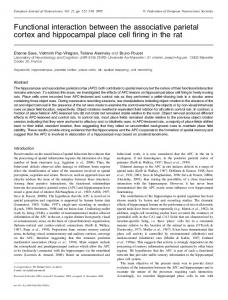
The partial purification of LEF-3 from 16 h p.i. NE using ssDNA–agarose has been previously reported (Evans & Rohrmann, 1997 ; Hang et al., 1995). LEF-3 elutes from the ssDNA–agarose resin in 700–1000 mM NaCl, resulting in 80–90 % purification of LEF-3. A second protein of approximately 140 kDa co-elutes with LEF-3 from the ssDNA– agarose (Fig. 1 a). This suggested that these proteins may elute
(a)
as a complex. The largest AcMNPV ORF encodes P143 (helicase) which is predicted to be 143 kDa (Ayres et al., 1994). To confirm whether both LEF-3 and P143 eluted in the same fractions from ssDNA–agarose, Western blot analysis was performed using either LEF-3 or P143 antisera. We observed a reaction with the LEF-3 antiserum (not shown) as we previously reported (Evans & Rohrmann, 1997). In addition, we demonstrated that P143 is also present in these fractions using an antiserum against P143 (Fig. 1 b, lane 3). Similar results were obtained by Wu & Carstens (1998) for protein purified by hydroxyapatite and subsequent ssDNA–cellulose chromatography although they employed step rather than continuous NaCl gradients. To confirm the interaction between LEF-3 and P143, immunoaffinity experiments were performed using anti-LEF-3 polyclonal antiserum. IgG from pre-immune or LEF-3 polyclonal antisera were purified by protein A–Sepharose chromatography and linked to CNBr-activated Sepharose 4B. The IgG bound to Sepharose beads was then incubated with 16 h p.i. NE or mock-infected NE, washed, and the retained proteins were visualized by Western blot analysis using anti-P143 polyclonal antiserum. When Sepharose–LEF-3 antiserum was used for immunoaffinity purification with 16 h p.i. NE, Western blot analysis with anti-P143 polyclonal antiserum revealed a
(b)
Fig. 1. Co-purification of LEF-3 and P143. (a) SDS–PAGE of 20 µl aliquots from 1 ml fractions eluted from the ssDNA–agarose column in 700–1000 mM NaCl. The gel was stained with Coomassie brilliant blue (Bio-Rad). (b) Western blot analysis of P143 purified by ssDNA–agarose chromatography using anti-P143 polyclonal antiserum. Lanes : 1, 16 h p.i. NE ; 2, 16 h mockinfected NE ; and 3, peak fraction from the ssDNA–agarose column. The same analysis was performed with anti-LEF-3 polyclonal antiserum and indicated that LEF-3 was present in the same peak fractions as P143 (data not shown ; Evans & Rohrmann, 1997). The positions of molecular mass markers, in kDa, are shown on the left and the sizes of LEF-3 and P143 are shown on the right.
EJF
J. T. Evans and others
Table 1. Yeast two-hybrid liquid assays 16 hr pi NE mock NE
Activity of lacZ on ONPG in nmol\min\mg protein is shown for each two-hybrid assay.
anti-LEF-3
pACTLEF-3
pACTP143
35n3p9n9 2n4p0n4 1
17p3n4 No growth 4n3p0n3
pACTII
pre-immune sera
pASLEF-3 pASP143 pASLEF-1
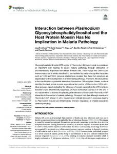
Fig. 2. Western blot analysis of P143 affinity-purified by LEF-3 antiserum bound to CNBr-activated Sepharose 4B. Lane 1 shows the affinity purification of P143 from 16 h p.i. NE with Sepharose–LEF-3 antiserum. Lanes 2 and 3 are negative controls with 16 h p.i. NE treated with Sepharose bound to IgG from pre-immune sera (lane 2) and mockinfected NE treated with Sepharose–LEF-3 antiserum (lane 3). Lane 4 shows the position of P143 from 16 h p.i. NE. The Western blot was developed with P143 antiserum as described in Methods. The positions of selected molecular mass markers, in kDa, are shown on the left and the position of P143 is shown on the right.
band of approximately 143 kDa (Fig. 2, lane 1) located in the same position as P143 from 16 h p.i. NE (lane 4), whereas immunoprecipitation of mock-infected NE with Sepharose– LEF-3 antiserum, or 16 h p.i. NE with Sepharose–pre-immune serum gave no bands corresponding to the size of P143 (lanes 2 and 3). Reciprocal immunoprecipitations with anti-P143 polyclonal antiserum and subsequent analysis with LEF-3 polyclonal antiserum were not possible due to the crossreaction of the secondary goat anti-rabbit IgG horseradish peroxidase conjugate with the IgG heavy chain from the P143 polyclonal antibody used in the immunoprecipitations. The IgG heavy chain has a molecular mass of approximately 50 kDa and produces a strong signal that obscures the reaction of the LEF-3 antiserum with LEF-3.
Yeast two-hybrid analysis of interaction
To confirm the interaction between LEF-3 and P143 suggested by co-purification and immunoaffinity studies, the yeast two-hybrid system was used. In this system, DNA segments encoding the GAL4 DNA-binding and activation domains have been separately cloned into two different EJG
1 1 1
plasmids (pAS1 and pACTII, respectively) (Durfee et al., 1993), each of which has different auxotrophic markers for selection of single or double recombinants in yeast (Chien et al., 1991 ; Fields & Song, 1989 ; Guarente, 1993 ; Keegan et al., 1986 ; Ma & Ptashne, 1987). Genes of interest can be cloned into these vectors and expressed as fusion proteins containing the GAL4 activation (pACTII) or GAL4 DNA-binding domain (pAS1) at their N termini. When co-transfected into yeast, interaction of the fusion partners allows the GAL4 activation domain to be brought into proximity with the lacZ and ura promoters causing transactivation of these genes. Interacting partners can be detected by both β-galactosidase assays (colorimetric colony lift assays and liquid assays) and by growth on plates lacking uracil. Using this system, an interaction between LEF-3 and P143 in both vector combinations was observed (Table 1). In these experiments, pASLEF-1 was used as a negative control because the vector pAS1 alone (without an insert) results in low levels of transactivation (Evans & Rohrmann, 1997). pASP143 expressed with pACTLEF-3 resulted in a transactivation of 2n4j0n4 nmol\min\mg, whereas control crosses between pASP143 and pACTII and between pACTLEF-3 and pASLEF1 showed no transactivation (Table 1). When pACTP143 was expressed with the control pASLEF-1, a transactivation of 4n3j0n3 nmol\min\mg (Table 1) was observed. We confirmed that pACTP143 alone exhibits transactivation (3–5 nmol\min\mg) by producing double recombinants of pACTP143 with a variety of pAS1 constructs that, by themselves, show no transactivation (data not shown). This level of activation was much less than that seen when pACTP143 was crossed with pASLEF-3 (17j3n4 nmol\ min\mg) (Table 1). Because pACTP143 was capable of transactivation by itself (although at much lower levels than when in the presence of pASLEF-3), this activation was taken as background and subtracted from the liquid assay results of all subsequent crosses involving pACTP143. Therefore, we observed positive interactions of LEF-3 with P143 (helicase) in both combinations of vectors. Although the levels of lacZ expression differed in the two combinations, it is welldocumented that the strength of an interaction cannot be deduced from values derived from yeast two-hybrid analyses (Estojak et al., 1995). Strengths of interaction can be influenced
Baculovirus SSB/helicase interactions
Fig. 3. Identification of LEF-3 domains that interact with full-length P143. The diagrams on the left show the portions of LEF-3 present in the mutants analysed. Clone 1 is the full-length LEF-3 (aa 1–385). The two right-hand columns show the levels of lacZ expression from yeast that contained both the LEF-3 deletion clones shown (in pAS1) and the full-length p143 gene cloned into pACTII, or pACTII with no insert (negative control). Expression of lacZ was calculated from three to four liquid assays from independent transformations (pSD). Specific activity is expressed as nmol/min/mg protein. The transactivation from pACTP143 (4n3 nmol/min/mg) (Table 1) has been subtracted from the pACTP143 crosses as background. Protein expression was confirmed by Western blot analysis using a monoclonal antibody (Babco) to the HA epitope.
by mRNA stability, protein stability, protein folding and the stability of the plasmid construct. Therefore, the values shown in Table 1 demonstrate the ability of fusion partners to interact, but do not necessarily represent relative interaction strengths between fusion partners. Yeast transformed with P143 (in pAS1 or pACTII) grew very slowly on plates and in liquid cultures used for the selection of the appropriate auxotrophic markers. In addition, no recombinants were found when P143 was crossed with itself, suggesting that the P143 fusions have an inhibitory effect on yeast growth which becomes lethal in double recombinants in which P143 is present in both vectors (Table 1). LEF-3 recombinants grow slowly, although they are not lethal (Evans & Rohrmann, 1997). It is possible that these proteins compete for components of the cellular replication apparatus, thereby inhibiting growth. Interaction of deletion clones with full-length LEF-3 and P143
To map the interaction domains of LEF-3 and P143, a set of N- and C-terminal deletions were constructed in the yeast twohybrid vectors pAS1 and pACTII (Durfee et al., 1993). These deletions were transfected into yeast with either full-length LEF-3 or P143 in the reciprocal vector. None of the N- or Cterminal deletions of P143 (aa 1–980, aa 1–738, aa 81–1221) interacted with full-length LEF-3 in pAS1 or pACTII, indicating that either multiple interaction domains exist, or the deletions we constructed disrupted the secondary structures required for the interaction between P143 and LEF-3 (data not shown).
When N- and C-terminal deletions of LEF-3 fused to the GAL4 DNA-binding domain (pAS1) were tested for interaction with full-length P143 fused to the GAL4 activation domain (pACTII), the C-terminal deletions retaining aa 1–165 interacted with full-length P143 (Fig. 3, rows 2–7), whereas a deletion containing only aa 1–77 failed to interact with fulllength P143 (Fig. 3, row 8). None of the LEF-3 N-terminal deletions tested were able to interact with full-length P143 (Fig. 3, rows 9–13). The same results were obtained when the LEF-3 deletions were tested as GAL4 activation domain fusions (pACTII) with P143 fused to the GAL4 DNA-binding domain (pAS1) (data not shown). We have previously reported that LEF-3 interacts with itself to form a homotrimer in solution (Evans & Rohrmann, 1997). The complete set of LEF-3 deletions shown in this report were tested for interaction with full-length LEF-3 (Evans & Rohrmann, 1997 ; data not shown). None of the deletions were able to interact with full-length LEF-3. In addition, selected 5h and 3h LEF-3 deletions were unable to support transient DNA replication (Evans & Rohrmann, 1997). However, as described above, the deletions containing aa 1–165 of LEF-3 are able to interact with full-length P143. These data indicate that the LEF-3 interaction with itself (trimerization) is not required for the interaction between LEF-3 and P143. Because of the recently reported evidence suggesting that LEF-3–P143 interaction is essential for transport of P143 into the nucleus (Wu & Carstens, 1998), the interaction between these two proteins that we have characterized is likely to be highly significant. In addition to being involved in nuclear EJH
J. T. Evans and others
transport, the association could be directly involved in DNA replication. In several other viral and non-viral systems (both prokaryotic and eukaryotic), helicases and SSBs have been shown to have a functional interaction in which the activity of the helicase is either stimulated or inhibited by the SSB (Biswas et al., 1995 ; Hamatake et al., 1997 ; Le Gac et al., 1996 ; Matson, 1991 ; Matson & Kaiser-Rogers, 1990 ; Seo & Hurwitz, 1993 ; Seo et al., 1991 ; Tsaneva & West, 1994 ; Umezu & Nakayama, 1993). Direct interaction of helicases and SSBs have been reported for herpes simplex virus type 1 (HSV-1), where the viral origin-binding protein (UL9), which also has helicase activity, has been shown to tightly associate with the HSV-1 SSB (ICP8) (Boehmer et al., 1994 ; Boehmer & Lehman, 1993) and in other systems where the heterotrimeric SSB, replication protein A (RP-A), interacts with the simian virus 40 large tumour antigen (Dornreiter et al., 1992) and co-purifies with the calf thymus DNA helicase F (Georgaki et al., 1994). Indirect interactions between SSBs and helicases have also been observed in the bacteriophage T4, where the gene 59 protein interacts with both the T4 helicase (gene 41 protein) and SSB (gene 32 protein) (Barry & Alberts, 1994 a, b ; Morrical et al., 1994 ; Yonesaki, 1994). The interaction between AcMNPV LEF-3 and P143 is consistent with the interactions observed in these other systems and may be required for the proper function of these proteins in DNA replication. In addition to nuclear transport, the function of the LEF-3–P143 interaction may be to assist in loading the helicase onto a partially melted origin of replication, or in the binding of the SSB to ssDNA after helicase denaturation, followed by coating of the ssDNA by co-operative binding of additional SSB. The authors thank Joel Funk for his suggestions and criticisms of this manuscript, and the Central Services Laboratory at OSU for assistance with this project. We would also like to thank Dr Bill Dougherty and Dr Steve Elledge for providing plasmid vectors used in this study. This project was supported by grants from the NSF (MCB-9630769) and ACS (SG-208). This is Technical Report No. 11432 from the Oregon State University Agricultural Experiment Station.
References Ayres, M. D., Howard, S. C., Kuzio, J., Lopez-Ferber, M. & Possee, R. D. (1994). The complete DNA sequence of Autographa californica
nuclear polyhedrosis virus. Virology 202, 586–605. Barry, J. & Alberts, B. (1994 a). Purification and characterization of
bacteriophage T4 gene 59 protein. Journal of Biological Chemistry 269, 33049–33062. Barry, J. & Alberts, B. (1994 b). A role for two DNA helicases in the replication of T4 bacteriophage DNA. Journal of Biological Chemistry 269, 33063–33068. Biswas, E. E., Chen, P.-H., Leszyk, J. & Biswas, S. B. (1995).
Biochemical and genetic characterization of a replication protein A dependent DNA helicase from the yeast, Saccharomyces cerevisiae. Biochemical and Biophysical Research Communications 206, 850–856. Boehmer, P. E. & Lehman, I. R. (1993). Physical interaction between the herpes simplex virus 1 origin-binding protein and single-stranded EJI
DNA-binding protein ICP8. Proceedings of the National Academy of Sciences, USA 90, 8444–8448. Boehmer, P. E., Craigie, M. C., Stow, N. D. & Lehman, I. R. (1994).
Association of origin binding protein and single stranded DNA-binding protein, ICP8, during herpes simplex virus type 1 DNA replication in vivo. Journal of Biological Chemistry 269, 29329–29334. Carson, D. D., Summers, M. D. & Guarino, L. A. (1991). Molecular analysis of a baculovirus regulatory gene. Virology 182, 279–286. Chien, C., Bartel, P. L., Sternglanz, R. & Fields, S. (1991). The twohybrid system : a method to identify and clone genes for proteins that interact with a protein of interest. Proceedings of the National Academy of Sciences, USA 88, 9578–9582. Choi, J. & Guarino, L. A. (1995). The baculovirus transactivator IE1 binds to viral enhancer elements in the absence of insect cell factors. Journal of Virology 69, 4548–4551. Clem, R. J., Fechheimer, M. & Miller, L. K. (1991). Prevention of apoptosis by a baculovirus gene during infection of insect cells. Science 254, 1388–1390. Dornreiter, I., Erdile, L. F., Gilbert, I. U., von Winkler, D., Kelly, T. J. & Fanning, E. (1992). Interaction of DNA polymerase α-primase with
cellular replication protein A and SV40 T antigen. EMBO Journal 11, 769–776. Durfee, T., Becherer, K., Chen, P.-L., Yeh, S.-H., Yang, Y., Kilburn, A. E., Lee, W.-H. & Elledge, S. J. (1993). The retinoblastoma protein
associates with the protein phosphatase type 1 catalytic subunit. Genes & Development 7, 555–569. Estojak, J., Brent, R. & Golemis, E. A. (1995). Correlation of two-hybrid affinity data with in vitro measurements. Molecular and Cellular Biology 15, 5820–5829. Evans, J. T. & Rohrmann, G. F. (1997). The baculovirus single-stranded DNA binding protein, LEF-3, forms a homotrimer in solution. Journal of Virology 71, 3574–3579. Evans, J. T., Leisy, D. J. & Rohrmann, G. F. (1997). Characterization of the interaction between the baculovirus replication factors, LEF-1 and LEF-2. Journal of Virology 71, 3114–3119. Fields, S. & Song, O.-K. (1989). A novel genetic system to detect protein–protein interactions. Nature 340, 245–246. Georgaki, A., Tuteja, N., Sturzenegger, B. & Hubscher, U. (1994). Calf thymus DNA helicase F, a replication protein A copurifying enzyme. Nucleic Acids Research 22, 1128–1134. Glocker, B., Hoopes, R. R., Jr, Hodges, L. & Rohrmann, G. F. (1993).
In vitro transcription from baculovirus late gene promoters : accurate mRNA initiation by nuclear extracts prepared from infected Spodoptera frugiperda cells. Journal of Virology 67, 3771–3776. Gordon, J. D. & Carstens, E. (1984). Phenotypic characterization and physical mapping of a temperature-sensitive mutant of AcMNPV defective in DNA synthesis. Virology 138, 69–81. Guarente, L. (1993). Strategies for the identification of interacting proteins. Proceedings of the National Academy of Sciences, USA 90, 1639–1641. Guarino, L. A. & Dong, W. (1991). Expression of an enhancer-binding protein in insect cells transfected with the Autographa californica nuclear polyhedrosis virus IE1 gene. Journal of Virology 65, 3676–3680. Guarino, L. A. & Summers, M. D. (1986). Interspersed homologous DNA of Autographa californica nuclear polyhedrosis virus enhances delayed-early gene expression. Journal of Virology 60, 215–223. Guarino, L. A. & Summers, M. D. (1987). Nucleotide sequence and temporal expression of a baculovirus regulatory gene. Journal of Virology 61, 2091–2099.
Baculovirus SSB/helicase interactions Hamatake, R. K., Bifano, M., Hurlburt, W. W. & Tenney, D. J. (1997). A
Matson, S. W. & Kaiser-Rogers, K. A. (1990). DNA helicases. Annual
functional interaction of ICP8, the herpes simplex virus single-stranded DNA-binding protein, and the helicase–primase complex that is dependent on the presence of the UL8 subunit. Journal of General Virology 78, 857–865. Hang, X., Dong, W. & Guarino, L. A. (1995). The lef-3 gene of Autographa californica nuclear polyhedrosis virus encodes a singlestranded DNA-binding protein. Journal of Virology 69, 3924–3928. Harlow, E. & Lane, D. (1988). Antibodies : A Laboratory Manual. Cold Spring Harbor, NY : Cold Spring Harbor Laboratory. Herschberger, P. A., Dickson, J. A. & Friesen, P. D. (1992). Sitespecific mutagenesis of the 35-kilodalton protein gene encoded by Autographa californica nuclear polyhedrosis virus : cell line-specific effects on virus replication. Journal of Virology 66, 5525–5533. Keegan, L., Gill, G. & Ptashne, M. (1986). Separation of DNA binding from the transcription-activating function of a eukaryotic regulatory protein. Science 231, 699–704. Kool, M. & Vlak, J. M. (1993). The structural and functional organization of the Autographa californica nuclear polyhedrosis virus genome : an overview. Archives of Virology 130, 1–6.
Review of Biochemistry 59, 289–329.
Kool, M., Ahrens, C., Goldbach, R. W., Rohrmann, G. F. & Vlak, J. M. (1994). Identification of genes involved in DNA replication of the
stimulates early expression from the Autographa californica nuclear polyhedrosis virus genome but is not required for virus replication. Journal of Virology 67, 5776–5785. Rodems, S. M. & Friesen, P. D. (1995). Transcriptional enhancer activity of hr5 requires dual-palindrome half sites that mediate binding of a dimeric form of the baculovirus transregulator IE1. Journal of Virology 69, 5368–5375. Sambrook, J., Fritsch, E. F. & Maniatis, T. (1989). Molecular Cloning : A Laboratory Manual, 2nd edn. Cold Spring Harbor, NY : Cold Spring Harbor Laboratory. Seo, Y.-S. & Hurwitz, J. (1993). Isolation of helicase a, a DNA helicase from HeLa cells stimulated by a fork structure and single-stranded DNAbinding proteins. Journal of Biological Chemistry 268, 10282–10295. Seo, Y.-S., Lee, S.-H. & Hurwitz, J. (1991). Isolation of a DNA helicase from HeLa cells requiring the multisubunit human single-stranded DNAbinding protein for activity. Journal of Biological Chemistry 266, 13161–13170. Smith, G. E. & Summers, M. D. (1978). Analysis of baculovirus genomes with restriction endonucleases. Virology 89, 517–527. Summers, M. D. & Smith, G. E. (1987). A Manual of Methods for Baculovirus and Insect Cell Culture Procedures. Texas Agricultural Experiment Station Bulletin no. 1555. Tomalski, M. D., Wu, J. & Miller, L. K. (1988). The location, sequence, transcription, and regulation of a baculovirus DNA polymerase gene. Virology 167, 591–600. Tsaneva, I. R. & West, S. C. (1994). Targeted versus non-targeted DNA helicase activity of the RuvA and RuvB proteins of Escherichia coli. Journal of Biological Chemistry 269, 26552–26558. Umezu, K. & Nakayama, H. (1993). RecQ DNA helicase of Escherichia coli. Journal of Molecular Biology 230, 1145–1150.
Autographa californica baculovirus. Proceedings of the National Academy of Sciences, USA 91, 11212–11216. Kovacs, G. R., Choi, J., Guarino, L. A. & Summers, M. D. (1992).
Functional dissection of the Autographa californica nuclear polyhedrosis virus immediate-early 1 transcriptional regulatory protein. Journal of Virology 66, 7429–7437. Krappa, R. & Knebel-Mo$ rsdorf, D. (1991). Identification of the very early transcribed baculovirus gene PE-38. Journal of Virology 65, 805–812. Kunkel, T. A., Roberts, J. D. & Zakour, R. A. (1987). Rapid and efficient site-specific mutagenesis without phenotypic selection. Methods in Enzymology 154, 367–403. Laufs, S., Lu, A., Arell, K. & Carstens, E. B. (1997). Autographa californica nuclear polyhedrosis virus p143 gene product is a DNAbinding protein. Virology 228, 98–106. Le Gac, N. T., Villani, G., Hoffmann, J.-S. & Boehmer, P. E. (1996). The UL8 subunit of the herpes simplex virus type-1 DNA helicase-primase optimizes utilization of DNA templates covered by the homologous single-strand DNA-binding protein ICP8. Journal of Biological Chemistry 271, 21645–21651. Leisy, D. J. & Rohrmann, G. F. (1993). Characterization of the replication of plasmids containing hr sequences in baculovirus-infected Spodoptera frugiperda cells. Virology 196, 722–730. Leisy, D. J., Rasmussen, C., Kim, H.-T. & Rohrmann, G. F. (1995). The Autographa californica nuclear polyhedrosis virus homologous region 1a : identical sequences are essential for DNA replication activity and transcriptional enhancer function. Virology 208, 742–752. Lu, A. & Carstens, E. B. (1991). Nucleotide sequence of a gene essential for viral DNA replication in the baculovirus Autographa californica nuclear polyhedrosis virus. Virology 181, 336–347. Lu, A. & Miller, L. K. (1995). The roles of eighteen baculovirus late expression factor genes in transcription and DNA replication. Journal of Virology 69, 975–982. Ma, J. & Ptashne, M. (1987). A new class of yeast transcriptional activators. Cell 51, 113–119. Matson, S. W. (1991). DNA helicases of Escherichia coli. Progress in Nucleic Acid Research and Molecular Biology 40, 289–326.
Mikhailov, V. S., Ataeva, J. O., Marlyev, K. A. & Kullyev, P. K. (1986 a).
Changes in DNA polymerase activities in pupae of the silkworm Bombyx mori after induction with nuclear polyhedrosis virus. Journal of General Virology 67, 175–179. Mikhailov, V. S., Marlyev, K. A., Ataeva, J. O., Kullyev, P. K. & Atrazhev, A. M. (1986 b). Characterization of 3h to 5h exonuclease associated with
DNA polymerase of silkworm nuclear polyhedrosis virus. Nucleic Acids Research 14, 3841–3856. Miller, L. K., Jewell, J. E. & Browne, D. (1981). Baculovirus induction of a DNA polymerase. Journal of Virology 40, 305–308. Morrical, S. W., Hempstead, K. & Morrical, M. D. (1994). The gene 59 protein of bacteriophage T4 modulates the intrinsic and single-stranded DNA-stimulated ATPase activities of gene 41 protein, the replicative DNA helicase. Journal of Biological Chemistry 269, 33069–33081. Pearson, M. N., Bjornson, R. M., Pearson, G. D. & Rohrmann, G. F. (1992). The Autographa californica baculovirus genome : evidence for
multiple replication origins. Science 257, 1382–1384. Rodems, S. M. & Friesen, P. D. (1993). The hr5 transcriptional enhancer
Vaughn, J. L., Goodwin, R. H., Tompkins, G. J. & McCawley, P. (1977).
The establishment of two cell lines from the insect Spodoptera frugiperda (Lepidoptera : Noctuidae). In Vitro 13, 213–217. Wang, X. & Kelly, D. C. (1983). Baculovirus replication : purification and identification of the Trichoplusia ni nuclear polyhedrosis virus-induced DNA polymerase. Journal of General Virology 64, 2229–2236. Wang, X., Lescott, T., DeClercq, E. & Kelly, D. C. (1983). Baculovirus EJJ
J. T. Evans and others replication : inhibition of Trichoplusia ni multiple nuclear polyhedrosis virus by [E]-5-(2-bromovinyl)-2h-deoxyuridine. Journal of General Virology 64, 1221–1227. Wu, Y. & Carstens, E. B. (1998). A baculovirus single-stranded DNA binding protein, LEF-3, mediates the nuclear localization of the putative helicase P143. Virology 247, 32–40.
FAA
Yonesaki, T. (1994). The purification and characterization of gene 59
protein from bacteriophage T4. Journal of Biological Chemistry 269, 1284–1289.
Received 26 August 1998 ; Accepted 13 October 1998