Planta DOI 10.1007/s00425-014-2047-9
Original Article
Chromium distribution in shoots of macrophyte Callitriche cophocarpa Sendtn. Joanna Augustynowicz · Paweł Wróbel · Bartosz J. Płachno · Grzegorz Tylko · Zbigniew Gajewski · Dariusz We˛grzynek
Received: 20 December 2013 / Accepted: 7 February 2014 © The Author(s) 2014. This article is published with open access at Springerlink.com
Abstract The aim of the study was the analysis of Cr distribution in shoots of the macrophyte Callitriche cophocarpa by means of two X-ray-based techniques: micro X-ray fluorescence (μXRF) and electron probe X-ray microanalysis (EPXMA). Plants were treated with 100 μM (5.2 mg l−1) chromium solutions for 7 days. Cr was introduced independently at two speciations as Cr(III) and Cr(VI), known for their diverse physicochemical properties and different influence on living organisms. A comparative analysis of Cr(III)-treated plants by EPXMA and μXRF demonstrated high deposition of Cr in epidermal glands/hairs localized on leaves and stems of the plant shoots. Cr in Cr(III)-treated plants was recorded solely in glands/hairs, and the element was not present in any other structures. On the other hand, Cr in Cr(VI)-treated group
J. Augustynowicz (*) · Z. Gajewski Unit of Botany and Plant Physiology, Faculty of Horticulture, Institute of Plant Biology and Biotechnology, University of Agriculture in Kraków, al. 29 Listopada 54, 31‑425 Kraków, Poland e-mail:
[email protected] P. Wróbel · D. We˛grzynek Department of Medical Physics and Biophysics, Faculty of Physics and Applied Computer Science, AGH University of Science and Technology, al. Mickiewicza 30, 30‑059 Kraków, Poland B. J. Płachno Department of Plant Cytology and Embryology, Faculty of Biology and Earth Sciences, Institute of Botany, Jagiellonian University, Gronostajowa 9, 30‑387 Kraków, Poland G. Tylko Department of Cell Biology and Imaging, Faculty of Biology and Earth Sciences, Institute of Zoology, Jagiellonian University, Gronostajowa 9, 30‑387 Kraków, Poland
of plants was rather found in vascular bundles. Moreover, the concentration of Cr in Cr(VI)-treated plants was significantly lower than in plants incubated in Cr(III) solution. The results obtained in this work suggest differences in chromium uptake, transport and accumulation dependent on the oxidative state of the element. Keywords Callitriche · Chromium · EPXMA · Glands · Macrophytes · X-ray
Introduction Excess of heavy metal ions in plant environment has a negative impact on the plant metabolism at different levels of plant functioning. In contrast to terrestrial plants, mechanisms related to the uptake or accumulation of heavy metal ions by aquatic ones are far less known. Due to the water environment of aquatic vascular plants (macrophytes), the availability of heavy metal compounds to their cells or tissues is much higher than in the case of terrestrial vegetation. Aquatic plants obtain minerals from both aquatic and sediment reservoirs. The uptake of metallic compounds by macrophytes depends on the chemical form of ions, and on the life form of particular plants: floating, emergent, submersed, well rooted or rootless (Malec et al. 2011). In the group of aquatic species, there are macrophytes that efficiently remove Cr contaminants, e.g.: Eicchornia crassipes, Polygonum hydropiperoides, Nymphaea spontanea, and Leersia hexandra (Choo et al. 2006; Qian et al. 1999; Zayed and Terry 2003; Zhang et al. 2007). Similarly to terrestrial plants, the Cr levels in shoots of aquatic species are in most cases lower than in roots, since root–shoot translocation of Cr is limited (Zayed and Terry 2003). For example, in the case of Borreria scabiosoides treated with
13
Planta
Cr(III), the element is preferentially accumulated in cell walls and in some vacuoles of cortical parenchyma (Mangabeira et al. 2006). Moreover, species submersed in water may have a higher accumulating potential than floating or emergent ones due to the increased contact area with the surrounding environment (Rai et al. 1995). Thus, these species are of great interest for phytoremediation purposes. In previous studies, we discovered the unusual ability of Callitriche cophocarpa Sendtn. to extract Cr from water solutions when the ions of the element were at Cr(VI) and Cr(III) oxidation states (Augustynowicz et al. 2010, 2013b). Callitriche cophocarpa belongs to the genus Callitriche (water starworts) that consists of about 50 globally distributed species, classified to the Callitrichaceae family. Species of Callitriche are aquatic, amphibious or terrestrial (Erbar and Leins 2004). The submersed C. cophocarpa is one of the most common Callitriche species in Europe (Schotsman 1972). Similarly to other species of the Callitriche genus, this plant is interesting due to its geitonogamy—a unique self-fertilization system (Philbrick and Bernardello 1992) and the potential utility of C. cophocarpa in phytoremediation of aquatic reservoirs (Favas et al. 2012; Pratas et al. 2010, 2012). Cr(VI) and Cr(III) forms are the most stable and common in the environment. The main source of chromium relates to anthropogenic activity (e.g., metal and alloy manufacturing, brick lining, chrome plating, production of pigments and leather tanning), since the erosion of Crrich rocks is relatively low (Kabata-Pendias and Mukherjee 2007). Cr(VI) and Cr(III) differ in their physiochemical properties and, in consequence, in activities related to living organisms. Although Cr(III) at low concentrations is an essential microelement necessary for the glucose metabolism in mammals, its function in plants is not clear. Cr(VI), however, is a strong oxidizing agent toxic to biota (Saha et al. 2011; Zayed and Terry 2003). In aquatic systems, the levels of both speciations are often significantly over-limited (Kyzioł-Komosin´ska and Kukułka 2008) being harmful to aquatic life. Therefore, Cr(VI) and Cr(III) are treated by the Environmental Protection Agency (USA) as prioritytoxic pollutants. The concentration of Cr ions used in the present study induced some stress symptoms to C. cophocarpa, but did not cause serious physiological disorders (Augustynowicz et al. 2010, 2013b). Still, it was more than a hundred times higher than the concentration of Cr acceptable by Polish Ministry of the Environment regulations (Regulation, 9th of Nov 2011). The experimental conditions were environmentally relevant in the case of medium composition—filtered water from natural C. cophocarpa habitat and light intensity. The objective of this work was to determine the distribution of chromium in leaves and stems of submersed
13
macrophyte C. cophocarpa when Cr was administrated to plant environment as either Cr(VI) or Cr(III) forms. The distribution of the element was analyzed by means of two X-ray fluorescence-based methods. Micro X-ray fluorescence (μXRF) spectroscopy was used to determine the pattern of Cr accumulation in micrometer scale, whereas X-ray microanalysis combined with scanning electron microscopy (electron probe X-ray microanalysis; EPXMA) revealed submicrometer Cr distribution. Micro X-ray fluorescence is a non-destructive technique that allows studies of elements with a very narrow beam of X-rays at concentration levels in μg g−1 range (for review see Punshon et al. 2009). Electron probe X-ray microanalysis, however, enables elemental analysis with much higher level of detection (mg g−1), but gives the possibility of concomitant observation of specimen morphology (Goldstein et al. 2003). Until now, there were no available data dealing with chromium distribution in plants of the Callitriche genus. The obtained results will contribute to the knowledge concerning organ/ tissue structures responsible for element uptake by aquatic phytoremediators.
Materials and methods Plant material and incubation in Cr media Callitriche cophocarpa was collected from the Dłubnia river, Southern Poland (50º16′N/19º56′E), during the vegetation season of 2012. Mature shoots about 10 cm long were rinsed with tap water several times followed by three times rinsing in distilled water. The Cr solutions were prepared using water derived from the natural environment of plants. River water was filtered (Supelco filters, 0.2 μm pore size) to prevent growth of microorganisms. The chemical composition of water was analyzed by means of inductively coupled plasma mass spectrometry (ICP-MS; ELAN 6100, Perkin Elmer, Waltham, MA, USA) (PN-EN ISO 9963-1:2001) and titration methods (PN-ISO 9297:1994, PN-EN ISO 17294-1:2007). The quantitative results were obtained with ICP multi-element standard (Merck). The concentrations of ions (mg l−1) present in water were the following: 4.24 Na+, 1.75 K+, 69.65 Ca2+, 5.01 Mg2+, 2·10−3 Fe2+, 5·10−3 Mn2+, 5·10−3 Zn2+, 6·10−4 Cu2+, 10−3 Mo6+, 16.50 Cl−, 10.20 SO42−, 189.00 HCO32−, 13.50 NO32−, 0.15 PO43−, 0.08 BO33−. The level of Pb, Hg, and Cd did not exceed 0.2 μg l−1 and Cr content was lower than 0.02 μg l−1. The electrical conductivity of water was equal to 0.335 mS cm−1, pH 7.8 and Eh = 180 mV. The solutions containing 100 μM (5.2 mg l−1) of Cr(VI) or Cr(III) were prepared from K2Cr2O7 and Cr2(SO4)318H2O, respectively (POCh Gliwice, Poland). 1.5 g of shoots were cultured in 300 ml of the aforementioned Cr solutions or in the control
Planta
solution (without Cr salts) for 7 days in the phytotron under the 16 h of light intensity at 35 μmol m−2 s−1 (LF 36 W/54, Piła, Poland) and 8 h of darkness, at 23 °C. The light intensity was comparable to the one detected in the natural Callitriche environment. The analysis of chromium distribution was performed on mature leaves and stems. μXRF of chromium The plant samples were prepared according to a freezedrying protocol to avoid dehydration and redistribution of Cr ions during prolonged μXRF measurements. After treatment with Cr(VI)- and Cr(III)-containing media, the shoots were thoroughly washed in distilled water, gently dried with filter paper to remove access of water and immediately plunged-frozen in liquid nitrogen. Then, the samples were transferred to lyophilizer chamber (Alpha 1-4 Martin Christ Gefriertrocknungsan-lagen GmbH lyophilizer, Germany) and left for 24 h at 1.03 mbar and −20 °C. After drying, the temperature of the specimen holder was gradually increased to achieve room temperature and the plant samples removed. All specimens were finally mounted between two 2.5 μm mylar films stretched on the plastic holder and positioned on the motorized stage of μXRF machine. Two-dimensional distribution maps of chromium or potassium (as a vascular bundle indicator) (Thompson and Zwieniecki 2005) were performed with a laboratory setup consisting of low-power X-ray tube (XOS, East Greenbush, USA) with molybdenum anode and SDD detector (Ketek, Munich, Germany) (Wróbel et al. 2012). The angle between the impinging beam and the sample normal was 45° and the angle between detector axis and the sample normal was 45°. The X-ray tube voltage and current were 50 kV and 1 mA, respectively. Primary radiation from the X-ray tube was focused with polycapillary lens into Gaussian-shaped beam (We˛grzynek et al. 2008). The size of the focal spot was 16.4 μm at full width of half maximum and the size of irradiated area was 380 μm2. The mapping of chromium was performed for the area of 1–1.5 mm2 with step size in X–Y direction equal to 20 μm and dwell-time 1–1.5 s. The average time of imaging of single sample was 4.5 h. Thus, the qualitative maps of Cr distribution obtained with μXRF present X-ray intensity (count per second) recorded by SDD detector from irradiated area. The intensity of X-ray emission is proportional to element content. Three (control) or eight (Cr-treated samples) independent leaves and stems were mapped. EPXMA of chromium Leaves of C. cophocarpa were cut out from the stem, transferred to a drop of plant culture medium and divided into two parts perpendicularly to their long axis. Then,
one group of leaf specimens was prepared for chromium analyses in their epidermal structures only—glands/hairs and stomata, whereas the second group was prepared for chromium investigation in mesophyll and vascular tissues. The specimens from the first group were gently dried with filter paper to remove excess of water and attached to aluminum specimen carriers (no. 16701950, Leica Microsystems, Germany) covered with a thin layer of tissue freezing medium (OCT Compound, Leica Microsystems, Germany). There, the specimens were positioned onto the carrier doubly to expose the upper and lower surface of the leaves. The specimens from the second analytical group were carefully enclosed in tissue freezing medium before fixation at low temperature. All samples were quickly plunged-frozen in solidified nitrogen (slash) at a temperature around −210 °C and stored in liquid nitrogen for further processing. The first group of frozen samples was transferred directly to the tissue dryer (Edwards ETD4, Edwards High Vacuum International, UK) in cold gas nitrogen atmosphere and lyophilized overnight at 0.01 mbar and −30 °C. After drying, the temperature of the specimen holder was gradually increased to achieve room temperature and the specimens removed. The second group of samples was transferred to a cryostat chamber (CM1850 UV, Leica Microsystems, Germany), attached perpendicularly to the surface of cutting holders with freezing medium and trimmed until the internal structure of the leaves was exposed; normally 0.5 mm of the leaf was trimmed to visualize mesophyll and vascular tissues in scanning electron microscope (SEM). Then, the samples were transferred to the tissue dryer and lyophilized as mentioned above. Dried leaves were additionally attached to the aluminum specimen holders with current conductive carbon glue (SPI Supplies, USA), coated with a thin carbon layer (~15 nm) in a JEE 4B evaporator (JEOL, Tokyo, Japan), and analyzed in a JSM-5410 scanning electron microscope (JEOL) with a NORAN 679ASES energy-dispersive spectrometer (EDS) equipped with a NORVAR thin-window (Noran Instruments, Middletown, WI, USA). The EDS detector was positioned at take-off angle of 25° and 30 mm away from the beam interaction volume (solid angle 0.0333 sr). Preliminary qualitative measurements to determine minimum detection limit for Cr were performed at 15 keV accelerating voltage with the beam size of 80 nm and the probe current of 250 pA as measured by means of the Faraday cup. It made analyses possible with the count rate of 2,000 quanta per second for the deadtime value of ~20 %. Point analyses of mesophyll and vascular tissues were performed to ascertain the presence of chromium. Mapping of chromium distribution was performed for leaf regions with glands/hairs and stomata at the same geometry of the analytical system. However, to
13
Planta
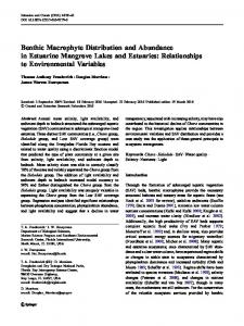
Fig. 1 Representative μXRF maps of chromium and potassium distribution in leaves of Cr(III)- and Cr(VI)-treated as well as control group of C. cophocarpa. a Cr(III)-treated leaf shows spot-like structures with high concentration of Cr; b K distribution in Cr(III)-treated leaf that visualizes vascular bundle region; c Cr(VI)-treated leaf with Cr accumulated in the region of vascular bundle; d K distribution
Table 1 The median as well as minimal and maximal intensities of chromium characteristic X-rays emitted from leaves and stems not exposed to Cr (control) or exposed to Cr(III) or Cr(VI) Specimen
Cr(III) Cr(VI) Control Cr(III) Cr(VI) Control
X-ray intensity (counts per second)
Leaf
Stem
Median
Min.
Max.
91.3 (d) 4.7 (b) 1.1 (a) 153.1 (e) 12.7 (c)
1.7 0.3 0.2 2.2 0.6
1,802.5 301.9 4.7 2,167.8 144.6
1.2 (a)
0.2
3.3
The median value relates to X-ray intensity of fluorescence of Cr signal (counts per second) at the scanned area (380 μm−2 ). The letters indicate statistically significant differences between treatments (Kruskal–Wallis non-parametric ANOVA and Dunn’s test; α = 0.05)
13
in Cr(VI)-treated leaf that visualizes vascular bundle region; e the level of background intensity of X-rays characteristic for chromium emission in a leaf from the control group of plants; f K distribution in a leaf of the control group of plant that visualizes vascular bundle region. Grayscale indicates intensity of X-ray signal (cps) characteristic for chromium emission energy
obtain sufficient intensity of characteristic X-rays for chromium, the beam size of 130 nm was used. Thus, the probe current increased to 800 pA and 4,500 counts per second were registered by EDS detector. Maps of Cr distribution were created for all experimental groups when 100 frames were accumulated by the system at the resolution of 512 × 512 pixels. Three samples of each experimental group were mapped. Plant morphology examination—light microscopy Fresh as well as ethanol-fixed (70 % ethanol solution) plant material was hand-sectioned with a razor-blade and examined under an Olympus BX60 (Olympus Corporation, USA) microscope equipped with differential interference contrast (DIC). Image-Pro PLUS ver.4.0 (Media
Planta
Fig. 2 Representative μXRF maps of chromium and potassium distribution in stems of Cr(III)- and Cr(VI)-treated as well as control group of C. cophocarpa. a Cr(III)-treated stem shows spot-like structures with high concentration of Cr; b K distribution in Cr(III)-treated stem that visualizes vascular bundle region; c Cr(VI)-treated stem with Cr accumulated in the region of vascular bundle; d K distribu-
tion in Cr(VI)-treated stem that visualizes vascular bundle region; e the level of background intensity of X-rays characteristic for chromium emission in a stem from the control group of plants; f K distribution in a stem of the control group of plant that visualizes vascular bundle region. Grayscales indicate intensity of X-ray signal (cps) characteristic for chromium emission energy
13
Planta
Cybernetics Inc., Rockville, MD, USA) software was applied to measure distances between epidermal glands/ hairs in microscopic images. At least six independent leaves/stems were used for analysis. Statistics Three independent sets of experiments were conducted, with each set comprising several independent replicates. Results were statistically verified based on STATISTICA 10 software. The statistical tests were chosen according to the distribution of results. Non-parametric Kruskal– Wallis/Mann–Whitney U tests were applied to compare differences between objects. Following the rejection of null hypothesis, non-parametric multiple comparison test (Dunn’s test) was performed to determine statistical significance of results at α = 0.05.
Results We found significant differences in Cr distribution in both groups of Cr-treated plants, i.e., in the plants exposed to Cr(III) and Cr(VI). Figure 1a and c shows representative maps of Cr accumulated in leaves of C. cophocarpa. The plants treated with Cr(III) ions revealed spot-like chromium distribution (Fig. 1a), whereas leaves obtained from plants treated with Cr(VI) (Fig. 1c) showed homogeneous accumulation of Cr with significantly higher Cr deposition in the region of vascular tissue as indicated by K pattern (Fig. 1b, d, f). The amount of Cr deposited in leaves in both Cr-treated plants differed significantly. The median X-ray intensity registered in Cr(III)-treated leaves was around 19-times higher in relation to leaves from Cr(VI)-treated plants (Table 1). There was no chromium registered in leaves obtained from the control group of plants (Fig. 1e). Similarly to the leaves, mapping of stems from both groups of Cr(III)-treated plants showed the presence of spot-like structures (Fig. 2a). The maps of Cr(VI)-treated stems revealed Cr-rich areas (Fig. 2c) in the region of vascular bundle as indicated by K (Fig. 2b, d, f). Similarly to the leaves, the median X-ray intensity of Cr signal in stems subjected to Cr(III) was 12-times fold higher than in stems treated with Cr(VI). There was no chromium registered in stems obtained from the control group of plants (Fig. 2e). In the next step of the study, distances between spotlike structures in leaves subjected to Cr(III) were measured on the basis of the μXRF maps and compared to distances obtained from light microscopic photographs of leaf surfaces (Fig. 3). The measurements were carried out only on flat surfaces of leaves because the geometry of stems (round in shape) made the measurements inaccurate. Many epidermal multi-cellular hairs were observed on
13
Fig. 3 Light microscopy photograph of C. cophocarpa leaf epidermis with well visible glands/hairs. The table represents median as well as minimal and maximal distances (in μm) between epidermal glands/hairs measured on the basis of light micrographs and those measured between spot-like structures registered in μXRF maps. No statistical differences between the values were found (Mann–Whitney U test; α = 0.05)
both lower and upper leaf epidermis (Fig. 3). The median distance between investigated structures observed in light microscope and in μXRF maps was not statistically different. It suggested that Cr-contained spot-like structures were probably glands/hairs present on the upper or lower epidermis. To confirm that glands/hairs are responsible for Cr accumulation in Cr(III)-treated plants, SEM equipped with X-ray energy-dispersive detector was applied. Concomitant SEM examination and X-ray analysis of Cr in C. cophocarpa stomata or glands/hairs confirmed the concentration of chromium below the limit of detection for EPXMA analysis (