3929
Development 124, 3929-3941 (1997) Printed in Great Britain © The Company of Biologists Limited 1997 DEV1199
Cis-acting elements conserved between mouse and pufferfish Otx2 genes govern the expression in mesencephalic neural crest cells Chiharu Kimura1,2, Naoki Takeda1, Misao Suzuki3, Mitsuo Oshimura2, Shinichi Aizawa1,* and Isao Matsuo1 1Department
of Morphogenesis and 3Laboratory of Transgenic Technology, Institute of Molecular Embryology and Genetics (IMEG), Kumamoto University School of Medicine, 2-2-1 Honjo, Kumamoto 860, Japan 2Department of Molecular and Cell Genetics, School of Life Sciences, Faculty of Medicine, Tottori University, Nishi-machi 86, Yonago 683, Tottori, Japan *Author for correspondence (e-mail:
[email protected])
SUMMARY Previous studies suggested that the Otx2 gene plays an essential role in the development of cranial skeletons and nerves of mesencephalic neural crest origin. To clarify this role, we have identified the cis-acting elements in mouse and pufferfish Otx2 genes responsible for the expression in the crest cells using a transgenic approach with the lacZ reporter gene. In mouse, 49 bp sequences in the proximal 5′ region upstream were essential and sufficient to direct the transgene expression in the cephalic mesenchyme. In pufferfish, the 1.1 kb distal region, located far downstream (from +14.4 to +15.5 kb), had almost identical activity. Between them, several DNA sequences were conserved, and mutational analyses indicated that motif A was critical for the transgene expression in the premandibular region while motif B was critical in both premandibular and mandibu-
INTRODUCTION Otx1 and Otx2 genes are the mouse cognates of the Drosophila head gap genes, orthodenticle (otd) (Finkelstein and Perrimon 1991; Simeone et al., 1992). The cognates have also been identified in human, chick, Xenopus and zebrafish (Simeone et al., 1993; Ang et al., 1994; Li et al., 1994; Bally-Cuif et al., 1995; Bliz and Cho, 1995; Pannese et al., 1995; Mercier et al., 1995). Common expression among these vertebrate cognates has suggested that (1) Otx2 may function as a head organizer component at the primitive streak stage, and (2) at a subsequent neurula to pharyngula stage those genes may be involved in regional patterning of the forebrain and midbrain (Puelles and Rubenstein, 1993; Simeone et al., 1992). Indeed, Otx2 homozygous mutants failed to develop a structure anterior to rhombomere 3 (Matsuo et al., 1995; Acampora et al., 1995; Ang et al., 1996), as do mutants of Lim1, another head organizer component (Shawlot and Behringer, 1995). At the same time, the Otx2 mutation displayed a craniofacial defect, designated otocephaly by haplo-insufficiency. Affected structures correspond to the most anterior and posterior parts of Otx2 expression at the pharyngula stage, where Otx1 is not or only weakly expressed (Matsuo et al., 1995). Defects were subtle in the Otx1 mutation, while those exhibited by the
lar regions. Motif B, CTAATTA, contains the core motif for binding of homeodomain proteins while motif A, TAAATCTG, does not match any known consensus binding sequences for transcriptional factors. The cephalic mesenchyme that expressed β-galactosidase under these ciselements is most likely to correspond to mesencephalic crest cells. Thus the molecular machinery regulating Otx2 expression in these cells appears to be conserved between mouse and fish, implying a crucial role of the Otx2 gene in development of the neural-crest-derived structures of the gnathostome rostral head. Key words: cephalic mesenchyme, neural crest cell, cis-acting element, Otx2, homeobox, pattern formation, transgenic analysis, pufferfish
Otx1/Otx2 double heterozygous mutant mice were marked through fore- and midbrains, where abnormalities were never found with a single mutation alone; Otx1 and Otx2 cooperate in the development of the rostral brain (Suda et al., 1996). Otx2 mutant defects caused by haplo-insufficiency were the particular focus of the present study (Matsuo et al., 1995). The major affected structures are the mandible and premandibular skull elements, and the ophthalmic branch of the trigeminal nerve and mesencephalic trigeminal neurons, all of which originate from mesencephalic neural crest and its closely related derivatives. These structures become highly modified with the transition from agnatha to gnathostome states, which is one of the most important innovations in the entire history of vertebrates, implying some evolutionary significance of the Otx2 gene. Furthermore, the defects are intimately related to defects by Hoxa-2 mutation and are consistent with the idea first presented by Huxley (1874; reviewed by Goodrich, 1930; de Beer, 1931, 1937) that the trabecula is a premandibular component of the viscerocranium (Matsuo et al., 1995; Kuratani et al., 1997). To elucidate the role of the Otx2 gene in mesencephalic neural crest cells, we attempted to determine the factors that specify the Otx2 expression in these cells. The expression of homeobox genes is generally regulated at the level of transcription (Krumlauf,
3930 C. Kimura and others 1994). In this study, therefore, we identified cis-regulatory elements that direct Otx2 expression in the crest cells using a transgenic approach with mouse and pufferfish Otx2 genomes. The tetraodontoid fish, Fugu rubripes (Fugu), has a compact genome of approximately 400 Mb, nine times smaller than the mouse genome, although the two genomes have a similar number of genes (Brenner et al., 1993). This is due to smaller intergenic and intronic sequences and fewer repetitive DNA sequences in the Fugu genome. Utilizing this fact, the transcriptional cis-elements of Hoxb-1 and Hoxb-4 genes were identified in intergenic and intronic regions as sequences highly conserved between the mouse and Fugu genomes (Marshall et al., 1994; Aparicio et al., 1995; Pöpperl et al., 1995). A similar comparative analysis of the Otx2 gene in this study also defined the genomic regions in both species that were capable of directing the expressions in mesencephalic crest cells. Among several DNA sequences highly conserved between them we have identified two DNA motifs that are essential for driving the expression. MATERIALS AND METHODS Transgene construction for mouse Otx2 regulatory elements All constructs used in this study were generated by standard molecular cloning techniques (Sambrook et al., 1989). The genomic fragments containing the mouse Otx2 locus were isolated from the mouse TT2 ES cell genomic library (Matsuo et al., 1995). The pCHβGAL vector was constructed by inserting a 2.5 kb SacI-BamHI fragment of the plasmid pFRTβGAL (Stratagene) into SacI and BamHI sites of the plasmid pCH110 (Pharmacia). The pBSlacZ vector was constructed by inserting a 3.0 kb HindIII-BamHI fragment of the plasmid pBS0CAT (Matsuo et al., 1991) into HindIII and BamHI sites of the pCHβGAL. To construct the pBSlacZII vector, a BglII linker was inserted into the HindIII site of the multicloning sites located in the 5′ end of the lacZ gene of the plasmid pBSlacZ and a SalI linker into the ApaI site located in the 3′ end of the lacZ gene. Construct 1 was made by inserting a 5′ flanking region from the BglII site to the translational start site of mouse Otx2 gene into the BglII site and the translational start site of the lacZ gene in the pBSlacZII, using polymerase chain reaction (PCR)-based mutagenesis (detailed procedures for in vitro mutagenesis are available upon request). Construct 2 was generated by removing the BglIIHindIII fragment from construct 1. Constructs 3, 4 and 5 were generated by deleting a 5′ flanking region of Otx2 gene in construct 2 using exonuclease III. The 5′ endpoints of these deletion plasmids were determined by DNA sequencing. Constructs 21 and 22 have three tandem repeats of 49 and 84 bp fragments, which correspond to the 5′ endpoint of construct 4-5 and the presumptive TATA box, respectively (Fig. 2A). These fragments were obtained by PCR; the primers used were 5′-AAAAGATCTTAAATCTGTTACTTACTTCGA-3′ (the sense strand primer of construct 21 and 22), 5′TCAGGATCCCCTAATTAGTAGTCTGGATAA-3′ (the antisense primer of construct 21) and 5′-CACGGATCCCCCACATTTGAAGTCATTTCC-3′ (the antisense primer of construct 22). The 49 bp and 84 bp PCR fragments were digested with BglII and BamHI, and the three respective tandem repeats were inserted into the BamHI site of pBluescript (KS−). The repeats were confirmed by DNA sequencing. An EcoRV-NotI fragment containing the 49 bp or 84 bp tandem repeats was then inserted into the NotI and SmaI sites of construct 11, respectively. Constructs 23, 24, 25 and 26 were similarly generated using four kinds of mutant 84 bp fragments in place of the wild-type fragment. The mutated nucleotide sequences were CTAGCGG in place of ATCTGTT for two motifs, snail and
the first A (construct 23), ATCAC and CCCG in place of TAAAT and AAAT for two motif A sites (construct 24), GCTAGC and ATCAGT in place of TAATTA and TAATTA for two motif B sites (construct 25) and AGCTC in place of CCAGA for motif C (construct 26), respectively (cf. Fig. 8A,B). Transgene construction for Fugu Otx2 regulatory elements A Fugu genomic phage library in the λFIXII vector (Stratagene) was constructed from MboI partially-digested Fugu genomic DNAs as described (Sambrook et al., 1989). Isolation of the Fugu rubripes Otx2 (Fotx2) gene will be described elsewhere. The pBSlacZIII was constructed by inserting the SacII-HindIII fragment of a modified pBluescript (KS−), having the BglII site in place of the BamHI site, into the SacII and HindIII sites of the plasmid pBSlacZII. Construct 11 was made by inserting a 2.4 kb 5′ flanking region from the NspV site to the translational start site of the Fotx2 gene into the EcoRV site and the translational start site of the lacZ gene in the pBSlacZIII using PCR-based mutagenesis (detailed procedures for in vitro mutagenesis are available upon request). Construct 12 was generated by cloning a 3′ XbaI-BamHI genomic fragment into the BglII and XbaI sites of construct 11. Construct 13 was generated by inserting a 3′ XbaI-BglII genomic fragment into BglII and XbaI sites of construct 11. Construct 14 was generated by inserting a BamHIBglII genomic fragment into the BglII site of construct 11. Constructs 15, 16 and 17 were generated by inserting the BglII-HindIII, HindIII-BanII and BanII-BamHI fragments, each blunted by T4 DNA polymerase, into the SmaI site of construct 11. Production and genotyping of transgenic mice Transgenic mice were generated by microinjection of DNAs into fertilized eggs from ICR mice as described (Hogan et al., 1994). Transgenic embryos were identified by PCR analyses of yolk sac DNAs. Briefly, yolk sacs were carefully dissected, avoiding cross contamination between littermates, then washed once in cold phosphatebuffered saline (PBS). After 2 hours incubation at 55°C in 50 µl Proteinase K solution, 1 µl of the heat-inactivated digest was subjected to PCR analysis specific to lacZ and SV40 t intron. The oligonucleotides, 5′-CATTACCAGTTGGTCTGG and 5′-TTATGTTTCAGGTTCAGG, amplify a 740 bp transgene-specific product. The mice were housed in environmentally controlled rooms of the Laboratory Animal Research Center of Kumamoto University School of Medicine under University guidelines for animal and recombinant DNA experiments. DNA sequencing DNA sequences were determined on both strands of DNAs using the dideoxy chain termination method (Sambrook et al., 1989). X-gal staining and histological analysis of embryos Transgenic embryos at 7.5 to 12.5 days post coitum (dpc) were fixed in 1% formaldehyde, 0.8% glutaraldehyde and 0.02% NP-40 in PBS for 10 minutes, followed by three washes with PBS for 10 minutes at room temperature. Staining was carried out overnight at 37°C in PBS containing 1 mg/ml X-Gal, 5 mM K3Fe(CN)6, 5 mM K4Fe(CN)6 and 2 mM MgCl2 in PBS. Stained embryos were washed twice with PBS and immediately stored in 10% formaldehyde in PBS. For histological analysis, embryos were embedded in Paraplast. Serial sections (10 µm thick) were stained with 1% eosin. In situ hybridization Whole-mount in situ hybridizations were performed with digoxigenin probes by the protocol of Wilkinson (1993). Probes for the Otx2 gene (Matsuo et al., 1995) were prepared with an RNA labeling kit (Boehringer Mannheim). Anti-digoxigenin antibodies conjugated with alkaline phosphatase were purchased from Boehringer Mannheim. The samples were sectioned using a D.S.K. microslicer (100 µm thick).
Conserved cis-elements for Otx2 expression 3931 RESULTS
Otx2 expression in cephalic mesenchyme Our previous studies suggested the role of Otx2 in mesencephalic neural crest cells (Matsuo et al., 1995). Little is known, however, about Otx2 expression in these cells, although expression in the neural tube has been examined in detail (Simeone et al., 1992, 1993; Ang et al., 1994). In order to identify the cis-elements responsible for Otx2 expression in the crest cells, we first confirmed the expression in the cephalic mesenchyme of 9.5 dpc mouse embryos. Although in wholemount embryos Otx2 expression in cephalic mesenchyme could not be discerned on top of the strong expression in the rostral neural tube, optic vesicles and otic vesicles (Fig. 1A), in sections Otx2 expression was found throughout the mesenchymal cells at the level of the dorsal mesencephalon (Fig. 1B). It was also found in the mesenchyme of the ventral region (Fig. 1C,D) and of the mandibular arch but not of the hyoid arch (Fig. 1D). Thus, the Otx2 gene was indeed expressed in
Fig. 1. In situ hybridization analyses of Otx2 mRNA expression in 9.5 dpc mouse embryos. (A) Whole-mount in situ RNA hybridization showing a lateral view of the Otx2 expression in the rostral brain with the posterior boundary at mesen/metencephalic junction (arrowhead). (B-D) Transverse sections of an embryo stained by whole-mount in situ RNA hybridization (100 µm thickness). (B) Otx2 is expressed in the entire cephalic mesenchyme at the level of the mesencephalon (m). (C) The expression is also seen in the cephalic mesenchyme near head veins (hv), but is not found in the most rostral mesenchyme nor in the mesenchyme caudal to mesencephalon (arrowheads). (D) Otx2 is also expressed in the mandibular arch (ma), but not in the hyoid arch (ha). Abbreviations: op, optic vesicle; ot; otic vesicle; t, telencephalon; V, trigeminal ganglion. Scale bars, 400 µm.
cephalic mesenchyme. The expression appeared to distribute caudally to optic vesicles; it was not apparent in the mesenchyme at the levels of telencephalon and rhombencephalon, nor in the trigeminal ganglion (Fig. 1C), nor was it found in the surface ectoderm. It was difficult, however, to determine the precise extent of Otx2 expression in cephalic mesenchyme, since the signal was not strong enough. Mouse Otx2 cis-element directing expression in cephalic mesenchyme The genomic region of mouse Otx2 gene encompassing −10.2 to 16.4 kb (0 is the translation start site) was surveyed for enhancer activity by generating transgenic mouse embryos with the reporter Escherichia coli lacZ gene (Fig. 2A). Screening was made in transgenic embryos at 10.5 dpc immediately after the emigration of mesencephalic neural crest cells had been completed. It identified the enhancer activity in the 5′ flanking region encompassing −1.8 to 0 kb (fragment II in Fig. 2A; construct 1 in Fig. 2C); no activity for the expression in cephalic mesenchyme was found in other regions (data not shown). The 10.5 dpc transgenic embryos harboring construct 1 exhibited β-gal expression in the cephalic mesenchyme at the levels of diencephalon, mesencephalon and mandibular arch (Fig. 3E,F). Weak expression was also found in the heart (Fig. 3E). The expression with construct 1 was then examined in detail to determine whether and how these β-gal-positive cells are related to mesencephalic crest cells. No β-gal expression was detected in the cephalic mesenchyme of transgenic embryos at 7.5 dpc (data not shown). The expression was first found in the cephalic mesenchyme at 8.5 dpc (Fig. 3A,B). It was never detected in the most anterior region of cephalic mesenchyme nor in the neural plate (Fig. 3A,B). The β-gal expression in the cephalic mesenchyme was restricted to the levels of diencephalon, mesencephalon, metencephalon and mandibular arch at 9.5 dpc (Fig. 3C,D). Later, at 10.5 dpc the expression persisted in the ventral or distal parts of these regions (Fig. 3E,F). The transgene expression was greatly decreased at 11.5 dpc, even in ventral cephalic mesenchyme (Fig. 3G). Still later at 12.5 dpc, β-gal expression was not found at all in the cephalic mesenchyme (Fig. 3H). β-gal expression was further analyzed in the sections. In the 8.5 dpc embryo the transgene began to be expressed in the cephalic mesenchyme but not in the neural plate, neural crest or the surface ectoderm (Fig. 4A). At 9.5 dpc, β-gal-positive cells were distributed throughout the entire dorsal mesenchyme at the level of the mesencephalon (Fig. 4B); however, most of the trigeminal ganglion cells did not express β-gal (Fig. 4C). The anterior limit of the positive cells was located caudal to the optic vesicles, and no positive cells were found at the telencephalic level (Fig. 4C). The β-gal expression was also found in the mandibular arch (Fig. 4D). Besides mesenchymal cells, the expression was found in the ventral diencephalon (Fig. 4C, D). At 10.5 dpc, β-gal-positive cells were distributed, as at younger stages, in the cephalic mesenchyme at the level of diencephalon and mesencephalon and were absent rostrally to the optic vesicles (Fig. 4E-G). The β-gal expression, however, was decreased towards the dorsal side (Fig. 4E,F). β-galpositive cells were rarely seen in the trigeminal ganglia, but were found in their most rostral portion (Fig. 4F); this expression was lost by 11.5 dpc (data not shown). β-gal-
3932 C. Kimura and others
A) Phage genomic clone #36
Phage genomic clone #12 RV
RV
10 kb
BII
X
Sh
(0)
5 kb
SII +5 kb
B
+10 kb
+15 kb
2 kb I II
III
V IV
B) AGATCTTGGAAGAAGGAACACAGAATATAAAGTGTTCTTCGCAAAAGTAACTAGTTTCACTCGTTAACAATCCAGTTCTCTTAGAAGGACATTTATAAAAATCACATTTTAGGGTGGGGA 120 #1 BglII AATAAGACTTGTGTTCTGAATGTAAATCTGGAAGAAATCACAGCTGTTGAGTTTTGTTTGTGAAGCTTTGCCTTTGTCTCTTAAGCAAGGTAAATATGAAAACCTAAGGTCTTAATTTTT 240 #2 HindIII TCATTAATGAACTTTATGAAGTGACAGAGAACTCAGTCTTGTATCCGCCATTTTGAGGAAGTTTCTCAGAGTGTCAAAGTTTCAAAGCAGAGAGAGAGAGAGAGAGAGAGAGAGAGAGAG 360 AGAGAGCTTTAAGTGCCTTATTTTTGGAAAATAAAAGCCAGCCTTACACACATTGCCTGCCTGTATTAACATCGATGGGGGGAGGGGTTCAGTAAAGGAACACAGATGACTTTTCTTTTT 480 CTCCCCCCACCCAGCTCCTTTTCTTACAAAATTGTAGGAAATTTAAAAAGCAGAGAGAAGTGCAGTTAGTTCCATTGCTTTGGATGTTTATTGACTTCAAACCTAAATATATGGGAAAGG 600 TGGCTCCCGTTGCAGCCCCCTCTCCCCCCAACCCAAAATCCAAGAAAAAGTGTTTCCCGCTGGATCTGTAGCCTCCTGACATAAAAAACATGGAAGCGGCTTGCTGTAGGAATGCACCCC 720 TCTGGACTTGCAAAATGTGGGGGCAGCAGTTTATCTGACTTTACTTCAGTTAGGATTCTAACTTAAGCATTTCTGTCAAGCCCTGTGCTAGTCTTGAAGAAAAGACTTTTTTTTCCCTCT 840
#3 CAGTATGTTTAAATTAAATATTTGATGGGTGTCGCTTAAAAGTTAAATAGTTGTTTGTTTAGCTATAGTTCTTTAGACACATGTAATCAAACTCCTCCAAAACCCTAATAAATCTGTTAC 960
#4 TTACTTCGAAATCTAATTATCCAGACTACTAATTAGGTGAAAATGATTACTGGAAATGACTTCAAATGTGGGATAATAGTGATTGGGCAGTTTTCACAAGTTTGTTGTTCAGCAAGTTGG 1080 primer 1 #5 TGTTTTAAATGTTGAATGCGTTTACATTTTAGGTTCACAGCAGTTGAAAGTGGTAATAATTTTAAAAAAAAAAATCAGCTAAGATAATTAAAACCTCTGCCAGGGAAAGGCAACAGTCTG 1200 primer 2 GGAAGAGGTAGATTTCTTGAATTGTTTCTTTGTTTCTCACCAATGGGCTTTGTTATCAGCATTATTTATTTAGCCAAAGAGTTCTTTGTCTCTGCAAATGGCCCCAATCAAGTTTTGTTT 1320 GAGACAATTAGCTAGTGCCAGCCAATGAGTCCTATGCAAATGTTGGGTTAGGAATGTAGAGCGTGCATTTGGAGGCGTGGGGTTTTGTTCTTGGGGAAGGCAGATTGTAATTGCTTTCCT 1440 CGGGTCGGTTTTCTTTCTTTCTTTCTTTTTTTAAGTTTAGGGTGGGGGTGGGGAAGACAGGTTTATCTGGTCTCACTCCATCCCCTCTAGTTTTGGAGCTGCTGGGGGGTGGGGGGGACG 1560 GCGGGGGTGGGGGACGCATCTGCAACTCCTTTAAAAGCCTGTGCCCAGCGTCTCCCGGGTTCTTTTTAGTTAGTGCTGGAACGTGGAGGAAGCTGCTCCCTCCGAAGCAGTAAACCAGCA 1680 TTTCTGTTTGTTTGTTTGTTTTGCCCTTAGTTCCGTCACTCCAAATCTACCCACCAAGGACCCTGACCCTGTCCACTCCAGGCGAATCGAGACCGTCCGGCTGGGTCCCCCCAATTTGGG 1800 CCGACTTTGCGCCTCCAAACAACCTTAGCATG primer 3 M
1832
C) BII
H 200 bp
(0) #1
17 / 19
#2
2/ 3
#3
4/ 5
#4
9 / 13
#5
0/ 6
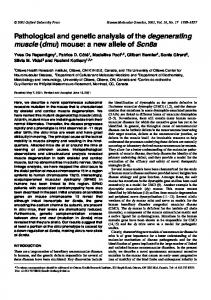
Conserved cis-elements for Otx2 expression 3933 Fig. 2. Identification of the mouse cis-element critical for the transgene expression in the cephalic mesenchyme. (A) The restriction map of the mouse Otx2 genomic locus surveyed in this study is shown along with the phage clones used on the top and coding regions (filled boxes) underneath. The translational start site is indicated by (0). Each fragment examined is identified by bars marked I-V. (B) Nucleotide sequences of 5′-flanking region of the mouse Otx2 gene. Restriction enzyme sites, putative TATA box and three primers used for RT-PCR analyses are underlined. The 5′ end of each deletion construct (2-5) is indicated by filled triangles with the number of constructs (see Fig. 2C). The translational start codon is indicated by M at the end of the nucleotide sequences. Gene bank accession no. U96488. (C) Schematic diagram of the deletion constructs used to identify the mouse cis-acting region within fragment II in A. The number of β-gal-positive embryos in the cephalic mesenchyme among transgenic embryos is indicated on the right. In all these embryos, the expression patterns were virtually the same although their levels were variable. The transgenic embryos were counted as β-gal negative, possibly because of the effects of integration sites and others, when the expression was not found in cephalic mesenchyme. Abbreviations of restriction enzyme sites: B, BamHI; BII, BglII; H, HindIII; RV, EcoRV; S, SalI; SII, SacII; Sh, SphI; X, XbaI.
positive cells also appeared to converge into and halt at the sites that may correspond to a future cartilaginous mesenchyme or peripheral nerves in the premandibular and mandibular regions (Fig. 4G-I; see Discussion). Actually, β-gal-positive cells were found in the cells migrating into the mandibular arch (Fig. 4I). At the same time, β-gal-positive cells were present more posteriorly and distally, in the mesenchymal cells of hyoid arch and the myocardium (Fig. 4J; data not shown). At this stage βgal expression was found in the ventral diencephalon, and the expression shifted to the neurohypophyseal bud (Fig. 4K); it was not found in Rathke’s pouch or in the oral ectoderm. Later, by 11.5 dpc, the transgene expression was limited to the ventral
Fig. 3. Developmental changes in β-gal expression (blue) with construct 1. Lateral (A) and frontal (B) views of an 8.5 dpc embryo showing the onset of expression. Lateral (C) and ventral (D) views of a 9.5 dpc embryo showing increase of the β-gal expression in the cephalic region and the mandibular arch. (E,F) Transgene expressions in 10.5 dpc embryos. (G) Lateral view of a 11.5 dpc embryo showing a great decrease in the transgene expression. (H) Lateral view of a 12.5 dpc embryo showing no β-gal expression. Abbreviations: m, mesencephalon; ma, mandibular arch; h, heart; np, neural plate; op, optic vesicle; t, telencephalon. Scale bars, 400 µm.
part of the cephalic mesenchyme, the floor of the diencephalon and neurohypophysis (Fig. 4L, data not shown). In summary, most of the expression under the control of the 1.8 kb DNA fragment appeared to correspond spatially and temporally with the distribution of mesencephalic neural crest cells, except for the ventral part of diencephalon (see Discussion). Regulatory elements essential for the transgene expression The nucleotide sequences of the 1.8 kb 5′ flanking region were determined (Fig. 2B). The major transcription start site of the Otx2 gene was preliminarily reported with cultured cells to locate at 3140 bp downstream of the 5′ XbaI site (Simeone et al., 1995). We confirmed this by reverse-transcription and polymerase chain reaction (RTPCR) experiments with 9.5 dpc mouse embryos. Primer 2 and primer 3 amplified the 713 bp fragment but primer 1 and primer 3 did not amplify the predicted 845 bp fragment (data not shown). This suggests that the first transcription start site locates between primer 1 and primer 2. The canonical TATA box sequence is present between these two primers (Fig. 2B). To define the critical cis-element for the expression in the cephalic mesenchyme, a series of deletions was introduced into construct 1 (constructs 2, 3, 4 and 5; Fig. 2B,C). Transgenic embryos with the three deletion constructs 2, 3 and 4 showed almost the same pattern of β-gal expression in the cephalic mesenchyme (Fig. 5A-C). Analysis of sections confirmed that the β-gal-positive cells were distributed in the cephalic mesenchyme, ventral diencephalon and mandibular arch (Fig. 5E; data not shown). Finally, we made construct 5 that had a 49 bp deletion from the 5′ end of construct 4 (Fig. 2B,C). It sent out no β-gal signals in the cephalic mesenchyme of transgenic embryos (Figs 2C, 5D). In contrast, the transgene expression in ventral diencephalon was preserved even after this 49 bp deletion (Fig. 5F). Thus it was concluded that the major regu-
3934 C. Kimura and others latory element for the transgene expression in the cephalic mesenchyme was located in the 49 bp sequence between the 5′ ends of constructs 4 and 5. Pufferfish cis-element for expression in cephalic mesenchyme We have speculated that the cis-element that controls Otx2 expression in mesencephalic crest cells may be conserved among all gnathostomes (Matsuo et al., 1995). In parallel with the analysis in mouse, we thus have pursued characterization of the regulatory region in the Japanese pufferfish, Fugu rubripes. Among gnathostomes, the sequences of Otx2 cognates are highly conserved, while those of Otx1 cognates are relatively diverged. The deduced amino acid sequences of the Fugu Otx gene that we isolated showed 96% homology with that of zebrafish zOtx2 and 92% with mouse Otx2; 60% homology with zOtx1, 57% with zOtx3 and 57% with mouse Otx1 genes. Thus, we concluded that the gene isolated is the Otx2 cognate in Fugu, and we called it Fotx2. Details of its genomic structure and DNA sequences will be reported elsewhere. On the basis of the result in mouse that construct 1 gave βgal expression in cephalic mesenchyme, we first tested the enhancer activity of the 2.4 kb 5′ flanking region with the lacZ reporter (construct 11; see Fig. 6). The construct, however, did not give any β-gal expression in transgenic embryos (Fig. 6 and data not shown). Next, we added the 3′ DNA fragment between the XbaI and BamHI sites, located at +10.5 to +16 kb, to construct 11 (Fig. 6; construct 12). The transgenic embryos harboring this construct 12 exhibited β-gal expression in the cephalic mesenchyme as good as with the mouse cis-acting element (Figs 7A, 3E,F). Subdivision of this
Fig. 4. Analyses of the β-gal expression (blue) with construct 1 in sections. (A) Transverse section through the cephalic region of an 8.5 dpc transgenic embryo showing the onset of the β-gal expression in the cephalic mesenchyme. (B-D) Transverse sections through a 9.5 dpc transgenic embryo. (B) The entire cephalic mesenchyme is stained at the level of mesencephalon (m). (C) The mesenchyme caudal to the optic vesicle (op) and the ventral diencephalon (vd) are stained, but the trigeminal ganglion (V) or the cephalic mesenchyme at the level of telencephalon (t) is not. (D) β-gal is expressed in the mandibular arch (ma) and the ventral diencephalon. Transverse (E-H and J) and sagittal (I,K) sections through 10.5 dpc transgenic embryos. (E,F) Dorsally the transgene expressions are downregulated in mesencephalic mesenchyme, but the most rostral part of the trigeminal ganglion is stained (F, arrowheads). (G) Ventral β-gal staining remains in the mesenchyme bilaterally (arrowheads); it may contain the procartilagious mesenchyme in the cranial base. The transgene is also expressed in the floor of diencephalon (fd). (H) β-gal expression in the mandibular arch converges in the center as spots (arrowheads). It probably contains the precartilage condensation of prospective mandibular bone. (I) The transgene expression in neural crest cells migrating into the mandibular arch (ma) located caudal to the telencephalic vesicle (v) (arrowheads). (J) β-gal is also expressed in a part of the myocardium (h). (K) β-gal is expressed in the floor of diencephalon and the posterior lobe (pl) but not in Rathke’s pouch (rp). (L) A transverse section through an 11.5 dpc transgenic embryo. β-gal is expressed in the floor of diencephalon (fd) and neurohypophysis (nh) but not in the pars intermedia (pi). Abbreviations: ha, hyoid arch; hv, head vein; np, neural plate; nt, neural tube; ot, otic vesicle; se, surface ectoderm; 3v, third ventricle. Scale bars, 200 µm.
Conserved cis-elements for Otx2 expression 3935
Fig. 5. Otx2/lacZ transgene expressions with deletion constructs. (A-D) Lateral views of the transgene expressions in 10.5 dpc embryos harboring constructs 2 (A), 3 (B), 4 (C) and 5 (D) (cf. Fig. 2). β-gal expressions are virtually identical among constructs 24, being high in the cephalic mesenchyme (m) and mandibular arch (ma). In contrast, no β-gal expression is found in the cephalic mesenchyme with construct 5 (D). (E) Transverse section through a 10.5 dpc transgenic embryo containing construct 4. Expression of the transgene in the cephalic mesenchyme and the ventral diencephalon (vd) is almost identical to that seen in transgenic embryos with construct 1 (Fig. 4). (F) Transverse section of a 10.5 dpc transgenic embryo harboring construct 5. The transgene expression is lost in the cephalic mesenchyme but preserved in the ventral diencephalon. Abbreviations: h, heart; m, mesencephalon; mt, metencephalon; op, optic vesicle; t, telencephalon; V, trigeminal ganglion. Scale bars, 400 µm.
N
X
B
(0)
B
BII
+10 kb
+15 kb II
I
III
2 kb BII
IV
HIII
Bn 500 bp
V
VI
B VII
Number of -gal positive embryos / transgenics
Construct #
Construction scheme
#11
I
lacZ
0/ 6
#12
II
I
lacZ
5/7
#13
III
I
lacZ
0/9
#14
IV
I
lacZ
10 / 13
#15
V
I
lacZ
0/5
#16
VI
I
lacZ
11 / 15
#17
VII
I
lacZ
0/8
Fig. 6. Schematic diagram of the Fotx2/lacZ transgene constructs used to identify Fugu cis-acting elements. The restriction map of the Fotx2 genomic locus surveyed in this study is shown. Coding regions are indicated by filled boxes underneath, and the translational start site by (0). Each fragment examined is identified by bars marked IVII. The table gives a summary of the transgenic analyses. In all βgal-positive embryos, the patterns of expressions were virtually the same though their levels were variable. Abbreviations: B, BamHI; BII, BglII; Bn, BanII; HIII, HindIII; N, NspV; X, XbaI.
5.5 kb fragment at the BglII site further localized the activity to the 2.3 kb BglII-BamHI fragment (construct 14, Figs 6, 7B). The XbaI-BglII fragment did not give any β-gal expression (construct 13, Fig. 6; data not shown). In 9.5 dpc whole-mount embryos harboring construct 14, transgene expression was found in the cephalic mesenchyme at the levels of diencephalon, mesencephalon, metencephalon and in the mandibular arch, but not in the most anterior region of cephalic mesenchyme or the neural tube (Fig. 7C). Some of the β-gal-positive cells were distributed further posteriorly and distally to the mesenchyme of hyoid arch and the myocardium (Fig. 7C; and data not shown). In sections, βgal-positive cells were distributed throughout the entire dorsal mesenchyme at the mesencephalic level, but not in the trigeminal ganglia (Fig. 7G,H). The β-gal-positive mesenchyme extended caudally to the optic vesicle but not rostrally (Fig. 7H,I), and it was also found in the mandibular arch (Fig. 7I). This distribution pattern of β-gal expression in cephalic mesenchyme was almost identical to that seen with the mouse cis-element. In contrast to the latter, however, no β-gal expression was observed in the neurohypophysis or ventral diencephalon (Fig. 7H). The 2.3 kb region was then further subdivided into BglIIHindIII 0.7 kb, HindIII-BanII 1.1 kb and BanII-BamHI 0.5 kb fragments; these were combined with construct 11 to generate constructs 15, 16 and 17, respectively (Fig. 6). Neither construct 15 nor construct 17 gave any β-gal staining (Fig. 7D,F), but construct 16 containing the 1.1 kb subfragment yielded the same pattern of β-gal expression in the cephalic mesenchyme (Fig. 7E; data not shown). These results suggest that the 1.1 kb subfragment is responsible for the Fotx2 expression in the cephalic mesenchyme.
3936 C. Kimura and others Critical DNA motifs conserved between mouse and Our previous study indicated the essential role of th Otx2 gene Fugu cis-elements in the cephalic crest cells, and we have attempted to determine A question remains as to whether the mouse 49 bp sequences the regulatory mechanism of Otx2 expression in these cells. corresponding to the 5′ endpoint of constructs 4 and 5 is sufThe current study specified the 49 bp 5′ flanking sequences of ficient for the transgene expression in cephalic mesenchyme mouse Otx2 gene that are essential and sufficient for the β-gal (Fig. 8A). The three tandem repeats of the 49 bp cis-element expression in the mesencephalic neural crest cells of transgenic were then linked to the Fotx2 promoter construct 11 (construct embryos. Furthermore, the 1.1 kb DNA fragment of Fotx2 21), and the β-gal expression in transgenic embryos harboring gene, a Fugu homologue of Otx2 gene, had almost the same this construct was analyzed. Four out of seven transgenic activity. Two DNA motifs, A and B, conserved between these embryos exhibited β-gal expression in the cephalic mestwo species, were crucial for the expression. This is the first enchyme as good as with the mouse 1.8 kb cis-acting element cis-element identified for gene expression in mesencephalic (Fig. 9A). Thus it was concluded that the mouse 49 bp cis-element is not only essential but also sufficient for driving the transgene expression in cephalic mesenchyme. The result also indicates that the mouse 49 bp cis-element is functionally equivalent to the Fugu 1.1 kb element. To determine the DNA motif(s) essential to the expression in the cephalic mesenchyme, our strategy is to find DNA sequences conserved between mouse and Fugu cis-elements. The comparison of nucleotide sequences between mouse 49 bp and pufferfish 1.1 kb DNA fragments revealed four candidate sequences (Fig. 8A): the consensus sequences for binding of snail transcription factor and three DNA motifs, A, B and C. To determine whether these DNA motifs were indeed crucial for the enhancer activity, we introduced mutations in each of them (Fig. 8B). The mutation in two motifs, snail and the first A (construct 23; Fig. 8B), gave β-gal expression that was indistinguishable from the expression by the control construct 22 (Fig. 9B,C). With the mutation in both motif A sites (construct 24; Fig. 8B) transgene expression in the premandibular region was completely lost but that in the mandibular region was retained (Fig. 9D). With construct 25, introducing the mutation into both of two B motifs(Fig. 8B), no transgene expression was found at all (Fig. 9E). The mutation in motif C (construct 26; Fig. 8B) did not affect the expression (Fig. 9F). These results indicate that motifs A and B are crucial for the transgene expression in the cephalic mesenchyme. Motif B, CTAATTA, is essential for expression in both premandibular and mandibular mesenchyme while motif A, TAAATCTG, is essential for expression only in premandibular mesenchyme. Fig. 7. Expression patterns of Fotx2/lacZ reporter constructs. (A-F) Lateral views DISCUSSION Gans and Northcutt (1983) proposed the vertebrate head rostral to otic vesicles as a ‘New Head’ that has no homologues in protochordates. According to their proposal, the cranial neural crest, a novel feature in vertebrates, plays an essential role in development of this region. The role of cephalic neural crest in the patterning of rostral head, however, has remained uncertain.
of β-gal expression in transgenic embryos with constructs 12 (A), 14 (B, C), 15 (D), 16 (E) and 17 (F) (cf. Fig. 6). (A,B) Transgenic embryos at 10.5 dpc. (C-F) Transgenic embryos at 9.5 dpc. β-gal expression patterns are virtually identical among constructs 12, 14 and 16, being high in the cephalic mesenchyme and in the mandibular arch (ma), but neither construct 13 (data not shown), 15 (D) nor 17 (F) gives any expression. (G,H) Transverse and (I) sagittal sections through 9.5 dpc transgenic embryos containing construct 14. (G) The entire cephalic mesenchyme is stained at the level of mesencephalon. (H) β-gal is expressed in the cephalic mesenchyme caudal to optic vesicles (op) but not in the trigeminal ganglions (V) nor in the ventral diencephalon (vd). (I) The transgene is expressed in crest cells migrating into the mandibular arch (ma) (arrowhead). Abbreviations: h, heart; ha, hyoid arch; m, mesencephalon; t, telencephalon. Scale bars, 400 µm.
Conserved cis-elements for Otx2 expression 3937 crest cells, and it will be useful to analyze the complex genetic controls that regulate Otx2 gene in these cells and thereby to understand the role of Otx2 gene in patterning of the rostral head. The cis-acting element for mesencephalic crest cells The mouse and Fugu Otx2 regulatory elements exhibited almost identical expression in cephalic mesenchyme. However, this is not true in all the mesenchymal cells, which raises the question of which cell populations they correspond to. The cephalic mesenchyme comprises the neural crest cells and mesoderm (Noden, 1988). No marker is available at present that distinguishes neural crest cells and cephalic mesoderm, so that no definite conclusion can be drawn. The distribution pattern of β-gal-positive cells, however, was quite similar to that of neural crest cells, as revealed by transplantation of avian neural crest and by dye-labeling experiments in mammals (Noden, 1984; Lumsden et al., 1991; Imai et al., 1996). In mouse, the emigration of crest cells first commences in the mesen/metencephalic regions at the 4-somite stage, and is completed by the 7- to 14-somite stage (Nichols, 1981; Serbedzija et al., 1992). The βgal-positive mesenchymal cells were first detected at the mesencephalic level at 8.5 dpc. The strong β-gal expression at 9.5 dpc at the level of dorsal mesencephalon, its disappearance in dorsal and its shift to ventral at 10.5 dpc, coincide well with migration of the crest cells. Migration is far less extensive in the mesodermal mesenchyme cells (Noden, 1984), and the possibility is less likely that these cells express β-gal at 9.5 dpc but not at 10.5 dpc, though such expression in the mesodermal mesenchyme cannot be excluded. The precise cell lineages of cephalic mesenchyme are still not known in mouse, but cephalic crest cells are believed to yield most of the craniofacial skeleton including mandible, cephalic connective tissues and cranial nerves in avian species (Le Douarin, 1982; Noden, 1984; Couly et al., 1993). These are structures primarily affected in Otx2 heterozygous mutants (Matsuo et al., 1995), although in our previous study it remained uncertain
A) mouse Otx2
ATAAATCTGTTACTTACTTCGAAATCTAATTATCCAGACTACTAATTAG snail #5 A #4 A B C B
49
Fotx2
AAGCTTGTGGCTGCCACACACTGACAAGCACACACATTAAGAGTGACAGCAAACACAAAC CTCAGACAAGTCAATCACATCATCATGTCAACACAGACAGGCGTTAATGGGCCTAATTCC GCCCAATACACATCGGTAAATCTATAGGAAAATATTTATCTTCATTAGCAAAACTCTCAA A A CACATATGGGTTGGGTTTTGCCTGTTGAGAGTTGCGGTCTCAGGTAGGAGCTGCAACTTA ATCCAGAAGAATAACAACAACCCTGAACTGAGAAATGTGTGTGGAGTTTAGCGGCAGGCA C A TTGGCAGTCTGCACAAAAGATGGGTGGCACCTCGTAAACATCCTTGAAACAGGTTTCTCA
60 120 180 240 300 360
snail
TGCTTAAAAGGGGATCTGCAGGCAAAGTTTTTTCCAGAGTGTGAGATTCTGCAAAATCTG C A AGGCCAGCTCAGTATATCATGCCTCTCACATCCTCTATTCTCTTCCCAAACACTCACCAG CGTCCCCCCCACCATCCCCACACCCTGCCCTCCACCCTCTCCACTCCAAGCTCCCATCCA C GCCAGGGAGAGCAGACGAGAGGAAGAGAAAGAGGAGATAGGCCCAGTTGCTAAAGCTGCC A TTTTCAGCCACTGCTCCCGCTCAGTCTATCTGAGATACACAGCAAGCGGAGGTGTCGCTT TTCCGCTGGCATTTCGGACAGTATCTACTTATAGACAGGAGAGAAAGAGAAAAGAAAAAC B AGAAGGGGTATTATCAGGGAGGGATGAGAGCAGCAAAGGCATTTTGTCTTTTTATTCCCC TCCTAAGCCCTGGGCTTTGCTCCCAATCCGATTGGTCGATACGGCCAGGGATGTAAATGA AATGCAAAGAGGAATAAGGCTGGCTTCCCATCTCCAGTGTGCACCCCCCCTAAGTCCTCC ACCCCTCTTTCTCTGCCTCACTCTGTCTTGCTCTCGCAGTTGTTTATGGTGTTGTGTTGT GTTGTGTGCGTCTCCATTCACAGACTTGCTGCTCTCCCCGCTTCCGTCCCCTTCCTCACT CTGCCACCCCCCCCCCCCACACACACACACACACACACACTCTCGCTCCATCCCTCCCCT CTCCCTGGCTGGGCTC
420 480 540 600 660 720 780 840 900 960 1020 1080
B) A
snail
A
B
C
B #22
9 / 13
#23
13 / 18
TAAATCTGTT TAACTAGCGG
TAAATCTG
GAAATCTA
ATCACCTG
GCCCGCTA
TCTAATTATC
ACTAATTAGG
TCGCTAGCTC
ACATCAGTGG
#24
2 / 12 *
#25
0 / 18
#26
6/9
ATCCAGA ATAGCTC
Fig. 8. DNA motifs conserved between mouse and pufferfish Otx2 cis-elements. (A) Nucleotide sequences of 49 bp mouse Otx2 (top) and 1.1 kb Fotx2 (below) cis-acting region. Homologous DNA sequences conserved between them are indicated: motifs A (green color), B (blue) and C (red) as well as the consensus binding site for snail (yellow; Kasai et al., 1992). Gene bank accession no. U96489. (B) Schematic diagram of the mutation constructs. The number of β-gal positive embryos in the cephalic mesenchyme among transgenic embryos with each construct is indicated on the right. In all β-gal-positive embryos, the patterns of the expressions were virtually the same except for construct 24. *The transgene expression by construct 24 was restricted to the mandibular mesenchyme (see Fig. 9D). Analyses with constructs 22-26 were performed in transgenic embryos carrying the mouse 84 bp cis-element, which corresponds to the sequences from the 5′ end of construct 4 to that of the presumptive TATA box (Fig. 2B), in three tandem repeats. Construct 22 is wild type, construct 23 has mutations in motifs, snail and the first A, construct 24 in both A sites, construct 25 in both B sites and construct 26 in motif C. The wild-type and substituted sequences of motifs for the mutational analyses are underlined.
3938 C. Kimura and others
Fig. 9. Roles of DNA motifs conserved between mouse and pufferfish Otx2 cis-elements for expression in cephalic mesenchyme. (A) Lateral view of the transgene expression at 10.5 dpc with construct 21, in which the three tandem repeats of the 49 bp ciselement are linked to the upstream region of Fotx2 promoter construct 11. High expression in the cephalic mesenchyme and in the mandibular arch (ma) demonstrates the sufficiency of the 49 bp ciselement for expression in cephalic mesenchyme. (B-F) Mutation analyses of the DNA motifs; lateral views of the transgene expressions at 10.5 dpc are given. (B) Wild-type construct 22; (C) construct 23 bearing mutations in motifs snail and the first A; (D) construct 24 in both A sites; (E) construct 25 in both B sites; (F) construct 26 in motif C (see Fig. 8b). No change in β-gal expression is found with constructs 23 (C) and 26 (F) from wild-type construct 22; both are high in the cephalic mesenchyme and in the mandibular arch. In contrast, with construct 24 no β-gal expression is found in the cephalic mesenchyme at the level of mesencephalon, while the expression at the level of mandibular arch is preserved (arrowhead, D). Construct 25 shows no β-gal expression at all (E). Constructs 22, 23 and 26 sometimes give ectopic expression in the forelimb. Abbreviations: m, mesencephalon; t, telencephalon. Scale bars, 400 µm.
whether the defects reside in the Otx2 function in these neural crest cells themselves or are secondary to abnormal neuroectoderm. Endogenous Otx2 is expressed in cephalic mesenchyme, and the sites of β-gal expression identified under the cis-element correspond well with the mesencephalic neural crest cells. Thus the Otx2 heterozygous defects may relate primarily to the Otx2 function in cephalic crest cells rather than secondarily to the Otx2 function in the midbrain neuroectoderm. The next question is the spectrum of neural crest cell lineages in which the cis-element is active. Most dorsal mesenchymal cells were β-gal-positive at the level of mesencephalon at 9.5 dpc. The β-gal-positive cells extended from diencephalon to the mesen/metencephalon boundary. No transgene expression was found rostral to the optic vesicles. No crest cells arise at the telencephalic level, and the majority of rostral crest cells are known to migrate from the most anterior edge of the diencephalon and to end up in the frontonasal region (Nichols, 1981; Noden, 1988; OsumiYamashita et al., 1994). The cis-element may thus not direct expression in the crest cells from the anterior diencephalon. β-gal was not expressed in most trigeminal ganglion cells by the cis-element, but the most rostral part of the cells expressed β-gal at 10.5 dpc. The trigeminal ganglia consist of crest cells originating from at least two different axial levels (Lumsden et al., 1991). Rhombencephalic crest cells fill the maxillo-mandibular lobe. The ophthalmic lobe, however, is likely to be composed of mesencephalic crest cells which may migrate into the rostral part of the trigeminal ganglia. In Otx2 heterozygous trigeminal ganglion, the maxillar and mandibular branches developed normally while the ophthalmic branch was specifically affected (Matsuo et al., 1995). This suggests that most, if not all, of the neural
crest cells at the level of mesencephalon express β-gal under the cis-element. The last question concerns the developmental stages of mesencephalic neural crest cells when the cis-element directs the expression. The mouse cis-acting element that we identified was unable to direct the expression in neural fold or neural crest, so that its activity apparently commences when the crest cells leave the neural crest, indicating that the regulatory mechanism of the Otx2 gene expression in mesencephalic neural crest cells was independent of that in the premigratory neural crest and neural plate. A similar situation is known in Hoxa-2 expression at the level of rhombomere 4 (Prince and Lumsden, 1994; Nonchev et al., 1996). At the same time, most of the transgene expression disappeared until 11.5 dpc when the crest cells began to differentiate into tissues. Mesencephalic crest cells contribute to the viscerocranium, peripheral nerves and connective tissues, as stated above (Noden, 1975, 1984; Couly et al., 1993, 1996; Köntges and Lumsden, 1996). β-gal expression apparently converged into the procartilaginous mesenchyme or peripheral nerves of premandibular and mandibular regions at 10.5 dpc, where it ceased; nor were expressions found in other differentiated neural crest-derived structures. βgal expression generally lasts longer than RNA expression, and it is most likely that the activity of the cis-element is characteristic of the migratory phase of mesencephalic crest cells. Besides the cephalic mesenchyme, the 1.8 kb mouse Otx2 region mediated the expression in the ventral diencephalon at 9.5 dpc. Later, the β-gal-positive cells were distributed to the posterior lobe but not in the placodal ectoderm of stomodeal roof, Rathke’s pouch. The former subsequently forms the neurohypophysis and the latter the adenohypophysis of the pituitary gland. Consistent with this, Otx2 heterozygous mutant mice showed defects in development of neurohypophysis
Conserved cis-elements for Otx2 expression 3939 (Matsuo et al., 1995). The 2.3 kb Fugu regulatory region, however, did not have the activity for expression in the ventral diencephalon. In addition, even in mouse, this activity appears to reside in sequences different from those for cephalic mesenchyme; the activity persisted even after deletion of 49 bp sequences that were essential for activity in the mesenchyme. At this point, however, we are not able to conclude the exact cell populations in which the cis-acting element is active and therefore how structures originating from these cells correspond to structures affected in Otx2 heterozygous mutants. To deduce more conclusively the cephalic mesenchyme cell lineages in which the cis-element is active, the production of mice that lack this proximal cis-element is eagerly awaited. The mice should coincidentally reveal the role of the Otx2 gene in mesencephalic crest cells and, more clearly the role of these crest cells in development of the rostral head. Activity in branchial arches β-gal expression under the cis-element appeared to largely coincide with endogenous Otx2 expression, but there were a few discrepancies. This was observed in the cells that had emigrated into the 2nd branchial arch and heart by both mouse and Fugu Otx2 cis-elements, though they apparently arise in the mesencephalic region. No endogenous Otx2 expression was found in the hyoid arch or heart. Since the β-gal expression is generally sustained longer than endogenous RNA and protein expression, it is possible that a minor population of mesencephalic crest cells emigrates into the 2nd arch and myocardial wall of the heart and suppresses the Otx2 expression when it enters these regions. The migration of the mesencephalic crest cells into the hyoid arch has also been demonstrated with chick/quail chimera analyses (Couly et al., 1996). The strong β-gal staining in the mandibular arch probably reflects the crest-derived population, which corresponds to the prospective Meckel’s cartilage and mandible. Mapping experiments in chick have suggested that the distal elements of the mandibular arch derive from the mesencephalic crest and proximal elements from the rhombencephalic crest (Köntges and Lumsden, 1996; Couly et al., 1996). Indeed, in Otx2 heterozygous mutants, distal elements of mandibular arch skeletons are lacking or severely affected but proximal elements develop normally (Matsuo et al., 1995). In contrast, the Hoxa-2 mutant mice duplicated only the proximal portions of the mandibular arch skeletons, and no duplication occurred in the distal element, the mandible (Rijli et al., 1993). Thus, the mandibular arch ectomesenchyme is composed of heterogeneous crest cell components at different neuraxial levels with different genetic codes (Matsuo et al., 1995); the distal part of this arch is under the control of the Otx2 gene, which is directed by the cis-element identified in this study. Motifs A and B are conserved between mouse and fish cis-elements In addition to snail consensus sequences, three DNA sequences were conserved between mouse and Fugu Otx2 cis-elements (A, B and C sequences in Fig. 8A). Mutational analyses of these sequences indicated that motifs A and B were essential for the transgene expression in cephalic mesenchyme, but
B)
A) mouse
Fugu
TAAATCTG GAAATCTA TAAATCTA TAAATATT GAAATGTG AAAATCTG TAAAGCTG
A motif TAAATCTG
mouse TCTAATTATC ACTAATTAGG
Fugu B motif
TCTACTTATA CTAATTA
GGTAATTACA G GGATTAG otd C T ATTAT T A TA MHox A G Msx1 CAATTA C
En
Fig. 10. Comparison of the A and B sequences in 49 bp mouse and 1.1 kb pufferfish Otx2 cis-elements. The sequences substituted in the mutational analyses (Figs 8B,9) are underlined. Binding sites of en, otd, MHox and Msx1 homeodomain products are also listed for comparison (Gehring et al., 1994; Cserjesi et al., 1992; Catron et al., 1993). The ATTA core motif of homeodomain proteins is shaded.
motifs snail and C were not (Figs. 8B, 9). Motif B is required for the cephalic mesenchyme at the level of premandibular and mandibular regions, while motif A is not required for the expression at the level of mandible. As stated above, our previous study has suggested that the Otx2 gene, or Otx2positive mesencephalic neural crest cells, might have played an essential role in the establishment of the masticating system upon transition from agnatha to gnathostome. It is our interest to examine the enhancer structure, especially motif B, in extant agnatha lamprey Otx2 for its expression in mesencephalic neural crest cells. Motif B, CTAATTA, contains the core motif, ATTA, for binding of homeodomain proteins (Fig. 10A,B; Gehring et al., 1994). The 5′ TA dinucleotide preceding the ATTA core is known to bind with a higher affinity to the Gln residue at position 50 in the recognition helix of such homeodomains as those of en, MHox and Msx1 products. In contrast, 5′ GG dinucleotide is known to bind with a higher affinity to the Lys residue at position 50, as seen in otd and Otx homeodomains (Gehring et al., 1994; Cserjesi et al., 1992; and references therein; Fig. 10B). Among the homeodomain proteins that bear the Gln side chain at position 50 in the recognition helix, MHox, Cart-1 and the Msx and Dlx families are expressed in the cephalic mesenchyme (Cserjesi et al., 1992; Zhao et al., 1994; Bulfone et al., 1993; Dollé et al., 1992; MacKenzie et al., 1991). In addition, mutations in these mouse genes are reported to cause defects in cephalic mesenchymederived structures such as cranial skeletons (Satokata and Maas, 1994; Qiu et al., 1995; Martin et al., 1995; Zhao et al., 1996). The potential role of motif A, TAAATCTG, cannot be discussed here since it does not match any known consensus binding sequences for transcriptional factors. In addition, the present mutational analyses do not exclude the possibility that motifs snail and/or C also regulate the expression in cooperation with other motifs. To test this possibility, transgenic analyses are required in combination with a series of detailed mutations. Further analysis, including in vitro binding experiments on these motifs, will be important steps towards the elu-
3940 C. Kimura and others cidation of the role of the Otx2 gene in development of the gnathostome rostral head. We are grateful to Dr Shigeru Kuratani for discussion and critical reading of the manuscript, to Miss Kazuko Kuroda for technical assistance and to the Laboratory Animal Research Center of Kumamoto University School of Medicine for housing of the mice. This work was supported in part by grants-in-aid from the Ministry of Education, Science and Culture of Japan (Specially Promoted Research), the Science and Technology Agency, Japan and the Ministry of Public Welfare, Japan.
REFERENCES Acampora, D., Mazan, S., Lallemand, Y., Avantaggiato, V., Maury, M., Simeone, A. and Brûlet, P. (1995). Forebrain and midbrain regions are deleted in Otx2−/−mutants due to a defective anterior neuroectoderm specification during gastrulation. Development 121, 3279-3290. Ang, S.-L., Conlon, R. A., Jin, O. and Rossant, J. (1994). Positive and negative signals from mesoderm regulate the expression of mouse Otx2 in ectoderm explants. Development 120, 2979-2989. Ang, S.-L., Jin, O., Rhinn, M., Daigle, N., Stevenson, L. and Rossant, J. (1996). A targeted mouse Otx2 mutation leads to severe defects in gastrulation and formation of axial mesoderm and to deletion of rostral brain. Development 122, 243-252. Aparicio, S., Morrison, A., Gould, A., Gilthorpe, J., Chaudhuri, C., Rigby, P., Krumlauf, R. and Brenner, S. (1995). Detecting conserved regulatory elements with the model genome of the Japanese puffer fish, Fugu rubripes. Proc. Nat. Acad. Sci. USA 92, 1684-1688. Bally-Cuif, L., Gulisano, M., Broccoli, V. and Boncinelli, E. (1995). c-otx2 is expressed in two different phases of gastrulation and is sensitive to retinoic acid treatment in chick embryo. Mech. Dev. 49, 49-63. Bliz, I. L. and Cho, K. W. Y. (1995). Anterior neuroectoderm is progressively induced during gastrulation: the role of the Xenopus homeobox gene orthodenticle. Development 121, 993-1004. Brenner, S., Elgar, G., Sandford, R., Macrae, A., Venkatesh, B. and Aparicio, S. (1993). Characterization of the pufferfish (Fugu) genome as a compact model vertebrate genome. Nature 366, 265-268. Bulfone, A., Kim, H-J., Puelles, L., Porteus, M. H., Grippo, J. F. and Rubenstein, J. L. R. (1993). The mouse Dlx-2 (Tes-1) gene is expressed in spatially restricted domains of the forebrain, face and limbs in midgestation mouse embryos. Mech. Dev. 40, 129-140. Catron, K. M., Iler, N. and Abate, C. (1993). Nucleotides flanking a conserved TAAT core dictate the DNA binding specificity of three murine homeodomain proteins. Mol. Cell. Biol. 13, 2354-2365. Couly, G. F., Coltey, P. M. and Le Douarin, N. M. (1993). The triple origin of skull in higher vertebrates: a study in quail-chick chimeras. Development 117, 409-429. Couly, G., Grapin-Botton, A., Coltey, P. and Le Douarin, N. M. (1996). The regeneration of the cephalic neural crest, a problem revisited: the regenerating cells originate from the contralateral or from the anterior and posterior neural fold. Development 122, 3393-3407. Cserjesi, P., Lilly, B., Bryson, L., Wang, Y., Sassoon, D. A. and Olson, E. N. (1992). MHox: a mesodermally restricted homeodomain protein that binds an essential site in the muscle creatine kinase enhancer. Development 115, 1087-1101. deBeer, G. R. (1931). On the nature of the trabecula cranii. Q. J. Microsc. Sci. 74, 701-731. deBeer, G. R. (1937). The Development of the Vertebrate Skull. Oxford University Press, Oxford. Dollé, P., Price, M. and Duboule, D. (1992). Expression of the murine Dlx-1 homeobox gene during facial, ocular and limb development. Differentiation 49, 93-99. Finkelstein, R. and Perrimon, N. (1991). The molecular genetics and head development in Drosophila melanogaster. Development 112, 899-912. Gans, C. and Northcutt, R. G. (1983). Neural crest and the origin of vertebrates: A new head. Science 220, 268-274. Gehring, W. J., Affolter, M. and Bürglin, T. (1994). Homeodomain proteins. Annu. Rev. Biochem. 63, 487-526. Goodrich, E. S. (1930). Studies on the Structure and Development of Vertebrates. Macmillan, London.
Hogan, B., Beddington, R., Costantini, F. and Lacy, E. (1994). Manipulating the Mouse Embryo: A Laboratory Manual, second edn. Cold Spring Harbor Laboratory Press, Cold Spring Harbor, NY. Huxley, T. H. (1874). On the structure of the skull and of the heart of Menobranchus lateralis. Proc. zool. Soc. London 186-204. Imai, H., Osumi-Yamashita, N., Ninomiya, Y. and Eto, K. (1996). Contribution of early-emigrating midbrain crest cells to the dental mesenchyme of mandibular molar teeth in rat embryos. Dev. Biol. 176, 151165. Kasai, Y., Nambu, J. R., Lieberman, P. M. and Crews, S. T. (1992). Dorsalventral patterning in Drosophila: DNA binding of snail protein to the singleminded gene. Proc. Nat. Acad. Sci. USA 89, 3414-3418. Köntges, G. and Lumsden, A. (1996). Rhombencephalic neural crest segmentation is preserved throughout craniofacial ontogeny. Development 122, 3229-3242. Krumlauf, R. (1994). Hox genes in vertebrate development. Cell 78, 191-201. Kuratani, S., Matsuo, I. and Aizawa, S. (1997). Developmental patterning and evolution of the mammalian viscerocranium: Genetic insights into comparative morphology. Dev. Dynam. 209, 139-155. Le Douarin, N. (1982). The neural crest. In Development and Cell Biology series; 12. Cambridge University Press, Cambridge. Li, Y., Allende, M. L., Finkelstein, R. and Weinberg, E. S. (1994). Expression of two zebrafish orthodenticle-related genes in the embryonic brain. Mech. Dev. 48, 229-244. Lumsden, A., Sprawson, N. and Graham, A. (1991). Segmental origin and migration of neural crest cells in the hindbrain region of the chick embryo. Development 113, 1281-1291. MacKenzie, A., Leeming, G. L., Jowett, A. K., Ferguson, M. W. J. and Sharpe, P. T. (1991). The homeobox gene Hox-7.1 has specific regional and temporal expression patterns during early murine craniofacial embryogenesis, especially tooth development in vivo and in vitro. Development 111, 269-285. Marshall, H., Studer, M., Pöpperl, H., Aparicio, S., Kuroiwa, A., Brenner, S. and Krumlauf, R. (1994). A conserved retinoic acid response element required for early expression of the homeobox gene Hoxb-1. Nature 370, 567-571. Martin, J. F., Bradley, A. and Olson, E. N. (1995). The paired-like homeo box gene MHox is required for early events of skeletogenesis in multiple lineages. Genes Dev. 9, 1237-1249. Matsuo, I., Kitamura, M., Okazaki, K. and Yasuda, K. (1991). Binding of a factor to an enhancer element responsible for the tissue-specific expression of the chicken αA-crystallin gene. Development 113, 539-550. Matsuo, I., Kuratani, S., Kimura, C., Takeda, N. and Aizawa, S. (1995). Mouse Otx2 functions in the formation and patterning of rostral head. Genes Dev. 9, 2646-2658. Mercier, P., Simeone, A., Cotelli, F. and Boncinelli, E. (1995). Expression pattern of two otx genes suggests a role in specifying anterior body structures in zebrafish. Int. J. Dev. Biol. 39, 559-573. Nichols, D. H. (1981). Neural crest formation in the head of the mouse embryos as observed using a new histological technique. J. Embryol. Exp. Morph. 64, 105-120. Noden, D. M. (1975). An analysis of the migratory behavior of avian cephalic neural crest cells. Dev. Biol. 42, 106-130. Noden, D. M. (1984). The use of chimeras in analyses of craniofacial development. In Chimeras in Development (ed. N. Le Douarin and A. McLaren), pp. 241-280. Academic Press, London. Noden, D. M. (1988). Interactions and fates of avian craniofacial mesenchyme. Development 103, suppl. 121-140. Nonchev, S., Vesque, C., Maconochie, M., Seitanidou, T., ArizaMcNaughton, L., Frain, M., Marshall, H., Sham, M. H., Krumlauf, R. and Charnay, P. (1996). Segmental expression of Hoxa-2 in the hindbrain is directly regulated by Krox-20. Development 122, 543-554. Osumi-Yamashita, N., Ninomiya, Y., Doi, H., and Eto, K. (1994). The contribution of both forebrain and midbrain crest cells to the mesenchyme in the frontonasal mass of mouse embryos. Dev. Biol. 164, 409-419. Pannese, M., Polo, C., Andreazzoli, M., Vignali, R., Kablar, B., Barsacchi, G. and Boncinelli, E. (1995). The Xenopus homologue of Otx2 is a maternal homeobox gene that demarcates and specifies anterior body regions. Development 121, 707-720. Pöpperl, H., Bienz, M., Studer, M., Chan, S.-K., Aparicio, S., Brenner, S., Mann, R. S. and Krumlauf, R. (1995). Segmental expression of Hoxb-1 is controlled by a highly conserved autoregulatory loop dependent upon exd/pbx. Cell 81, 1031-1042. Prince, V. and Lumsden, A. (1994). Hoxa-2 expression in normal and
Conserved cis-elements for Otx2 expression 3941 transposed rhombomeres: independent regulation in the neural tube and crest. Development 120, 911-923. Puelles, L. and Rubenstein, L. R. (1993). Expression patterns of homeobox and other putative regulatory genes in the embryonic mouse forebrain suggest a neuromeric organization. Trends Neurosci. 16, 472-479. Qiu, M., Bulfone, A., Martinez, S., Meneses, J. J., Shimamura, K., Pedersen, R. A. and Rubenstein, J. L. R. (1995). Null mutation of Dlx-2 results in abnormal morphogenesis of proximal first and second branchial arch derivatives and abnormal differentiation in the forebrain. Genes. Dev. 9, 2523-2538. Rijli, F. M., Mark, M., Lakkaraju, S., Dierich, A., Dollé, P. and Chambon, P. (1993). A homeotic transformation is generated in the rostral branchial region of the head by disruption of Hoxa-2, which acts as a selector gene. Cell 75, 1333-1349. Satokata, I. and Maas, R. (1994). Msx1 deficient mice exhibit cleft palate and abnormalities craniofacial and tooth development. Nature Genetics 6,348355 Sambrook, J., Fritsch, E. F. and Maniatis, T. (1989). Molecular Cloning: A Laboratory Manual, second edn. Cold Spring Harbor Laboratory Press, Cold Spring Harbor, NY. Serbedzija, G. N., Bronner-Fraser, M. and Fraser, S. T. (1992). Vital dye analysis of cranial neural crest cell migration in the mouse embryo. Development 116, 297-307. Shawlot, W. and Behringer, R. R. (1995). Requirement for Lim1 in headorganizer. Nature 374, 425-430. Simeone, A., Acampora, D., Gulisano, M., Stornaiuolo, A. and Boncinelli,
E. (1992). Nested expression domains of four homeobox genes in developing rostral brain. Nature 358, 687-690. Simeone, A., Acampora, D., Mallamaci, A., Stornaiuolo, A., D’Apice, M. R., Nigro, V. and Boncinelli, E. (1993). A vertebrate gene related to orthodenticle contains a homeodomain of the bicoid class and demarcates anterior neuroectoderm in the gastrulating mouse embryo. EMBO J. 12, 2735-2747. Simeone, A., Avantaggiato, V., Moroni, M. C., Mavilio, F., Arra, C., Cotelli, F., Nigro, V. and Acampora, D. (1995). Retinoic acid induces stage-specific antero-posterior transformation of rostral central nervous system. Mech. Dev. 51, 83-98. Suda, Y., Matsuo, I., Kuratani, S. and Aizawa, S. (1996). Otx1 function overlaps with Otx2 in development of mouse forebrain and midbrain. Genes to Cells 1, 1031-1044. Wilkinson, D. G. (1993). In situ hybridization. In Essential Developmental Biology: A Practical Approach (ed. C. D. Stern and P. W. H. Holland), pp. 257-274. IRL Press, Oxford. Zhao, G-Q., Eberspaecher, H., Seldin, M. F. and de Crombrugghe, B. (1994). The gene for homeodomain-containing protein Cart-1 is expressed in cells that have a chondrogenic potential during embryonic development. Mech. Dev. 48, 245-254. Zhao, Q., Behringer, R. R. and de Crombrugghe, B. (1996). Prenatal folic acid treatment suppresses acrania and meroanencephaly in mice mutant for the Cart1 homeobox gene. Nature Genetics 13, 275-283. (Accepted 30 July1997)