(r = 0.997; n = 281) and a homogeneous enzyme immunoassay (r = 0.987; n = 93). The reference interval established on 121 healthy subjects was 8 â 57 U/l ...
Melzi d'Eril et al.: Continuous colorimetric lipase assay
439
Eur. J. Clin. Chem. Clin. Biochem. Vol. 30, 1992, pp. 439-444 © 1992 Walter de Gruyter & Co. Berlin · New York
TECHNICAL REPORT
Clinical Validity of a Continuous Colorimetric Method for Serum Lipase By G. V. Melzi d'Eril1, T. Bosoni2, R. Moratti2, M. Ventrucci3, A. Fumagalli4 and G. Tarenghi4 1 2 2 2
Servizio Analisi Chimico-Cliniche e Neurochimica, Fondazione "C. Mondino", Pavia Servizio Analisi Chimico-Cliniche, IRCCS Policlinico S. Matteo, Pavia Dipartimento di Gastroenterologia, Ospedale S. Orsola, Bologna Direzione Scienti ca, Bayer Diagnostici SpA, Cavenago Brianza (Milano), Italia
(Received July 2, 1991/March 16, 1992)
Summary: The clinical validity of a continuous colorimetric method for measuring pancreatic lipase was assessed. 1,2-Diacylglycerol containing long-chain fatty acid residues was used as substrate, and the method was adapted to a discrete analyser. The dynamic range was ascertained up to at least 30-fold the upper reference limit. Precision tests on three control sera yielded overall CVs of 4.6% (mean value 21 U/l), 2.4% (115U/1), and 1.0% (386 U/l), respectively. Using serum samples from normal subjects and patients with pancreatic and non-pancreatic disorders, the present method was compared with a turbidimetric method (r = 0.997; n = 281) and a homogeneous enzyme immunoassay (r = 0.987; n = 93). The reference interval established on 121 healthy subjects was 8 — 57 U/l (central 95th percentile, median 22 U/l). The sensitivity of this lipase assay in the diagnosis of acute pancreatitis (100%, median 5.6-fold the upper reference limit) was equal to that of the pancreatic isoamylase assay, and higher than that of the total oc-amylase assay (88.2%); the specificity for acute pancreatis with respect to a group of patients with acute and chronic non-pancreatic abdominal diseases (91%) was higher than that of both pancreatic isoamylase (76%) and total oc-amylase (71%).
Introduction
, ,
.
,
,
. ..
u t u methods haverlimited analytical reliability or practicSerum lipase appears to be a better indicator of pan- ability (2—4). creatic disorders than total α-amylase, which is ele- ReCently, a method, based on Imamura's principle (5) vated in numerous non-pancreatic abdominal and and using a natuml 1,2-diacylglycerol containing non-abdominal diseases (1). Nevertheless, the lipase iong.chain fatty acid residues as substrate, was made assay has not yet gained wide acceptance in the rou- available in kit form and thoroughly assessed with tine laboratory, particularly because the available regard to its performance characteristics (6). We adapted this method to a selective analyser and briefly checked its suitability. We then determined the refer(alpha- 1,4-glucanohydrolase; EC 3.2.1.1) ' ence interval on a healthy Population. In parallel Lipase (triacylglycerol acylhydrolase; EC 3.1.1.3) studies, we also determined the clinical sensitivity and Glycerol kinase (EC 2.7.1.30) specificity of this lipase assay, two other lipase assays Glycerol-3-phosphate oxidase (EC 1.1.3.21) ,f , . « . , . «. .. Α. Λ \ Λ 2-Monoglyceride lipase (EC no. not assigned) (turbidimetric and immunoenzymatic methods), and, Peroxidase (EC 1.11.1.7) the total oc-amylase and pancreatic isoamylase assays.
Eur. J. Clin. Chem. Clin. Biochem. / Vol. 30, 1992 / No. 7
440
Melzi d'Eril et al.: Continuous colorimetric lipase assay
Materials and Methods
The following comparison methods were used:
Determination of lipase
— turbidimetric method (7): reagent kit from Boehringer Mannheim GmbH, Germany, applied to Cobas Fara analyser (assay temperature 37 °C);
. The method under evaluation is based on the following reaction sequence: 1,2-Diacylglycerol + H20
Lipase (EC 3.1.1.3) ». (Colipase/ Deoxycholate/Ca2+) 2-MonoacylgJycerol lipase
2-Monoacylglycerol + H20 Glycerol + ATP
2-Monoacylglycerol -f- Fatty acids
Glycerol kinase
Glycerol-3phosphate + O2 2 H2O2 + 4-Aminophenazone + N-Ethyl-N-(2-hydroxy-3sulphopropyl)-m-toluidine
Glycerol + Fatty acids
— immunoactivation method (8): IMAC Lipase kit from E. Merck Co., Darmstadt, Germany, used manually according to the manufacturer's directions. Determination of total and pancreatic a-amylase
Glycerol3-phosphate + ADP
Total a-amylase activity was measured by an enzymatic colorimetric method using an ethyhdene-protected substrate (oc-Amylase EPS kit from Boehringer Mannheim).
Dihydroxyacetone phosphate + H2O2
Pancreatic α-amylase was determined after inhibition of the salivary fraction by monoclonal antibodies (Pancreatic a-Amylase EPS kit from Boehringer Mannheim).
Peroxidase
Quinone monoimine dye + 4 H2O
The reagents are available in a kit form (SERA-PAK® Lipase KC kit from Bayer Diagnostici SpA, Cavenago Brianza, Italy). The reagents were reconstituted according to the manufacturer's instructions to produce two solutions; all necessary components are contained in Solution 1, except deoxycholate and 4-aminophenazone, which are contained in Solution 2 (starter). The method was applied to a Cobas Fara discrete analyser (F. Hoffmann-La Roche Co. Ltd, Basel, Switzerland). The instrument's setting is shown in table 1.
Both methods were run at 30 °C following the manufacturer's directions. Specimens For precision studies, we used three commercial control sera at different lipase levels (SERA-CHEK™ Normal and Abnormal control sera from Bayer Diagnostics, and Monitrol HE from Baxter). For the comparison with the turbidimetric method, we collected 281 patient sera with lipase activities ranging from normal to markedly elevated. Lipase was also determined in 96 sera using the immunoactivation method.
Table 1 . Instrument setting COBAS FARA General conditions
Measurement mode Reaction mode Calibration mode Reagent blank Wavelength Temperature Decimal position Unit
Analysis
P
Calculation
Calibration
Control
Absorb P-T-IO-SR1-T-AO Factor REAG/DIL 550 nm 37 °C 0 U/l
4μ1 Sample Reagent 240 μΐ T Temperation delay 10s 180s IO Incubation SRI Start Rl 80 μΐ AO Readings Is first 60s interval
Conversion factor Reactive direction Sample limit Test range LO Normal range LO Calculation steps Calculation step A Readings first Reaction limit
1.0000 Increase No 0 8 1 Kinsearch 4 1.4000
Calibration interval Factor Reagent range LO Blank range LO
Each run 3472 0.0000 -0.0090
Diluent
: 10 μΐ
Diluent
10 μΐ
Number
11
Offs Check
0.0000 ON
HI HI
1200 U/l 57 U/l
Last
:7
HI HI
: 0.1500 A : 0.0200 DA
Definable by the operator Eur. J. Clm. Chem. Clin. Biochem. / Vol. 30,1992 / No. 7
441
Melzi d'Eril et al.: Continuous colorimetric lipase assay For the reference interval assessment, we collected sera from 121 blood donors (86 men and 35 women; range 20 — 60 years, median 39 years). They were recruited among clinically healthy subjects, and their blood concentrations of glucose, urea, alanine and aspartate aminotransferases were also within the respective reference limits.
Tab. 2. Imprecision data from control sera Mean U/l
Within-run SD, U/l
CV, %
SD, U/l
CV, %
To assess the diagnostic usefulness we studied 141 patients divided in the following groups:
20.6 114.6 386.1
0.81 2.16 2.85
3.94 1.89 0.74
0.49 1.67 2.32
2.39 1.46 0.60
Acute pancreatitis
For each level: 4 replicates for 9 runs (n = 36)
Seventeen (10 men and 7 women). Diagnosis was based on physical examination and imaging studies, without using laboratory data. Pancreatitis was haemorrhagic in one patient and oedematous in the remaining subjects. Blood samples were taken within 72 hours of the attack; serum specimens were kept frozen until analysis. Chronic pancreatitis Fifty-nine patients (50 men and 9 women). Twenty-eight out of these were studied during relapse and 31 during clinical remission. Diagnosis was made on the basis of pain history and confirmed by at least one of the following studies: abdominal X-ray, ultrasound, endoscope retrograde cholangiopancreatography, and computerized tomography. Pancreatic carcinoma Ten patients (7 men and 3 women). Diagnosis was based on the detection of morphological abnormalities by ultrasound, computerized tomography, or endoscope retrograde cholangiopancreatography. Non-pancreatic abdominal diseases Fifty-five patients (30 men and 25 women). Thirty-one out of these had acute abdominal conditions, due to intestinal occlusion, biliary disease, acute gastritis, acute appendicitis, or bowel perforation. The remaining 24 patients had chronic abdominal disease (chronic hepatitis in 6, irritable colon in 6, hepatic carcinoma in 5, biliary disease in 4, gastrectomy in 1, ischaemic colitis, sprue in 1). None of these patients had evidence of pancreatic disease.
Results Analytical performance Recently, the performance characteristics of this method have been thoroughly assessed by various authors (6, 9). We restricted our work to checking the validity of our application. We verified the dynamic range of the method with serum pools spiked with human pancreatic fluid; the response proved linear (deviations lower than 5% from the regression line calculated on the first four points) over the entire range tested (190-1900 U/l). Within-run and between-run imprecision studies were carried out by replicate analysis (36 assays in 9 runs, over a 30-day period) of three control sera with different lipase levels. Results are summarized in table 2; overall coefficients of variation ranged from 0.9 to 4.6%. Eur. J. Clin. Chem. Clin. Biochem. / Vol. 30, 1992 / No. 7
Between-run
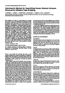
We compared the present method with the turbidimetric method by testing 281 human sera. The statistical parameters resulting from the non-parametric regression analysis according to Passing & Bablok (10) are given in figure la. The correlation between the methods was very high (r = 0.997); the approximately threefold lower enzyme activity values seen with the present method probably reflect the different method of expressing enzyme units, i. e. amount of glycerol released from 1,2-diacylglycerol per unit time in the present assay, compared with quantity of fatty acids cleaved from triolein per unit time in the turbidimetric method. We then compared the present method (measuring lipase as catalytic activity, U/l) with the enzyme immunoassay (measuring lipase as protein concentration, μg/l) on 96 human sera (fig. Ib). Again, a strong correlation was found (r = 0.987). Reference interval The histogram of lipase values obtained by the present method is shown in figure 2. Since the distribution was found to be non-Gaussian, the central 95th percentile range (0.90 confidence interval) was calculated by a non-parametric statistical technique (11); the following values were obtained: 8 (7 —10) —57 (36 — 68) U/l. Clinical sensitivity and specificity For each patient group, the numbers of values exceeding the upper reference limit and the ranges (expressed as a multiple of the upper reference limit) for all the enzymes tested are reported in table 3. The scattergrams of the individual values for lipase (present and turbidimetric methods), total α-amylase, and pancreatic isoamylase are shown in figure 3. All the 17 patients with acute pancreatitis had elevated serum lipase (with all the assay methods) and pancreatic isoamylase (100% sensitivity), whereas two of them (88%) showed normal activity for total a-amylase. The increased values ranged from 1.3 to 31.4-
442
Melzi d'Eril et al.: Continuous colorimetric lipase assay
fold the upper reference limit (median 5.6 x upper reference limit) with the present lipase assay (compared with 1.3 — 24.9 x upper reference limit, median 5.2 x upper reference limit, for the turbidimetric method); very similar values were found for pancreatic isoamylase.
1200
ΘΟΟ
400
1000 2000 3000 Lipase (turbidimetric method) [U/l]
200 400 600 Lipase (immunoactivation method) [μο/Ι]
Fig. 1. Correlation plots for lipase methods. Regression parameters for: a) continuous colorimetric method (y) vs turbidimetric method (x): slope = 0.316 (95% limits 0.3090.322); intercept = -0.79 U/l (from -1.28 to -0.01); median χ = 103 U/l; median y = 32 U/l; r = 0.997; n = 281 b) continuous colorimetric method (y) vs immunoenzymatic assay (x); slope = 0.950 (0.900-0.985); intercept = -1.50 μg/l (from -3.01 to 0.0); median x = 40 μ§/1; median y = 37 U/l; r = 0.987; n = 93) 40-,
Ο
6
12 18 24 30 36 42 48 54 60 66 72
Lipase (continuous colorimetric method) [U/l]
Fig. 2. Histogram of lipase values in healthy subjects (n = 121), as determined by the continuous colorimetric method. 2.5—97.5 percentiles (0.90 confidence interval): 8(7 _10) -57(36 -68) U/l, median 22 U/l.
As expected, the above enzymes are not totally specific for acute pancratitis. We found increased values in patients with chronic pancreatitis not only during relapse but also in clinical remission, as well as in patients with pancreatic carcinoma. However, the degree of elevation was considerably less for all the enzymes tested: highest values did not exceed 4.6-fold the upper reference limit in the patients with chronic pancreatitis during clinical remission and about 2.5fold the upper reference limit in patients with pancreatic carcinoma (except in one case for this last condition). In addition, enzyme increases were also observed in patients with non-pancreatic abdominal diseases, particularly in acute conditions (from about 13% to 19%); in chronic conditions, total α-amylase showed the highest percentage increase (46%), followed by pancreatic isoamylase (38%), and lipase (4 — 5%). Discussion
The evaluated method appears to fulfil the needs of routine clinical laboratories. The presence of appropriate concentrations of bile salts and colipase in the reagent favours full and specific action by pancreatic lipase, at the same time inhibiting serum esterases. The substrate used is a natural 1,2-diacylglycerol containing long-chain fatty acid residues, extracted from egg lecithin. The first position is taken mainly by palmitic and stearic acids; the second position (attacked by a specific 2-monoacylglycerol lipase) is occupied mainly by oleic and linoleic acids. The substrate solution is clear, stable, and lot-to-lot reproducible; therefore, the well-known difficulties in preparing uniform and long-term stable substrate emulsions have been overcome. The method is easily adapted to automated instruments. The absorbance value at the upper reference limit (under our reaction conditions 0.0165 ΔΑ/min) allows good analytical precision for usual activities. The dynamic range extends up to at least 30-fold the upper normal limit, a value much higher than those of the turbidimetric and immunoenzymatic assays (about 4-fold and 19-fold, respectively); this avoids the frequent dilution of samples. Our comparison studies show very good correlations with both the turbidimetric method and the immunoenzymatic assay. Eur. J. Clin. Chem. Clin. Biochem. / Vol. 30,1992 / No. 7
443
Melzi d'Eril et al: Continuous colorimetric lipase assay Table 3. Enzyme elevations in pancreatic and non-pancreatic abdominal diseases Diagnosis
Lipase
Total α-amylase
Continuous colorimetric
Pancreatic isoamylase
Turbidimetric
IMAC
17/17 (100.0%)
11/11 (100.0%)
5.2 (1.3-24.9)
5.0 (1.5-15.6)
4.6 (0.8-21.5)
6.4 (1.1-30.4)
Chronic pancreatitis During relapse No. > upper reference limit/ 15/28 (53.6%) total No. (%) median (range)* 1.2(0.1-20.1)
14/28 (50.0%)
15/21 (71.4%)
12/28 (42.8%)
12/28 (42.8%)
1.1 (0.2-18.1)
2.6(0.2-28.9)
0.8(0.2-14.1)
0.7(0.08-22.5)
During remission No. > upper reference limit/ 6/31(19.4%) total No. (%) median (range)* 0.5(0.1-4.6)
6/31 (19.4%)
3/19 (15.8%)
7/31 (22.5%)
5/31 (16.1%)
0.5 (0.1-3.9)
0.6 (0.2-3.7)
0.6 (0.2-3.1)
0.4 (0.09-4.2)
3/10 (30.0%)
1/4 (25.0%)
2/10 (20.0%)
2/10 (20.0%)
0.6 (0.1-24.9)
0.6 (0.3-2.8)
0.7 (0.2-10.1)
0.5 (0.07-16.9)
Acute abdomen No. > upper reference limit/ 4/31 (12.9%) total No. (%) median (range)* 0.5 (0.09 -17.2)
6/31 (19.4%)
2/14 (14.3%)
5/31 (16.1%)
4/31 (12.9%)
0.5 (0.07-15.7)
0.6 (0.2-16.9)
0.6 (0.2-10.1)
0.6 (0.08-17.3)
Chronic abdominal diseases No. > upper reference limit/ 1/24 (4.2%) total No. (%) median (range)* 0.5 (0.1-1.2)
1/24(4.2%)
1/19(5.3%)
11/24(45.8%)
9/24(37.5%)
0.5 (0.1-1.5)
0.6 (0.2-1.5)
0.9 (0.3-3.4)
0.8 (0.2-4.3)
Pancreatic Diseases Acute pancreatitis No. > upper reference limit/ 17/17 (100.0%) total No. (%) 5.6(1.3-31.4) median (range)*
Pancreatic carcinoma No. > upper reference limit/ 3/10 (30.0%) total No. (%) median (range)* 0.6(0.1-26.8)
15/17 (88.2%)
17/17 (100.0%)
Non-pancreatic abdominal diseases
* multiples of upper reference limit Upper reference limit = Continuous colorimetric lipase 57 U/l; Turbidimetric lipase 190 U/l; IMAC lipase 54 μg/l; Total oc-amylase 160 U/l; Pancreatic isoamylase 87 U/l
Acute pancreatitis (n=17) .«; 30-
A
Remission (n=31)
Non-pancreatic abdominal diseases Acute abdomen (n=31) Chronic (n=24)
α
•30^
0
1 20-A Δ 2
ϊ u
Relapse (n=28)
Pancreatic carcinoma (n=10)
ε
Δ
|25-
&15α. «Ξ 10 iS- 5 "
Chronic pancreatitis
Δ
ο
0
Α
^
* Φ
Δ
ο
-25g
Ο
0
α
°
α
α 0
a Δ
θ ° Q
I I I I
a
Δ
ο
Α
i *
Ο
$
α
η
ο
D A
ο
0
o B 8 Β
A
$
Q
- 10 «*-
α
g
§
D
^^ — "O Q.
ο
4-l-n -4-l-i-l- 4-I--.-I- -4- 4- -ί- *Α
-20 1 2 -15 fe α.
-*--t-8-i
_ f\
Fig. 3. Relative elevations of enzyme activities (Δ lipase (continuous colorimetric method), Ο lipase (turbidimetric method), ο total α-amylase, and D pancreatic isoamylase) in various disease groups.
In our patient population, we found 100% sensitivity therefore, all patients diagnosed as having acute panin diagnosing acute pancreatitis with the present lipase creatitis showed an increased serum lipase (from 1.3 assay, as well as with the other two lipase methods; to 31.4-fold the upper reference limit with the present Eur. J. Clin. Chem. Clin. Biochem. / Vol. 30,1992 / No. 7
444
Melzi d'Eril et al.: Continuous colorimetric lipase assay
assay). We found the same sensitivity for pancreatic isoamylase and a lower value for total a-amylase (88.2%).
the turbidimetric procedure. Measurements of pancreatic isoamylase and total a-amylase gave lower specificities (76.4% and 70.9%, respectively).
Specificity for detecting acute pancreatitis in control patients with both acute and chronic non-pancreatic abdominal diseases was 90.9% for the present lipase method and the enzyme immunoassay, and 87.3% for
In conclusion, the method proved to be specific for pancreatitis, as well as being reliable, simple and rapid. Since the method is also easily automated, it is convenient both for routine and emergency use.
References 1. Tietz, N. W. (1988) Amylase measurement in serum. Old myths die hard. J. Clin. Chem. Clin. Biochem. 26, 251 253. 2. Rick, W. & Hockeborn, M. (1982) Zur Bestimmung der Aktivität der Lipase mit dem sogenannten turbidimetrischen Test. J. Clin. Chem. Clin. Biochem. 20, 735-744. 3. Tietz, N. W., Shuey, D. F. & Astles, J. R. (1987) Turbidimetric measurements of lipase activity — Problems and some solutions. Clin. Chem. 33, 1624-1629. 4. Lott, J. A., Patel, S. T., Sawheney, A. K., Kazmierczak, S. C. & Love, Jr J. E. (1986) Assay of serum lipase: analytical and clinical considerations. Clin. Chem. 32, 1290-1302. 5. Imamura, S., Hirayama, T, Arai, T., Takao, K. & Misaki, H. (1989) An enzymatic method using 1,2-diglyceride for pancreatic lipase test in serum. (Abstract) Clin. Chem. 35, 1126. 6. Fossati, P., Ponti, M., Paris, P., Berti, G. & Tarenghi, G. (1992) Kinetic colorimetric assay of lipase in serum. Clin. Chem. 38, 211-215.
7. Ziegenhorn, J., Neumann, U, Knitsch, R. W. & Zwez, W. (1979) Determination of serum lipase. (Abstract) Clin. Chem. 25, 1067. 8. Drosdat, H., Dreher, M., Linxweiler, W. & Gunzer, G. (1989) Homogeneous immunoassay for determination of pancreatic lipase in routine use. (Abstract) Biochim. Clin. 73, 166. 9. Cattozzo, G., Franzini, C., Pagani, A. (1991) Evaluation of a new continuous-monitoring colorimetric procedure for the measurement of lipase activity in serum. Biochim. Clin. 75,1413-1417. 10. Passing, H. & Bablok, W. (1983) A new biometrical procedure for testing the equality of measurement from two different analytical methods. Applications of linear regression procedures for method comparison studies in clinical chemistry. Parti. J. Clin. Chem. Clin. Biochem. 27, 709730. 11. Solberg, H. E. (1983) The theory of reference values. Part 5. Statistical treatment of collected reference values. Determination of reference limits. J. Clin. Chem. Clin. Biochem. 27, 749-760. Prof. GianVico Melzi d'Eril Fondazione "C. Mondino" Clinica Neurologica Universita degli Studi Via Palestro 3 1-27100 Pavia
Eur. J. Clin. Chem. Clin. Biochem. / Vol. 30,1992 / No. 7