Computational Methods for Pigmented Skin Lesion Classification in Images: Review and Future Trends Roberta B. Oliveiraa, João P. Papab, Aledir S. Pereirac and João Manuel R. S. Tavaresa,* a
Instituto de Ciência e Inovação em Engenharia Mecânica e Engenharia Industrial, Departamento de Engenharia Mecânica, Faculdade de Engenharia, Universidade do Porto, rua Dr. Roberto Frias, 4200-465, Porto, Portugal
b
Departamento de Computação, Faculdade de Ciências, Universidade Estadual Paulista, av. Eng. Luiz Edmundo Carrijo Coube, 14-01, 17033-360, Bauru, SP, Brazil c
Departamento de Ciências de Computação e Estatística, Instituto de Biociências, Letras e Ciências Exatas,
Universidade Estadual Paulista, rua Cristóvão Colombo, 2265, 15054-000, São José do Rio Preto, SP, Brazil
Abstract Skin cancer is considered as one of the most common types of cancer in several countries and its incidence rate has increased in recent years. Melanoma cases have caused an increasing number of deaths worldwide, since this type of skin cancer is the most aggressive compared to other types. Computational methods have been developed to assist dermatologists in early diagnosis of skin cancer. An overview of the main and current computational methods that have been proposed for pattern analysis and pigmented skin lesion classification is addressed in this review. In addition, a discussion about the application of such methods, as well as future trends are also provided. Several methods for feature extraction from both macroscopic and dermoscopic images and models for feature selection are introduced and discussed. Furthermore, classification algorithms and evaluation procedures are described, and performance results for lesion classification and pattern analysis are given. Keywords: Pattern analysis; feature extraction and selection; classification methods; macroscopic and dermoscopic images. 1. Introduction Computational methods for skin cancer diagnosis have been proposed in order to aid dermatologists in early assessment of skin cancer and in the follow-up of pigmented skin lesions [1-3]. Such lesions represent *
Corresponding author. Tel.: +351 220413472; fax: +351 225081445 (João Manuel R. S. Tavares).
Email addresses:
[email protected] (Roberta B. Oliveira),
[email protected] (João P. Papa),
[email protected] (Aledir S. Pereira),
[email protected] (João Manuel R. S. Tavares).
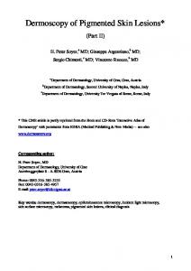
an abnormal production of melanocytes cells, which are mainly caused by excessive sun exposure. Melanocytes cells are responsible for creating the substance melanin, one of the functions of which is to provide pigmentation in the skin. Furthermore, the number of skin cancer cases has increased in the last years, and consequently, an increasing number of deaths caused by this disease has been reported, particularly due to melanoma cases (Figures 1c and d) [4-6]. Therefore, pigmented skin lesions have been a cause for global concern, since some types of benign lesions may become skin cancer, such as dysplastic nevi (Figures 1a and b).
Fig. 1 Two examples of macroscopic images (a and c) and dermoscopic images (b and d): (a) and (b) are images of a dysplastic nevus and, (c) and (d) are of an invasive melanoma (images publicly available from Bourne et al. [7]).
Image acquisition, pre-processing, segmentation, feature extraction, and classification are fundamental steps commonly found in computational systems for diagnosing skin lesions. Different non-invasive imaging techniques have been used to assist dermatologists [8]. Macroscopic images [9,10] and dermoscopic images [11,12] are examples of images acquired from such techniques that have been widely used in the diagnosis of pigmented skin lesions by computational methods. Macroscopic images (Figures 1a and c), commonly known as clinical images, are usually acquired from standard cameras or mobile devices. On the other hand, dermoscopic images (Figs. 1b and d), may be acquired from dermatoscope devices or specific cameras in order to better visualize the pigmentation pattern on the skin surface. However, their imaging conditions are frequently inconsistent; for example, macroscopic images can be acquired from variable distances and/or under different illumination conditions. Furthermore, the images may have poor resolution, which may be challenging when the lesion under study is small. An additional
2
problem with both macroscopy and dermoscopic images is related to the presence of artefacts, such as hair, reflections, shadows, skin lines and bubbles, which may hinder adequate analysis of the imaged skin lesions. The identification of the regions of the lesions in such images may be performed in order to assist in the process of classification [13]. Segmentation is an important step that allows the extraction of such regions of interest (ROI) from an image [14-34]. However, before the segmentation step, previous pre-processing methods are usually applied to reduce the effects of undesirable artefacts that may influence the outcome of the segmentation step. These methods can be based on colour space transformation [20,26,35], illumination correction [36,37], contrast enhancement [20,22,23,26,38-42], artefact removal [14,20,21,43,44] and approximate lesion localization [45]. In addition, hair removal methods are also used in pre-processing steps, since this artefact may considerably affect the detection of lesion borders [46-53]. Lee et al. [54] proposed a solution for hair removal, especially thick dark hairs, which is based on one of the first widely adopted methods for hair removal in dermoscopy images, and consists of identifying the hair location, replacing the values of the detected hair pixels in the original image by the values of the corresponding nearby non-hair pixels, and smoothing the thin lines. An overview of lesion border detection methods, including the pre-processing, segmentation and post-processing steps, is presented in Celebi et al. [55,56]. In addition, the authors also discuss performance evaluation issues and propose guidelines for future studies. Computational methods for pigmented skin lesion classification are usually based on the features of the pixels within the segmented ROIs. Therefore, the extraction of representative features of the ROIs under analysis is an important step for the efficient classification of the segmented lesions. In this step, common difficulties are: 1) identification of the features to be used; 2) to confirm that the number of selected features is sufficient to describe the classification problem; 3) the number of selected features is too large, which requires high computational resources; and 4) there are redundant and/or irrelevant features that should be removed from the feature set. Techniques to reduce the dimensionality of the data may be used to solve these problems according to one of the following reduction strategies: feature transformation (also known as feature extraction in literature concerning pattern recognition [57,58]), and feature selection [59]. The feature extraction strategy allows the modification of all the data of the image, in order to emphasize the most effective features, ensuring the correct separation of the classification classes [57]. Such strategy is based on the generation of a new feature space, which may expand or reduce, according to the adopted strategy. The new features may be extracted by means of discovery of missing information from relationships among the features, or even by means of searching for a new feature space with smaller dimensions through functional mapping. Contrary, new features are not created in the feature selection strategy, meaning that a subset from the original features is defined when using this approach. Both strategies may also be combined in order to achieve a better representation of the features. For example, in cases in which the feature extraction step increases the number of features, feature selection algorithms can 3
provide an automatic reduction of such excessive features. Furthermore, a larger feature space may include redundant or irrelevant data [60]. Several solutions [61-64] have been proposed for feature extraction and selection of pigmented skin lesions, in order to represent them according to a certain clinical criteria [65-67]. Such features may be used for the classification process, in order to provide dermatologists with a computer-aided diagnosis of pigmented skin lesions [2,12]. In this review, some of the most relevant solutions that have been developed to assist the skin lesion diagnosis from macroscopic and dermoscopic images are introduced, including those concerning the steps of feature extraction and selection, and image classification. Hence, this review is highly valuable for those wishing the design and/or implementation of competent expert systems for the automated classification of skin lesions in images. This paper is organized as follows: a review of the main computational methods that have been applied to extract and select features from macroscopic and dermoscopic images of pigmented skin lesions is presented in Section 2. The main focus of that section is on the feature extraction step according to several clinical criteria. In addition, the feature selection process is addressed. The current state-of-the-art concerning the pigmented skin lesion classification, including the advantages and disadvantages of the reviewed methods, evaluation measures, and performance results for pattern and lesion classification, is presented in Section 3. Finally, conclusions and future trends about the computational methods of pigmented skin lesion classification are pointed out in the last section. 2. Image analysis of pigmented skin lesions Computational methods regarding the feature extraction have been commonly developed based on the ABCD(E) rule, pattern analysis, seven-point checklist and Menzies’ method, which are examples of clinical approaches used for the diagnosis of skin cancer from images [67-69]. The first approach can be used to extract features from both macroscopic and dermoscopic images, whereas the other approaches are usually applied to dermoscopic images in order to identify more detailed pattern features on the surfaces of the lesions. The feature analysis based on these approaches, as well as the feature selection and extraction steps are presented with details in the following sections. 2.1. Feature analysis based on clinical approaches The ABCD(E) rule is based on asymmetry, border, colour, diameter (or differential structures in the case of dermoscopic images), and evolution (or elevation) features, according to the criteria presented in Table 1. Such rule has been widely used for the feature extraction and automatic diagnosis of pigmented skin lesions [10,70].
4
Table 1 Criteria of the ABCD(E) rule for the diagnosis of skin cancer from clinical and dermoscopy analysis. Dermoscopy analysis a
Clinical analysis Feature Asymmetry (A) Border (B) Colour (C) Diameter (D)
Benign lesion
Malignant lesion
Shape is symmetric
Shape is asymmetric
Border is regular or well-defined Colours are uniform
Border is irregular or ill-defined Colours are nonuniform
Size