J Neurophysiol 104: 3105–3112, 2010. First published July 28, 2010; doi:10.1152/jn.00697.2009.
Consistency of Angular Tuning in the Rat Vibrissa System Marie E. Hemelt,1 Ernest E. Kwegyir-Afful,2 Randy M. Bruno,2,3 Daniel J. Simons,2 and Asaf Keller1 1
Program in Neuroscience, Department of Anatomy and Neurobiology, University of Maryland School of Medicine, Baltimore, Maryland; Department of Neurobiology, University of Pittsburgh School of Medicine, Pittsburgh, Pennsylvania; and 3Department of Neuroscience, Columbia University, New York, New York 2
Submitted 3 August 2009; accepted in final form 24 July 2010
Hemelt ME, Kewgyir-Afful EE, Bruno RM, Simons DJ, Keller A. Consistency of angular tuning in the rat vibrissa system. J Neurophysiol 104: 3105–3112, 2010. First published July 28, 2010; doi:10.1152/jn.00697.2009. Each region along the rat mystacial vibrissa pathway contains neurons that respond preferentially to vibrissa deflections in a particular direction, a property called angular tuning. Angular tuning is normally defined using responses to deflections of the principal vibrissa, which evokes the largest response magnitude. However, neurons in most brain regions respond to multiple vibrissae and do not necessarily respond to different vibrissae with the same angular tuning. We tested the consistency of angular tuning across the receptive field in several stations along the vibrissato-cortex pathway: primary somatosensory (barrel) cortex, ventroposterior medial nucleus of the thalamus (VPM), second somatosensory cortex, and superior colliculus. We found that when averaged across the population, neurons in all of these regions have low (superior colliculus and second somatosensory cortex) or statistically insignificant (barrel cortex and VPM) angular tuning consistencies across vibrissae. Nevertheless, in each region there are a small number of neurons that display consistent angular tuning for at least some vibrissae. We discuss the relevance of these findings for the transformation of inputs along the vibrissa trigeminal pathway and for the detection of sensory cues by whisking animals. INTRODUCTION
Angular tuning, or directional selectivity, is the phenomenon whereby a neuron responds preferentially to a stimulus moving in a particular direction. One system in which neurons display angular tuning is the vibrissa system in the rodent. Neurons in all stations along this pathway have varying levels of angular tuning [trigeminal ganglion: Lichtenstein et al. 1990; principal trigeminal nucleus: Minnery et al. 2003; trigeminal nucleus interpolaris: Furuta et al. 2006; ventroposterior medial nucleus of the thalamus (VPM): Minnery et al. 2003; Simons and Carvell 1989; Timofeeva et al. 2003; barrel cortex: Bruno et al. 2003; Simons 1978; Simons and Carvell 1989; second somatosensory cortex: Kwegyir-Afful and Keller 2004; superior colliculus: Hemelt and Keller 2007]. Typically, analysis of angular tuning has been performed only on responses to deflections of the principal vibrissae, which evoke the largest magnitude responses in their associated neurons. However, many vibrissae in a receptive field may evoke spikes, and in some brain regions, these adjacent (nonprincipal) vibrissae can evoke response magnitudes similar to those evoked by the principal vibrissa (principal trigeminal nucleus: Veinante and Deschênes 1999; thalamus: Diamond et al. 1992; primary somatosensory cortex: Bruno and Simons 2002; second somatosensory cortex: Address for reprint requests and other correspondence: A. Keller, Dept. of Anatomy and Neurobiology, University of Maryland School of Medicine, 20 Penn St., Baltimore, MD 21201 (E-mail:
[email protected]). www.jn.org
Kwegyir-Afful and Keller 2004; superior colliculus: Hemelt and Keller 2007). This raises the question whether the angular tuning of the principal vibrissa is preserved across the receptive field, that is, if nonprincipal vibrissae within a receptive field respond with similar angular tuning to those produced by the principal vibrissa. A small number of earlier studies have addressed the issue of angular tuning consistency. Angular tuning is largely conserved across the receptive fields of neurons in the trigeminal interpolaris nucleus (Furuta et al. 2006). In the somatosensory (barrel) cortex, angular consistency is preserved when broad tuning angles (i.e., rostral vs. caudal) are considered (Kida et al. 2005; Shimegi et al. 2000). However, the use of only two test angles may overestimate the angular consistency, as it essentially groups all the directions into two groups. Here we improve on this approach by testing eight angles of deflection. In addition, to determine whether angular consistency characterizes all stations along the vibrissa trigeminal pathway, we tested for angular consistency in four brain regions involved in vibrissa sensory processing: primary somatosensory cortex, second somatosensory cortex, VPM, and the superior colliculus. Two of the regions, the second somatosensory cortex and the superior colliculus, are of particular interest due to the large receptive fields of their neurons and large magnitude responses of nonprincipal vibrissae (second somatosensory cortex: Kwegyir-Afful and Keller 2004; superior colliculus: Hemelt and Keller 2007). METHODS
Surgical procedures These data were collected from experiments using 56 female Sprague-Dawley rats weighing 200 –350 g. Other data from these experiments have been published previously (superior colliculus: Hemelt and Keller 2007; primary and second somatosensory cortex: Kwegyir-Afful and Keller 2004; VPM: Bruno and Simons 2002). All procedures strictly adhered to institutional and federal guidelines. As previously described, recordings were obtained under fentanyl (10 g · kg⫺1 · h⫺1) (Bruno and Simons 2002; Kwegyir-Afful and Keller 2004) or under isoflurane (superior colliculus) (Hemelt and Keller 2007). In preparation for recordings under fentanyl, rats were anesthetized with halothane (1.5–2.0%), and bone overlaying the area of interest was removed. After the animals received fentanyl, they were immobilized with pancuronium bromide (1.6 mg · kg⫺1 · h⫺1), and the animal was artificially respired (90 breath\min) using a positivepressure ventilator during recording. For recording under isoflurane, the anesthetic was delivered through a tracheal tube at 1.0 –2.0% during both surgery and recording. During all experiments, body temperature was maintained at 37°C with a servo-controlled heating blanket during recordings.
0022-3077/10 Copyright © 2010 The American Physiological Society
3105
3106
M. E. HEMELT, E. E. KWEGYIR-AFFUL, R. M. BRUNO, D. J. SIMONS, AND A. KELLER
Recording Recording procedures and data used here have been previously described in detail (superior colliculus: Hemelt and Keller 2007; primary and second somatosensory cortex: Kwegyir-Afful and Keller 2004; VPM: Bruno and Simons 2002). Briefly, we obtained extracellular unit recordings with either quartz-insulated platinum electrodes (2–12 M⍀) or glass-insulated tungsten electrodes (2– 4 M⍀). Electrodes were advanced perpendicular to the brain surface. For recordings in barrel cortex, second somatosensory cortex, and superior colliculus, waveforms recorded from well-isolated units were digitized at 40 kHz through either an AlphaLab (Alpha Omega, Nazareth, Israel) or a Plexon data acquisition system (Dallas, TX). These units were isolated off-line with a combination of threshold and waveform component analysis using off-line sorter (Plexon). VPM recordings were digitized at 32 kHz using a 1 GHz personal computer equipped with a PCI-MIO-16E-1 board (National Instruments, Austin TX) and data acquisition software custom written in LabView version 5.1.1 (National Instruments). These units were isolated off-line using MClust version 2.0 (A. David Redish, University of Minnesota, Minneapolis, MN) and custom-written programs. In total, we report data from 160 well-isolated neurons. Following electrolytic lesions to mark the recording site, the animals were deeply anesthetized with sodium pentobarbital (60 mg\kg) and perfused transcardially with buffered saline followed by 4% buffered paraformaldehyde. Recording sites were identified in histological sections.
Vibrissa stimulation Vibrissae were inserted into a small tube—approximately 10 mm from their base—attached to a computer-controlled piezoelectric stimulator that can be deflected in eight different directions (see Simons and Carvell 1989). We applied ramp-and-hold stimuli, 200 ms in duration, having onset\offset velocity between 100 and 140 mm\s. To reduce mechanical ringing, the trapezoid ramp-and-hold waveforms were filtered with a Bessel filter. The peak onset and offset velocity were measured as the slope of the linear portion of the deflection ramp. We calibrated the stimulator with a photodiode device. Vibrissae were deflected in eight different directions (in 45° increments) in a pseudo-random sequence at 0.5 or 1 Hz, for a total of 15 deflections at each angle. The receptive field was determined by deflecting vibrissae with a hand-held probe, and responses were recorded for as many vibrissae as possible within the receptive field. All of these nonprincipal vibrissae are referred to as “adjacent vibrissae.” In VPM, only the four immediately adjacent vibrissae were tested (i.e., those rostral, caudal, dorsal, and ventral to the principal vibrissa). Neurons that responded to only one vibrissa are not reported in this analysis.
Data analysis
response onset and offset. In determining the response magnitude, stimuli from all eight directions were pooled. In all brain regions, the vibrissa that evoked the largest response magnitude was defined as the principal vibrissa. We constructed tuning curves by calculating the spike\stimulus of a neuron in response to deflections of a vibrissa at each of the eight angles (see Fig. 1A). The polar plot displays response magnitude at each angle of deflection. We repeated this analysis for each of the vibrissae within the receptive field. To determine if the angular tuning was conserved across the receptive field, we compared the tuning of responses evoked by each adjacent vibrissa to tuning evoked by the principal vibrissa. For this comparison, we calculated the similarity index, which has been described previously (see Bruno et al. 2003). This index, the correlation between the two tuning curves, compares the tuning of the neuronal response to the deflection of two different vibrissae using the Pearson product-moment correlation. A similarity index of 1 indicates that the angular tuning curves are perfectly correlated (identically tuned), while a similarity index of 0 indicates that the angular tuning curves are uncorrelated. A similarity index of ⫺1 indicates the angular tuning curves are inversely correlated (oppositely tuned). A similarity index was generated for every adjacent vibrissa, in comparison with its principal vibrissa. Statistical analyses were performed in STATA and Microsoft Excel. Because the distributions of similarity indices were nonnormal, we present medians and nonparametric comparisons of the data. We use the Wilcoxon matched-pairs signed-ranks test to compare the similarity indices in each region with the randomized indices, because these two groups are drawn from the same population. Other comparisons between brain regions use Mann-Whitney U. RESULTS
Recording sites and data selection Responses to vibrissa deflections were recorded from 160 well-isolated neurons in 53 animals. Individual vibrissae were deflected at eight different angles, as previously described (see METHODS). Recordings are reported from four brain regions: primary somatosensory cortex (layer IV, 34 units), second somatosensory cortex (layers II\III to VI, 38 units), superior colliculus (intermediate and deep layers, 18 neurons), and the ventroposterior medial nucleus of the thalamus (VPM, 69 units). Recording sites were confirmed histologically. We present data only from units with regular spiking waveforms, the vast majority of which represent excitatory neurons (Connors and Gutnick 1990). We also excluded any neurons that only responded to a single vibrissa; all 160 neurons presented here respond to at least two vibrissae. Similarity of angular tuning
Time stamps of well-isolated units and of stimulus triggers from recordings were exported to Matlab (MathWorks, Natick, MA) for analyses using custom written software. Peristimulus time histograms (PSTHs, 1 ms bins) were constructed, and significant stimulus-evoked responses were defined as PSTH bins with response magnitudes significantly exceeding (99% confidence interval) spontaneous activity levels, computed from a 200 ms period preceding the stimuli. Response onset was defined as the first two consecutive bins (poststimulus) that displayed significant responses (above the 99% confidence interval), and response offset was defined as three consecutive bins in which the response duration fell below the 99% confidence interval. The magnitude of the response was defined as the total number of spikes (exceeding the confidence interval) per stimulus occurring between the onset and offset of the significant response. The duration of the significant response was defined as the time between J Neurophysiol • VOL
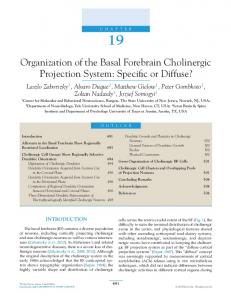
We sought to determine whether the angular tuning of a neuron’s response to its principal vibrissa was preserved across the receptive field. In each neuron, we recorded responses to computer-controlled deflections of several vibrissae with each vibrissa deflected in eight different directions in a pseudorandom sequence (see METHODS for details). The average number of vibrissae per neuron for which data were collected was 4.2 in primary somatosensory cortex, 3.2 in second somatosensory cortex, 6.4 in superior colliculus, and 2.8 in VPM. All nonprincipal vibrissae in the receptive field are referred to as adjacent vibrissae. Figure 1A illustrates our procedure for computing the tuning curve using a representative neuron from the superior collicu-
104 • DECEMBER 2010 •
www.jn.org
CONSISTENCY OF ANGULAR TUNING
A
B
3107
Primary Somatosensory Cortex
Second Somatosensory Cortex
90
90
90
Probability
0
180
0
180
1 0
0 0
180
.02 .04
Seconds
270
270
VPM
Superior Colliculus
90
90
270
0
180
270
0
180
270
FIG.
1. A: an angular tuning plot of a representative superior colliculus neuron. Rasters and peristimulus time histographs (PSTHs) depict responses to vibrissa displacements in eight different directions (0° is rostral, 90° is dorsal). The polar plot in the center shows the response magnitude in each direction, with the hatch marks on the axes representing 2 spike\stimulus. B: examples of angular tuning plots from each of the brain regions studied. Blue angular tuning plots were produced from responses to the principal vibrissae. Red angular tuning plots were produced from responses to an adjacent vibrissa with high similarity indices (0.82 in primary somatosensory cortex, 0.80.in VPM, 0.72 in second somatosensory cortex, and 0.95 in superior colliculus). Green angular tuning plots were produced from responses of the same neurons to another adjacent vibrissae. These responses have low similarity indices (0.10 in primary somatosensory cortex, 0.10 in VPM, 0.23 in second somatosensory cortex, and 0.12 in superior colliculus). Radial axes indicate response magnitude, and hatch marks indicate 2 spike\stimulus.
lus. PSTHs depict the neuron’s response to deflection of the principal vibrissa in eight directions. The polar plot displays the magnitude of responses to each of these directions (see METHODS for details). Figure 1B shows tuning curves computed from individual neurons recorded in four brain regions. For each neuron, we present tuning curves computed from responses to deflection of three different vibrissae: the principal vibrissa (blue) and two adjacent vibrissae (red and green). To quantify the similarity between two angular tuning curves obtained from responses of a neuron to deflection of different vibrissae, we computed the similarity index using Pearson’s correlation (see METHODS for details). For each neuron, we computed similarity indices comparing responses to each adjacent vibrissa with the responses evoked by the principal vibrissa. A similarity index of 1 indicates that the angular tuning curves are perfectly correlated (identically tuned), while a similarity index of 0 indicates that the angular tuning curves are uncorrelated. A similarity index of ⫺1 indicates the angular tuning curves are inversely correlated (oppositely tuned). Each neuron in Fig. 1B depicts one tuning curve with a high similarity index (red) and one with a low similarity index (green). Variability in the responses of individual neurons are expected to affect their angular tuning and hence the computation of similarity indices. To determine the potential impact of this variability we presented, to the same neuron, two identical sequences of multi-directional stimuli and correlated, using the J Neurophysiol • VOL
similarity index, the tuning curves computed for these two stimulus sets. Because responses to adjacent vibrissae are typically weaker than those to the principal vibrissa, we present medians (and ranges) for adjacent and principal vibrissae separately. In the superior colliculus (n ⫽ 7 neurons), the median similarity index for adjacent vibrissae was 0.90 (0.31– 0.98), and for principal vibrissae, it was 0.86 (0.80 – 0.90). In VPM (n ⫽ 8) the medians were 0.65 (⫺0.46 – 0.85) and 0.79 (0.60 – 0.80) for adjacent and principal vibrissae, and in primary somatosensory cortex (n ⫽ 8), they were 0.66 (⫺0.22– 0.92) and 0.63 (0.13– 0.78) for adjacent and principal vibrissae. These relatively high values represent the upper bounds of similarity indices expected from recordings in these brain regions, and they provide a measure of the reliability of these computations. Individual similarity indices for every adjacent vibrissa in every neuron are shown in Fig. 2, where each data point represents the comparison between the adjacent and the principal vibrissa. Similarity indices computed for the same neuron are arranged in a column. These analyses demonstrate that there exists a wide range of similarity indices, not only among neurons, but also within neurons in each brain region. Most neurons that respond to several adjacent vibrissae show a wide range of similarity indices across the receptive field. Thus neurons in which angular preference is conserved for a given pair of vibrissae are likely to display no angular consistency for a different pair of vibrissae (Fig. 2).
104 • DECEMBER 2010 •
www.jn.org
3108
M. E. HEMELT, E. E. KWEGYIR-AFFUL, R. M. BRUNO, D. J. SIMONS, AND A. KELLER
Primary Somatosensory Cortex
Similarity Index
0.8 1.0 0.5
AV/PV:
0.4 0 -0.4 -0.8 5
10
15
20
25
30
35
VPM
Similarity Index
0.8 0.4
FIG. 2. Similarity indices for each adjacent vibrissa evoking responses in each neuron in the four brain regions. Similarity indices () computed for the same neuron are arranged in a column. Circle size represents the relative response magnitude of the adjacent vibrissa, expressed as a ratio of the response magnitude of the principal vibrissa (AV\PV). Neurons are arranged by average similarity index, from highest to lowest.
0 -0.4 -0.8 5
10
15
20
25
30
35
Second Somatosensory Cortex
40
45
50
55
60
65
70
Superior Colliculus
Similarity Index
0.8 0.4 0 -0.4 -0.8 5
10
15
20
25
30
35
5
10
15
Neuron: Highest to Lowest Average Similarity
Group data are shown in Fig. 3A as histograms of similarity indices for all nonprincipal vibrissae. Median similarity indices were low in all four regions: primary somatosensory cortex ⫽ 0.11, VPM ⫽ ⫺0.12, second somatosensory cortex ⫽ 0.31, and superior colliculus ⫽ 0.26. These values are well below the upper bounds for similarity indices computed in the preceding text to assess the reliability of these measures, indicating that these low similarity indices are not constrained by the reliability of estimating angular preferences. The distribution of similarity indices in VPM displayed a positive skew (0.25) while the skewedness in the other regions was negative (⫺0.21 in primary somatosensory cortex, ⫺0.34 in second somatosensory cortex, ⫺0.32 in superior colliculus). These negative skews may indicate a subpopulation of neurons in these brain regions where some adjacent vibrissae have similar angular tuning as the principal vibrissa. We considered the possibility that the calculation of angular consistency (i.e., similarity index) might be affected by the precision of a neuron’s tuning curve. As depicted in Fig. 3B, in none of the four regions examined did we find a strong correlation J Neurophysiol • VOL
between the strength of tuning (i.e., the angular tuning) of each principal vibrissa and the similarity index computed with each of their adjacent vibrissae. This suggest that the similarity index is not directly dependent on the precision of the tuning curves, strengthening our interpretation of the findings. We also considered the possibility that our analysis might be affected by an asymmetrical relationship between the tuning strength of a neuron’s principal versus adjacent vibrissae. As seen in Fig. 3C, there is no consistent correlations, in any of the four brain regions, between the angular selectivity index of principal versus adjacent vibrissae. As detailed in METHODS, recordings were obtained under fentanyl analgesia except for the superior colliculus recordings, which we obtained under isoflurane anesthesia. We cannot exclude the possibility that the choice of anesthetic affected the comparisons of results from the different brain regions. However, the finding that similarity indices of neurons from the colliculus and from the second somatosensory cortex were nearly identical and had similar distributions argues against this possibility.
104 • DECEMBER 2010 •
www.jn.org
CONSISTENCY OF ANGULAR TUNING
3109
Comparison with randomized index
A
Primary Somatosensory Cortex (0.11)
15
10
5
5 0
-0.5
(0.36)
15
10
-1
Percent of pairs
Second Somatosensory Cortex
0.5
1
10
5
5 -0.5
0.5
1
0
(0.26)
15
10
-1
0
Superior Colliculus
(-0.12)
15
-0.5
-1
VPM
0.5
1
-0.5
-1
0
0.5
1
Similarity index
B
Second Somatosensory Cortex
Primary Somatosensory Cortex 1.0
Responses to rostrocaudal deflections
0.6 2
0.2
R = 0.03
-0.2
R = 0.14
-0.6 -1.0
0
1
2
3
4
6
5
0
7
Similarity Index
1.0
2
3
4
5
6
7
R2 = 0.0004
0. 6 0. 2 -0.2
-0.6
R 2 = 0.14
-1.0
0
1
2
3
4
5
6
7
Angular tuning (PV)
0
1
2
3
4
5
6
7
Primary Somatosensory Second Somatosensory Cortex Cortex 4 80
VPM
1 6
Superior Colliculus 4 7
AV tuning index
1
Superior Colliculus
VPM
C
To determine if the similarity indices differ significantly from a random distribution— that is, if there is any similarity of tuning above chance levels—we compared the population of similarity indices with the population of randomized similarity indices. We computed randomized similarity indices for each neuron by comparing tuning curves of responses to the adjacent vibrissae with tuning curves generated by a different neuron, in the same brain region, in response to deflection of its principal vibrissa. This provides a randomized population of similarity indices for each brain region, where the similarity indices vary only by chance. Any deviation from zero in the randomized similarity index would reflect the overall preference of the entire brain region for a particular direction. Mean randomized similarity indices were 0.01 in barrel cortex, ⫺0.02 in VPM, 0.1 in second somatosensory cortex, and 0.03 in superior colliculus. We used Wilcoxon matched-pairs signedranks tests to compare the randomized similarity index with the nonrandomized similarity index for each brain region (Fig. 4). P values were 0.1 (primary somatosensory cortex), 0.92 (VPM), 0.02 (second somatosensory cortex), and 0.001 (superior colliculus; Fig. 4).
6 26
6 4 2 0 0 2 4 6 PV tuning index J Neurophysiol • VOL
Our results are in contrast to those reported by Kida et al. (2005) and Shimegi et al. (2000), who found that the principal vibrissae of primary somatosensory cortex neurons evoke responses with angular tuning similar to those evoked by adjacent vibrissae. However, Kida et al. report data from neurons in all layers of the primary somatosensory cortex, whereas we restrict our analysis in this area to neurons in layer IV. In addition, they tested eight angles of deflection in only 12 neurons, using only rostral and caudal deflections in all other neurons. The use of only two test angles may overestimate the similarity of tuning between vibrissae, as it essentially groups all the directions into two groups. To test this possibility, we used the same analytical approach described by Kida et al. and Shimegi et al. on our data from primary somatosensory cortex and second somatosensory cortex, using responses only to rostral and caudal deflections. As in their studies, we computed a bidirectional similarity index. For all vibrissae in the receptive fields (adjacent and principal vibrissae), we computed the difference in response magnitude evoked by rostral and caudal deflections divided by the sum of these magnitudes. This value for the principal vibrissa was compared with the mean of the
FIG. 3. A: histograms show the similarity indices for each adjacent vibrissa\ principal vibrissa pair recorded in the 4 brain regions. Median similarity index is shown in parenthesis with the location of the median marked with an arrow. n ⫽ 34 neurons and 110 adjacent vibrissae (primary somatosensory cortex); 69 neurons and 124 adjacent vibrissae (VPM); 38 neurons and 82 adjacent vibrissae (second somatosensory cortex); and 18 neurons and 97 adjacent vibrissae (superior colliculus). B: plots comparing the angular tuning of individual neurons (responding to deflections of the principal vibrissa, PV) against the similarity index computed relative to each of their adjacent vibrissae. C: relationship between the angular tuning index of each adjacent vibrissa\principal vibrissa pair recorded in the 4 brain regions. The number of overlapping data points is coded as a gray scale; the corresponding legend provided as an inset for each of the 4 plots depicts the lowest and highest values for each panel.
104 • DECEMBER 2010 •
www.jn.org
3110
M. E. HEMELT, E. E. KWEGYIR-AFFUL, R. M. BRUNO, D. J. SIMONS, AND A. KELLER p = 0.1
p = 0.92
p = 0.02
p = 0.001
Similarity Index
1 Similarity Indices Randomized Similarity Indices
0.5
FIG. 4. Box and whisker plots show the distributions for angular similarity indices and randomized similarity indices. Randomized similarity indices were computed by correlating responses to adjacent vibrissae with responses from a different neuron in the same brain region to its principal vibrissa. The faint gray line is at zero. Boxes represent the 25th and 75th percentile of distribution, horizontal lines are medians and whiskers show the 5th and 95th percentiles. P values are computed with the Wilcoxon matched-pairs signed-ranks test.
0
-0.5
-1 Primary Somatosensory Cortex
VPM
Second Somatosensory Cortex
Superior Colliculus
values for adjacent vibrissa in the receptive field. The correlation between the principal and mean of adjacent whiskers is computed for the entire population. This analysis revealed higher correlations between the tuning of principal and adjacent vibrissae for neurons in both primary somatosensory cortex (r ⫽ 0.45, P ⫽ 0.02) and in second somatosensory cortex (r ⫽ 0.86, P ⬍ 0.0001) (Pearson’s correlation, as in Kida et al.). This suggests that when only two greatly different tuning angles (i.e., rostral vs. caudal) are considered, angular consistency among adjacent vibrissae appears to be better preserved.
tween these populations (Man-Whitney U, P values range from 0.25 and 0.69). DISCUSSION
Angular consistency in multi-vibrissae receptive fields Our goal was to examine whether angular preferences are conserved across the receptive field of neurons that respond to multiple vibrissae. We computed similarity indices as a metric comparing angular tuning of each neuron’s principal vibrissa to the tuning of each of its adjacent vibrissae. When analyzed as a popu-
J Neurophysiol • VOL
1.2 0.8 0.4 0 -0.4 -0.8
Primary Somatosensory Cortex
Second Somatosensory Cortex
r = 0.11
r = 0.15
Superior Colliculus Similarity Index
Inputs from adjacent vibrissae can evoke different response magnitudes in central neurons with some evoking responses that are similar in magnitude to those evoked by the principal vibrissa and others producing negligible responses (trigeminal ganglion: Lichtenstein et al. 1990; principal trigeminal nucleus: Minnery and Simons 2003; Veinante and Deschênes 1999; trigeminal nucleus interpolaris: Furuta et al. 2006; VPM: Minnery et al. 2003; Simons and Carvell 1989; Timofeeva et al. 2004; Po thalamus: Diamond et al. 1992; Furuta et al. 2006; barrel cortex: Simons 1978; Simons and Carvell 1989; second somatosensory cortex: Kwegyir-Afful and Keller 2004; superior colliculus: Hemelt and Keller 2007). While overall similarity indices were low (⬍0.4) in all four brain regions, this may be of little functional consequence if the adjacent vibrissae were contributing little to the activity of the neuron in comparison to the activity evoked by the principal vibrissa. To address this issue, we first calculated the relative magnitude of responses evoked by each adjacent vibrissa by computing the ratio of the response magnitude of the adjacent vibrissa (AV) to that of the principal vibrissa (PV). We then asked whether the AV\PV ratio is correlated with the magnitude of the similarity index (Fig. 5; see also Fig. 2). None of the brain regions showed a significant correlation between these metrics. Pearson’s correlation coefficients were: 0.11 in primary somatosensory cortex, 0.08 in VPM, 0.15 in second somatosensory cortex, and 0.06 in superior colliculus. These findings suggest that even adjacent vibrissae that evoke large magnitude responses do not necessarily share angular preferences with the principal vibrissae. We confirmed this by comparing the similarity indices for vibrissa with small (lowest 20%) AV\PV ratios with those of vibrissae with large (greatest 20%) AV\PV ratios. We found no significant difference be-
Similarity Index
Effects of response magnitude
VPM r = 0.08
r = 0.06
1.2 0.8 0.4 0 -0.4 -0.8 0
0.4
0.8
AV/PV response magnitude ratio
0
0.4
0.8
AV/PV response magnitude ratio
FIG. 5. Correlations of similarity index with response magnitude of adjacent vibrissae are low. The response magnitude of adjacent vibrissae is expressed as a ratio of the response magnitude of the principal vibrissa. No significant correlation was found in any brain regions (all P ⬎ 0.1). Pearson’s correlation rs values are: primary somatosensory cortex, 0.11 (110 observations); VPM, 0.08 (124 observations); somatosensory cortex II, 0.15 (80 observations); superior colliculus, 0.06 (97 observations).
104 • DECEMBER 2010 •
www.jn.org
CONSISTENCY OF ANGULAR TUNING
lation in each brain region, we found modest (medians ⬍0.4) correlations between the angular tuning of response magnitudes evoked by adjacent and principal vibrissae. Although these correlations were modest, they were statistically significant for neurons in the superior colliculus (P ⫽ 0.001) and second somatosensory cortex (0.02). In contrast, VPM neurons had low similarity indices (median ⫽ ⫺0.12) that were statistically indistinguishable from randomized indices (P ⫽ 0.92). This is surprising because at least some of the response properties of layer IV barrel neurons are thought to be established primarily by inputs from VPM afferents (Kwegyir-Afful et al. 2005; Miller et al. 2001). Indeed we found that the similarity indices in barrel cortex are significantly higher than those in VPM (P ⫽ 0.04, Mann-Whitney U test). These findings suggest a transformation of angular preference properties along the thalamocortical pathway. Pertinent is the finding that the multivibrissae receptive field of an individual VPM neuron is not consistently reflected in the receptive field of excitatory barrel cortex neurons to which it connects (Bruno and Simons 2002). The increased angular consistency observed in barrel neurons thus suggests specificity of convergence onto a single barrel neuron of multiple VPM neurons having adjacent whisker angular tuning similar to one another and to the tuning of their respective principal vibrissae. To the extent that intra-cortical connections create angularly consistent barrel neuron receptive fields, such connection are likely to be similarly specific (Bruno and Simons 2002; Kwegyir-Afful et al. 2005; Simons and Carvell 1989). The second somatosensory cortex and superior colliculus were found to have the highest median similarity indices (0.36 and 0.26, respectively), which were significant when compared with randomized indices (P ⫽ 0.02 and 0.001, respectively). Both of these structures are considered part of the paralemniscal system in that both receive most of their trigeminal inputs from the spinal trigeminal interpolaris nucleus (SpVi) (Bruce et al. 1987; Huerta et al. 1983; Killackey and Erzurumlu 1981; Pierret et al. 2000; Zhang and Deschenês 1997). SpVi neurons display conserved angular preferences when the vectors for angular preference were compared using four angles of deflection (Furuta et al. 2006). This is consistent with the idea that the relatively high similarity indices in superior colliculus and second somatosensory cortex reflect the properties of input cells in SpVi. Functional implications
3111
et al. 2006), lower deflection velocities might elicit stronger tuning, though with lower response magnitudes (Lee and Simons 2004). 4) Angular preference and consistency might be encoded not in the magnitude of the responses of individual neurons but in the temporal relationships of firing in neuronal populations (Abeles 1991). Indeed our finding that the variance in angular tuning computed for individual neurons was relatively high supports the postulate that reliable angular tuning might require a population code. Further, while we examined the responses evoked by each vibrissa deflected alone, a behaving rat will typically experience many deflections simultaneously or near-simultaneously as they explore their surroundings. Deflecting multiple vibrissae simultaneously appears to have little effect on VPM neurons; they respond similarly to stimulation of their principal vibrissa and to stimulation of multiple vibrissa (Aguilar and Castro-Alamancos 2005; Hirata et al. 2006). In layer IV of the barrel cortex, simultaneous deflections of vibrissae results in sharper angular tuning of the principal vibrissa, a decrease in or loss of the response to adjacent vibrissae and a shortening of response latency (Boloori and Stanley 2006; Brumberg et al. 1996, 1999; Hirata and Castro-Alamancos 2008; Khatri and Simons 2007; Simons 1985). Thus in both VPM and in cortical barrels, responses to adjacent vibrissae are small during multiple vibrissae stimuli. Therefore the low angular consistency in these brain regions might have little or no functional significance. By contrast, neurons in the superior colliculus respond preferentially to co-activation of multiple vibrissae, and these neurons are highly sensitive to the degree of temporal dispersion and contact order of multi-whisker stimuli (Cohen et al. 2008). The relatively high angular consistency in these neurons might, therefore, be essential for reliable encoding of angular preferences. We are not aware of studies of interactions between adjacent and principal vibrissae in the second somatosensory cortex, the neurons of which also display a relatively high angular consistency. However, we have previously reported that the response of these neurons to their principal and adjacent vibrissae have similar latencies and magnitudes (Kwegyir-Afful and Keller 2004). Taken together, these findings suggest that angular consistency is more likely to be preserved in brain regions in which there is a large impact of adjacent vibrissae during behaviors that result in multi-vibrissae inputs. GRANTS
The low similarity indices in each of the four structures examined here do not necessarily imply that the direction of vibrissae deflections is not reliably encoded in the vibrissa pathway. Several mechanisms might provide accurate angular preference information, even in a system where angular preferences are not generally conserved. 1) Angular information might be encoded by the small population, in each brain region, of neurons with high angular consistency. 2) Downstream recipients may respond preferentially to the maximum response magnitude, that is, when the principal vibrissa is deflected at its preferred angle. 3) The directional selectivity of a neuron depends on the velocity and frequency of deflection (Khatri and Simons 2007; Lee and Simons 2004; Puccini et al. 2006). While the frequency of deflection we used (0.5–1 Hz) is sufficiently low that neurons display maximum tuning (Puccini J Neurophysiol • VOL
This work was supported by Natoinal Institute of Neurological Disorders and Stroke Grants NS-051799 and NS-035360 to A. Keller and NS-19950 to D. J. Simons and Fellowship F31-NS-060506 to M. E. Hemelt. DISCLOSURES
No conflicts of interest, financial or otherwise, are declared by the author(s). REFERENCES
Abeles M. Corticonics: Neural Circuits of The Cerebral Cortex. Cambridge, MA: Cambridge Univ. Press 1991. Aguilar JR, Castro-Alamancos MA. Spatiotemporal gating of sensory inputs in thalamus during quiescent and activated states. J Neurosci 25: 10990 – 11002, 2005. Boloori AR, Stanley GB. The dynamics of spatiotemporal response integration in the somatosensory cortex of the vibrissa system. J Neurosci 26: 3767–3782, 2006.
104 • DECEMBER 2010 •
www.jn.org
3112
M. E. HEMELT, E. E. KWEGYIR-AFFUL, R. M. BRUNO, D. J. SIMONS, AND A. KELLER
Bruce LL, McHaffie JG, Stein BE. The organization of trigeminotectal and trigeminothalamic neurons in rodents: a double-labeling study with fluorescent dyes. J Comp Neurol 262: 315–330, 1987. Brumberg JC, Pinto DJ, Simons DJ. Spatial gradients and inhibitory summation in the rat whisker barrel system. J Neurophysiol 76: 130 –140, 1996. Brumberg JC, Pinto DJ, Simons DJ. Cortical columnar processing in the rat whisker-to-barrel system. J Neurophysiol 82: 1808 –1817, 1999. Bruno RM, Khatri V, Land PW, Simons DJ. Thalamocortical angular tuning domains within individual barrels of rat somatosensory cortex. J Neurosci 23: 9565–9574, 2003. Bruno RM, Simons DJ. Feedforward mechanisms of excitatory and inhibitory cortical receptive fields. J Neurosci 22: 10966 –10975, 2002. Cohen JD, Hirata A, Castro-Alamancos MA. Vibrissa sensation in superior colliculus: wide-field sensitivity and state-dependent cortical feedback. J Neurosci 28: 11205–11220, 2008. Connors BW, Gutnick MJ. Intrinsic firing patterns of diverse neocortical neurons. Trends Neurosci 13: 99 –104, 1990. Diamond ME, Armstrong-James M, Ebner FF. Somatic sensory responses in the rostral sector of the posterior group (POm) and in the ventral posterior medial nucleus (VPM) of the rat thalamus. J Comp Neurol 318: 462– 476, 1992. Furuta T, Nakamura K, Deschenes M. Angular tuning bias of vibrissaresponsive cells in the paralemniscal pathway. J Neurosci 26: 10548 –10557, 2006. Hemelt ME, Keller A. Superior sensation: superior colliculus participation in rat vibrissa system. BMC Neurosci 8: 12, 2007. Hirata A, Aguilar J, Castro-Alamancos MA. Noradrenergic activation amplifies bottom-up and top-down signal-to-noise ratios in sensory thalamus. J Neurosci 26: 4426 – 4436, 2006. Hirata A, Castro-Alamancos MA. Cortical transformation of wide-field (multiwhisker) sensory responses. J Neurophysiol 100: 358 –370, 2008. Huerta MF, Frankfurter A, Harting JK. Studies of the principal sensory and spinal trigeminal nuclei of the rat: projections to the superior colliculus, inferior olive, and cerebellum. J Comp Neurol 220: 147–167, 1983. Khatri V, Simons DJ. Angularly nonspecific response suppression in rat barrel cortex. Cereb Cortex 17: 599 – 609, 2007. Kida H, Shimegi S, Sato H. Similarity of direction tuning among responses to stimulation of different whiskers in neurons of rat barrel cortex. J Neurophysiol 94: 2004 –2018, 2005. Killackey HP, Erzurumlu RS. Trigeminal projections to the superior colliculus of the rat. J Comp Neurol 201: 221–242, 1981. Kwegyir-Afful EE, Bruno RM, Simons DJ, Keller A. The role of thalamic inputs in surround receptive fields of barrel neurons. J Neurosci 25: 5926 –5934, 2005.
J Neurophysiol • VOL
Kwegyir-Afful EE, Keller A. Response properties of whisker-related neurons in rat second somatosensory cortex. J Neurophysiol 92: 2083–2092, 2004. Lee SH, Simons DJ. Angular tuning and velocity sensitivity in different neuron classes within layer 4 of rat barrel cortex. J Neurophysiol 91: 223–229, 2004. Lichtenstein SH, Carvell GE, Simons DJ. Responses of rat trigeminal ganglion neurons to movements of vibrissae in different directions. Somat Motor Res 7: 47– 65, 1990. Miller KD, Pinto DJ, Simons DJ. Processing in layer 4 of the neocortical circuit: new insights from visual and somatosensory cortex. Curr Opin Neurobiol 11: 488 – 497, 2001. Minnery BS, Bruno RM, Simons DJ. Response transformation and receptive-field synthesis in the lemniscal trigeminothalamic circuit. J Neurophysiol 90: 1556 –1570, 2003. Minnery BS, Simons DJ. Response properties of whisker-associated trigeminothalamic neurons in rat nucleus principalis. J Neurophysiol 89: 40 –56, 2003. Pierret T, Lavallee P, Deschenês M. Parallel streams for the relay of vibrissal information through thalamic barreloids. J Neurosci 20: 7455–7462, 2000. Puccini GD, Compte A, Maravall M. Stimulus dependence of barrel cortex directional selectivity. PLoS ONE 1: e137, 2006. Shimegi S, Akasaki T, Ichikawa T, Sato H. Physiological and anatomical organization of multiwhisker response interactions in the barrel cortex of rats. J Neurosci 20: 6241– 6248, 2000. Simons DJ. Response properties of vibrissa units in rat SI somatosensory neocortex. J Neurophysiol 41: 798 – 820, 1978. Simons DJ. Temporal and spatial integration in the rat SI vibrissa cortex. J Neurophysiol 54: 615– 635, 1985. Simons DJ, Carvell GE. Thalamocortical response transformation in rat vibrissa/barrel system. J Neurophysiol 61: 311–330, 1989. Timofeeva E, Lavallee P, Arsenault D, Deschenês M. Synthesis of multiwhisker-receptive fields in subcortical stations of the vibrissa system. J Neurophysiol 91: 1510 –1515, 2004. Timofeeva E, Merette C, Emond C, Lavallee P, Deschênes M. A map of angular tuning preference in thalamic barreloids. J Neurosci 23: 10717– 10723, 2003. Veinante P, Deschênes M. Single- and multi-whisker channels in the ascending projections from the principal trigeminal nucleus in the rat. J Neurosci 19: 5085–5095, 1999. Zhang ZW, Deschenês M. Intracortical axonal projections of lamina VI cells of the primary somatosensory cortex in the rat: a single-cell labeling study. J Neurosci 17: 6365– 6379, 1997.
104 • DECEMBER 2010 •
www.jn.org