Protein Science (1995), 4:872-884. Cambridge University Press. Printed in the USA. Copyright
0 1995 The Protein Society
Continuous and discontinuous domains: An algorithm for the automatic generation of reliable protein domain definitions
ASIM S. SIDDIQUI
AND GEOFFREY J. BARTON Laboratory of Molecular Biophysics, University of Oxford, The Rex Richards Building, South Parks Road, Oxford OX1 3QU, United Kingdom
(RECEIVEDDecember 14, 1994; ACCEPTED February 17, 1995)
Abstract An algorithm is presented for the fast and accurate definition of protein structural domains from coordinate data without prior knowledge of the number or type of domains. The algorithm explicitly locates domains that comprise one or two continuous segmentsof protein chain. Domains that include more than two segments are also located. The algorithm was applied to a nonredundant database of 230 protein structures and theresults compared to domain definitions obtained from the literature, or by inspection of the coordinates on molecular graphics. For 70% of the proteins, the derived domains agree with the reference definitions, 18% show minor differences and only 12%(28 proteins) showvery different definitions. Threescreens were applied to identify the derived domains least likely to agree with the subjective definition set. These screens revealed a set of 173 proteins, 97% ofwhich agree well with the subjective definitions. The algorithm represents a practical domain identification tool that can be run routinely on the entire structural database. Adjustment of parameters also allows smaller compact units to be identified in proteins.
Keywords: automatic domain definitions; contacts; domains database; protein structural domains
Rossmann and Liljas (1974) applied Phillips-Ooi Ca-Ca disThe concept of the domain has long been convenient to simplify and classify protein structure. Although thereis no strict, unitance maps (Phillips, 1970; Nishikawa et al., 1972; Nishikawa versally accepted definitionof a domain, domains are normally & Ooi, 1972) to locate domains. Theysuggested that a domain considered to be compact, local,semi-independent units (Richhas many shortresidue-residue distances within itself, butfew short distances with therest of the protein. Althougha powerardson, 1981). In a multidomain protein, the domains may In make up functionally and structurally distinct modules (Camp- ful abstraction, distance plots require human interpretation. an attempt to automate the identification of domains, Crippen bell & Baron, 1991; Baron & Campbell, 1991). Modules are usu(1978) applied hierarchical cluster analysis to protein fragment/ ally formed from a single continuous segmentof protein chain fragment contacts. This procedure generated a hierarchical tree (Fig. lA), and it is conceptually easy to see how such domains of protein fragmentsfrom small, locally compact regions through with similar three-dimensional structures mayhave arisen indifto the complete protein. Rather than build up from fragments, ferent proteins by exon shuffling (Patthy, 1994). However, exRose (1979) examined the complete protein to find the optimum amination of multidomain proteinsalso reveals compact regions point to cut the polypeptide chain based on the geometry of the that are built of two or more nonsequential segments asillusprotein. The procedure generateda hierarchy of fragments but trated in Figure 1B and C and Kinemages 1 and 2 (Russell, 1994). was only able todeal with single segment (continuous) domains. Although domains canbe identified subjectivelyby eye, their imInstead of considering cutting planes or simple distances,Woportance to protein architectureand their possible role as independak and Janin(1981) calculated the interface area between two dent nucleation sites in protein folding (Wetlaufer, 1973) prompted segments of the protein. They chose the minimum in the interseveral groups during the late1970s and early 1980s to investiface area as the domain boundary. The approach was extended gate more systematic techniques for domain identification. to deal with domains made of two segments, though thiswas computationally expensive and not fully automated. Rashin Reprint requests to: Geoffrey J. Barton, Laboratory of Molecular (1981), Go (1983), and Zehfus and Rose(1986) applied globuBiophysics, University of Oxford, The Rex Richards Building, South Parks Road, Oxford OX1 3QU, UK; e-mail:
[email protected]. larity or compactness as domain definitions, but their methods 872
Continuous and discontinuous domains
873
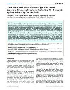
physical or geometric model to divide a protein into domains. Although domains defined in this waymay provide new insights about the protein structure, they do notalways agree with the domain definitions in the literature. Accordingly, the approach adopted in this paper is to start from a subset of known proDomain I Domain II tein structures forwhich the domain definitions havebeen well established, then derive a method that can reproduce the definitions automatically. The success of the method is evaluated by application to a larger test set of proteins. A domain referB ence set has been constructed from domain definitions described in the literature. Where definitions fora protein have notbeen described, assignmentshave been made by inspection. The new f”) method starts froma simple geometric modelsimilar to thatused Domain I Domain II by Wodak and Janin(1981) (a domain has more residue-residue contacts within than without). However, alone this is insufficient to reproduce the normally accepted domain boundaries. The method has been refined to take into account secondary structure content and other factors in order to improve the agreement with the training set. Finally, three simple rules that are applied to any domain definition obtainedby the method provide a ranking scheme to identify the definitions that are most likely to be correct. Domain II Domain I The method explicitly allows for two-segment domains and Fig. 1. Schematic diagram showing three possible paths that the poly- implicitly allows the formation of three- or more segment dopeptide chain may follow in a two-domain protein. A: The simple case in which the chain first passes through one domain and then the other. mains. It runs ina reasonable time on proteins of anysize and B: The chain runs from the first domain into the second and then back can optionally provide a hierarchical classification of compact into the first to complete it. C: Same as B except that, after the chain regions within the protein. completes the first domain, it passes back into the secondto complete it. A unique definition of the domains is presented for a set of 230 protein chains. Automatic screening of thisset picked out 173 proteins, ofwhich 97% agreed with the reference definitions.
- -
-
C
- -
could deal only with single segment domains. More recently, Results Zehfus (1994) used compactness as a measure of “domainness” and searched for compactunits in the structurecomposed of two Comparison of domain definitions noncontiguous sections of the chain. The technique resulted in Table 1 shows definitionsof the domains foundby the program a series of overlapping domain units, but did not provide a unique definition of the domainsin the protein. Furthermore, DOMAK with default parameters. Table 1 also illustrates the corresponding reference definitions obtained from the literature the method could not be run in a reasonable time on proteins that contained more than300 residues. Holm and Sander(1994) and visual inspection (see Materials and methods). In the following discussion the set of definitions obtained by the algorithm describe a method that searches for potential folding units using an eigenvalue analysisof contact maps. Although their elegant is referred to as the derived set. and fast method dealswith multiple segment domains, manyof For 161 of the proteins (Set A), the derived domains agree with those in the reference set (see Materials and methods sectheir published domain definitionsdisagree with those found in tion for definitionof reference set).This gives a confidence level the literature. With the current rapid growthin the number of known pro- of 70% for the method.Only 28 proteins (12%) (Set C) had all domains defined differently to the reference set. tein three-dimensional structures, there is a pressing need to identify systematically the domains.Knowledge ofdomain locaDomain definitions for 41 proteins (18%) (Set B) did notagree tions is important in any reference database of protein structure, closely with the reference domains, but either had one or more such knowledge is also needed for construction of representaidentically defined domains, orby inspection were split into what tive sets of protein structures for derivation of parameters in preone would subjectively term domains. The41 proteins in Set B diction. Prediction of protein structure by threading techniques highlight some of the difficulties with subjective definitionsof (Jones et al., 1992; Bowie & Eisenberg, 1993; Bryant & Lawdomains. For example, glycogen phosphorylase is split into two rence, 1993; for review see Wodak & Rooman, 1993) is best domains (Kinemage 2). However, 18 residues at the C-terminus approached at the domainlevel because this reduces the comcome back across the N-terminal domain. As the tail packs putational overhead. Furthermore, if effective methods are to loosely against the first domain, thereference definitions do not be developed to identify domain boundaries in proteins of unassign it as part of either domain. However, DOMAKassigns known three-dimensional structure, then a reliable library of doit to the C-terminal domain.A further example is actin, which mains is required to derive the necessary parameters. the authors of the structure classed as having two domains(KiA problem faced by all methods of domain definition is how nemage 3; Kabsch et al., 1990). The first domain consists of resto assess the quality of the domains that are identified. The ma-idues 1-144 and 338-375 (domain I in Fig. 2) and the second jority of the early techniques reviewed above apply a simple domain of residues 145-337 (domain I1 in Fig. 2). However, it
A . S. Siddiqui and G. J. Barton
874
Table 1. Domains found by DOMAKa ~
~
.
_
_
_
A
B
C
D
$ I aait
A
l
l 2 3 1 2 3 1 l 1 l 1 1 2 3 1 2 1 2 1 2 1 2 3 1 1 1 2 3 1 2 1 2
_
_
_
_
___.
laait
$laak $ 1aap $ 1aaq $laar Slaba 1ace
I ake
B
A B A -
A
3
1 l 1 l 1 3
2
1alc
-
2
$ lald
-
2
lamat
-
3
UaPk Slaps flatnt
A
1 1 3
$lam 1 avrt
$lbbh Slbbk
D -
A A
2 3
l 6
$lbbk SlbbP $lbbt Slbbt $1bbt $1biat
B A 1 2 3 -
1 l 1 1 1 3
1bic
-
2
$ I bmv $1bmk
1 2
1 2
$lbov SlbPk $lbrd $ lcaa 1cbx
A -
1 1 1 2
$Ice5
-
I
l
3 4 l 1 2 3 4 5 6 1 l 1 1 1 1 2 3 1 2 1 1 2 l 1 1 1 1 2 1
-
~
E 1-267
188-257 139-187, 258-262 1-138 1-150 1-56 10-99 1-76 1-87 4-3 15 346-399, 522-534 316-345, 400-484. 490-521 1-111, 174-214 112-173 38-104 1-37, 105-122 191-307, 342-363 1-190, 308-341 74-296 48-73, 297-324 13-47, 325-410 143-260 1-98 1-137, 358-372 138-185, 272-357 186-27 1 89-225 1-88, 226-260 161-225 3-14, 86-160, 226-304 15-85, 305-318 1-131 120-204 205-253 29-68 69-1 19 254-320 321-373 16-131 2-178 24-192 9-21 8 52-220 1-64 65-270 271-317 149-173, 217-241 3-148, 174-216, 242-261 1001-1185 3012-3181 3182-2192 1-69 261-379 8-225 1-53 1-127, 174-307 128-173 5-87
F G ~~_____
3
2
1 1
1 1
1 1
1-117 118-210 21 1-267 1-135 136-262 ALL ALL ALL ALL ALL ALL
A
~
C
2 1
ALL 38-104 1-37, 105-122 ALL
2
48-325 15-47, 326-410
1 1 2
ALL 1-98 1-144, 338-375 145-337
1 4
1 1
ALL 14-86 87- 160 161-346 247-3 18 ALL ALL
~~
E
1 2
I A A
1
~-
E
F
1
1-114 1-27, 122-235 28-121, 236-245 4-56 1-104 43-83, 114-146 1-42, 84-113, 147-151 5-201 5-43, 216-286 156-215, 324-381, 412-441 95-155, 287-323, 382-411,442-506 44-94 1-174 1-282, 378-423 283-377 8-70 53-120 2-42 43-85 86-130 131-171 1-5, 38-1 16 6-37, 117-186 1-136 2-138 1-53 5-200 1-142, 171-199 143-170, 200-298 6-183 48-98 1-107 1-79 19-24, 153-314 25-152 1-58 1-96 67-98, 176-210, 323-5 I7 3-66, 99-137, 211-322, 518-583
1 2
0-147, 314-333 148-313 1-70 1-32, 93-186 33-92 1-13, 245-431 14-244, 432-471 5-123 1-96 1-359 112-155 10-111, 156-193 482-83 1 19-481, 832-841 4-161 1-209 34-1 14 1-217
2
1
2 1 1
1 2
1
2 A -
1 1 2
1
4
A -
l 2
4 l 1 2
1
1
A
1 4
-
2
-
1 1 1 1 2
A -
1 1
1 2 3 4 1 2 1 1 1 1 1 2 1 l 1
-
1
1
-
2
A -
1
1 2 1 l 1
l
l 2
1
1
2 1 1 1 1 1
3
1
1 2
ALL ALL 31-189 ALL 42-214 1-60 61-271 272-3 17 ALL
1
ALL 3025-3 181 3182-2189 ALL ALL ALL ALL ALL
1
ALL
1 1
1 1
~~
D
3
1
~~ ~
B
0
2
-
1 2
-
2
A A A
l l 1 2
-
2
A A A
1
l l l 2
3 1 2 1 1 2 1 2 l l 1 1 2 1 2 1 l l l
1 1 1
.~
-
~~~~ ~
G
ALL 1-16, 124-233 28-123, 234-245 ALL ALL ALL
1 2
ALL 45-225, 317-461 5-44, 226-316, 462-506
1 2
ALL 1-274, 381-437 275-380 ALL ALL 2-41 42-88 89- 128 129-171 ALL
1
1 4
1
1 1
1 1
2 1 1
1 1
2 1 1
3
1 2 2 1 I 1
1
2 I 1 1 2
ALL ALL ALL ALL 1-135 136-298 ALL ALL ALL ALL 19-152 153-314 ALL ALL 3-108, 225-327 514-583 109-225 328-5 13 0-148, 318-333 149-3 17 ALL 1-30, 81-186 31-80 1-20, 227-432 21-226, 441-471 ALL ALL ALL ALL 485-813 19-484, 814-831 ALL ALL ALL 1-81 90-2 17 (continued)
Continuous and discontinuous domains
Table 1. Continued -_
~ "
A
_
_
B
C
-
2
-
3
A A A
1 1 l 1 1 l 2
B -
1 1 1 2
-
2
-
3
-
4
A
1 3
A A D A -
l 1 l l 1
l 4
A A A
l l 3
E
1 2
A
1 1
-
3
-
1 3
2
D 1 2 1 2 3 1 1 l 1 1 l 1 2 1 1 1 1 2 1 2 1 2 3 1 2 3 4 1 1 2 3 l 1 l 1 1 l 1 2 3 4 l l 1 2 3 4 5 6 1 1 2 1 1 1 2 1 2 3 1 1 2 3
_
875
~
E 1-71, 103-138 72-102, 139-147 130-294, 342-653 5-129 295-341 1-59 1-85 1-99 1-74 1-68 427-556 1-181 182-276 1-99 3-153 1-131 100-252 1-99, 253-345 1-13, 59-164 14-58 162-484 74-161 1-73 435-594 340-434, 595-691 1-91, 251-339 92-250 25-180 146-3 19 7-77 78-145 6-92 23-166 4-22, 60-156 1-103 1-146 1-58 161-224 225-299 120-160,300-348 3-119, 349-359 1-96 1-153 76-82, 176-265 266-406 83-175, 407-465
1-169 126-230 15A-125, 231-244 1-120 1-81 0- 138, 252-304 139-25 1, 305-3 19 21 1-253 181-210, 254-436 1-180 8-77 67-128, 170-204, 241-371 10-66 129-169, 372-414
G
B
C
1
ALL
-
2
3
1-177 178-400 40 1-663 ALL ALL ALL ALL 1-68 ALL 1-175 182-276 ALL ALL 1-131 ALL
-
1 2
-
1 1 2
c
1
H M
I I
1 1 1 1
1 1
2 1 1 1 1
2 3
4
1
2
1 1 1 1 1
1 2
1 1
6
1 2 1 1
2 2
1-69 70-164 162-484 1-160 353-484 434-595 345-433, 596-663 1-90, 252-320 91-251 ALL 147-319 7-146 ALL ALL ALL ALL ALL ALL 3- I2 1,344-359 133-338
ALL ALL 108-173 174-214 215-267 267-3 14 315-394 395-459 ALL 118-230 15-117, 232-244 ALL 1-81 0-139, 256-304 140-255, 305-319 33-344, 438-466 345-437
1
ALL
2
10-101, 296-355 102-295, 355-414
D 1
-
1 1 2
-
1
2 1 1 2 1 1 1 2 1 2 1 1 2 3 1 1 1 2 1
-
1
1
-
3
A A A
1
1 2 3 1 l l 1 2
-
2
l l 2
1 2 1 1 A 2 1 2 1 1 1 1 I 1 1 3 1 2 3 1 1 - 1 1 - 1 1 2 3 1 1 1 A l l - 2 1 2 I 1 1 1 A 2 1 2 A 2 1 2 B 2 1 2 1 1 2 1 2 A l l A l l 2 1
-
E
F
1-45, 100-154 46-99, 155-394 3-63 1-255, 447-452 256-446 0-80 1-99 1-18, 112-207 19-111, 208-212 24-332
2
25-82, 117-243 1-323
1 3
1-280 1-63 1-49, 79-103 50-78, 104-124 1-174 1-129 157-181, 208-274 1-156 182-207,275-293 1-104 2-110 1-56 19-47, 139-164 2-18, 48-138, 165-245 16-22, 122-233 23-121, 234-245 7-141 114-178 179-264 13-133 1-112 1-56 238-279 132-205 1-131, 206-237 1-170 33-135 1-710
1 1
28-256 6-157 1 17-244 1-116, 245-315 1-289 1-70 1-33, 88-254 34-87, 255-362 173-232 1- 172, 233-265 9-27, 224-331 28-223, 332-393 2-387 34-86 -5-33, 87-103 1-106 1-129 34-95
G
~-
."
1
ALL
1 2
ALL 1-255 256-452 ALL ALL 1-9, 1 12-206 10-111 33-143, 315-332 1-32, 144-314 133-258 1-51 52- 190 191-323 ALL ALL ALL
1 1 2
1 1 1 2
ALL ALL 159-293 1-158
1 1 1 1
ALL ALL ALL ALL
2
16-22, 129-229 23-128, 230-245 ALL 114-177 178-264 ALL ALL ALL 1-127 128-208
1 2 1 1 1
2
1 1 3
1 1
ALL ALL 1-383 384-494, 649-733 495-648 31-250 ALL 117-244 1-1 16, 245-316 ALL ALL 1-32, 87-254 33-86, 255-362 1-188 189-268 1-52, 86-204 53-75, 205-397 2-317 ALL
1 1 3
ALL ALL 1-62
1 1 2 1
1 2 2 2
(continued)
A . S . Siddiqui and G.J. Barton
876
Table 1. Continued -.
-
~
~~
~
B
C
"
~
mcY $2cdv $2cpk
A
t2ctx 12CYP
-
1 2
2dpv
-
1
"
E
2 3 l
l 1
1
2
QfbJ
H
2
$2fbJ
L
2
5 -
1 1 1 1
$2hip $2hmq Uhpr $21iv
A A -
1 2
$21tn $2mcm $2mev $2msb 2npx
A 4 B -
l 1 1 1 5
A $2pab 1 $2PIV 4 $2PlV $ 2 ~ ~A 7
l l
l
1 2 1 1 2 1 2 3 1 2 1 2 1 1 1 1 2 l l 1 1 2 l 1 1 1 1 2 3 4 5 l
1
1
1 4
1 1 2 3 4 1 1 2 1 l l 1 1 1 l 1 2 3 1 2 I 1
$2por $2reb
-
1 2
$2rn2 $2rsp $2SCP $2~13 $2stv 2tmv $2trx 2tslt
A A P A -
1
1 1 1 l 3
2tsc
A
2
$2wrp 2yhx
R -
I
l l
3
2 3 $351~
-
1
E
D
2c2ct
$2fx2 $2fxb m ~ 2had
~
~
A
1
~
.~
F ~
~~
1-33, 96-112 2- 128 29- 107 122-340 15-121, 341-350 1-71 145-262 2-144, 263-294 37-584
1-1 18 119-220 1-106 107-213 2-148 1-81 1-87 1-310
1
1 2 1 1
3
2 2 1 1 1
2
~-
G
~~~
B
C
-~
~
63-95 95-112 ALL ALL 15-31, 126-317 33-126, 318-350 ALL ALL
$3b5c 3bcl $3cd4
-
I
-
1
$3chy 3cla
-
1 2
3dfrt
-
2
37-281, 336-410, 446-584 282-335 41 1-445 1-118 119-208 1-104 105-210 ALL ALL ALL 1-155, 230-310 156-229 ALL ALL ALL 1-120, 250-328 121-249, 329-344 ALL ALL ALL ALL 1-114 115-243 244-324 325-446
$3ebx 3enlt
-
1 2
Vfgf Ugap
A
I
3grs
-
1
1 1-71 1 1-113 1 2-88 2 1-118, 251-325 119-250, 326-344 1 1-181 I 1-1 12 1 13-70 1 109-22 1 4 78-115, 244-283 1-77 284-323 116-243 324-447 I ALL 10-123 1 83-202, 234-265 6-291 I ALL 2-69 4 420-562 420-561 1-188 1-188 189-297, 379-408 189-303, 388-419 298-378 304-387 1 ALL 1-301 2 23-268 27-269 269-328 270-328 1 ALL 1-155 ALL 1 1-124 I ALL 1-174 1 ALL 1-65 1 26-195 25- 195 1 ALL 1-154 1 ALL 1-108 2 248-319 223-319 1-220 1-130, 173-222 131-172 I ALL 1-56, 146-264 57-145 1 ALL 5-108 3 20-49, 190-282, 19-48, 188-285, 364-432 363-458 50-189.433-451 49- 187 2-19, 284-363 2-18, 286-362 1 ALL 1-82
1
A
1
2
2
~~~
~
1 2 1 1 2 1 2 1 1 2 1 1 2 1
~
~~~
~
~~~
~~
~
~~
3-87 3-358 1-97 98- 178 2-129 6-91, 141-219 92- 140 1-29, 96- 162 30-95 1-62 1-33, 11 1-436 34-110 20- 143 1-136 137-208 18-478
~~~~
F
E
D
~ "
~~
ALL 1 I 2
5-72 1-61
-
1
1
-
1
1
t3pgkt
-
3
3pgm
-
2
3psgt
-
4
$3rub 3sdp
s A
$3sic 4blm
1
1
1
A
2
1 2
$4bp2 $4fdl 4gcr
-
1
-
1 2
$4icb $4mdh
A
$4sbv Wsgb $4tnc
A 1 -
5fbPt
A
$5~21 5rubt
A
$6abp
-
$6ebx $7cat
A A
7th
A
1 2
2 1 2 3 1 2 1 2 3 4 1 1 2
1
1 1 2 I 1 3 1 2 3 l l 1 1 2 1 2 2 1 2 1 1 5 1 2 3 4 5 2 1 2 l l 2 1 2 3 3 1
I 1 1
ALL
1
ALL 1-142 143-420 ALL 1-125 126-208 18-57, 108-158, 293-363 50-107, 159-291 365-478 ALL 1-29 30-6 I 199-387 1-198, 388-478
2 1
3
I 2
2 193-235, 328-402 236-327 1-192, 403-415 2 1-88, 131-230 89-130 2 192-309 125-191, 310-326 lop-12 1P-9P, 13-124 I 10-123 1 5-74, 111-176 75-110, 177-190 1 7-113 2 31-86, 154-291 87- 153 1 1-123 1 1-106 2 1-83, 172-725 84-171 1 0-75 2 1-84 85-153 154-333 1 62-260 1 1-51 2 3-90 91-162 2 6-212, 240-320 213-239.321-335 ALL 1 1-166 2 393-457 2-137, 292-316 3 17-366 138-162, 367-392 163-291 2 109-254,286-306 2-108, 255-285 1 1-62 3 25-67 68-500 2-6, 125-230
~
ALL 1-97 98- I78 ALL ALL
2 3 $3i18 $3mt2t
G
1
1-88, 149-230 89- I48 1-170 180-327
ALL ALL ALL 31-70, 217-291 71-216 ALL ALL 1-80 83-174 ALL 1-151 152-333 ALL ALL 3-88 101-162 1-201 202-335 1-139 140-457
110-253, 295-306 2-109, 254-286 ALL 3-68 69-433 434-500 ALL (continued)
877
Table 1. Continued ~~~
B
C
D
E
~~
7tim 8acn
~~
2 3
-
5
1
~
.~
~ ~
A
~~
~
F
~
_
E
G
_ ~
~~
F
G
~
7-61, 23 1-248 62- I24 2-15, 63-197, 271-300, 505-529 198-270, 301-346 347-504 16-62 530-754
L
I 3
2-201 202-5 I 1 532-754
A
2
2
B$8atc
I 2 1
2
$8rxn
A
1
1
1-178, 318-374 179-3 17 131-291 1-130. 292-310 8-98 99-153 1-52
2
2
1-175, 319-374 176-3 I8 144-290 1-143, 291-310
2
8-100 101-153
I
ALL
'I Abbreviations used for column headings in this table: A, Brookhaven code; $ before the code indicates that the algorithm thinks that its definition is correct: t after the code indicates the definitionwas taken from the literature. B, chain. C, number of domains in derived definition. D, domain number. E, derived definition. F, number of domains in reference definition. G , reference definition. "ALL" indicates protein i F a single domain made up of all residues. t after the name indicates the definition was taken from the literature.
has alsobeen suggested that each of the domains candivided be into two subdomains(Kabsch et al., 1990). So residues I-32,70144, and 338-375 make up subdomain la, and residues 33-69 make up subdomain Ib. For the second domain, residues 145180 and 270-337 make up subdomain Ita andresidues 18 1-269 make up subdomain Ilb. DOMAK classes the protein into three domains, 1, Ila, Ilbwith thedefault parametervalues. Ifthedefault parameters are varied, it is possible to find all four subdomains or find only the twomain domains. Thus,there is a"gray area"of domain definition where one is not surei f a subunit of the protein structure should be classed as a separate domain or
Subdornain la
.,
Subdomain Ib
whether it is merely a lobe or local compact region. By choosing a set of parameters (principally the MSVvalue),fixed a subjective limit has been set and applied objectively to the whole set. After applying the three reliability screens described in the Materials and methods, domains from 57 proteins are found that are believed to be defined incorrectly by the algorithm. Twenty-three of the 57 proteins were incorrectly defined in comparison with the reference set. Twenty-five were from Set B. Nine definitions from Set A were picked out as incorrect. Hence, the list of definitions automaticallydefined as correct is reduced to 173 (75% of the original 230 proteins). Of these,
.
Subdomain Ilb
Subdomain Ila
Domain II
Fig. 2. Actin can be thought of consisting of two main domains, eachof which can be split into two smaller subdomains. Thisexample highlights the gray area of domain definition where one has to draw the line between what one calls a domain and what one termsa subdomain. Thealgorithm split thisproteinintothreedomainsmarked by shading (domain I , subdomain Ila, and subdomain Ilb). Figure was produced using MOLSCRIPT (Kraulis, 1991).
A . S. Siddiqui and G.J. Barton
878
Table 3. Table of domains listed as correct thaf have 88% match the reference set. Nine percent are from Set B and split the chain into what look like domains (Table 2). If one major difference to the defined definition " _ _ _ ~ ~ _ _ ~ _ __ _ __ ~_ ~ _ ~ . chooses to accept these definitions, the reliability of the algoBrookhaven rithm rises to 97%. The five (3oio) remaining structures that were Why code difference? isathere incorrectly defined are listed in Table 3, together with the rea- Chain sons why the algorithm gave different definitionswith the delaai A Is incorrectly classed single aas domain that fault parameters. The structures that are automatically defined made up of four segments. as correct are labeled with a in column A of Table 1. lgst A Is incorrectly classed single aas domain.
Analysis of the derived set
1ipd
-
1 wsy
B
.~
~
~
~
is
Is split into two "domain-like parts," except for the fact that a sheet is split. Both definitions split this into two domains, but the two definitions are quite dissimilar. This istwo-domain a protein with each domain containing about 30 residues (smaller than the minimum domain size, MDS).
The structures that the algorithm identified as correctly split can be divided on thebasis of the number of domains they contain. 3mt2 of an n-domain Table 4 summarizes the number of occurrences protein. Single-domain proteins are thelargest group at75% of the set. Over the entire set there is an average of 1.3 domains per protein. The number of occurrences of an n-domain protein falls off rapidly as n is increased, and 98% of the proteins contain three or fewer domains. tease. It is divided into two domains,with a single cut in the midExamples of a single-, two-, and four-domain proteins are dle of the chain and with both the N- and C-termini crossing showninFigure 3 (seealsoKinemage 4). Figure 3A and back over into opposite domains, making each domaina twoKinemage 1 show trypsin (Read & James, 1988), a serine prosegment domain, similar to the topology of the two domains shown in Figure 1C. Figure 3B illustrates the A chain of the protein phosphoglucomutase (Lin etal., 1986). It is split into four domains. The chain runs from thefirst domain into thefirst half Table 2. Table of domains listed as correct that have of the second domain, passes through the third domain, comes an acceptable difference to the reference definition back into the second domain to complete it, and finally makes Brookhaven What difference is the between up the fourth domain. code Chain reference thederived anddefinitions? Figure 4 shows the distributionof the number of residues in a domain. Most domains are made up of between 50 and 100 lald - Reference definition is a single domain. Derived residues. Ninety percent of the domains are comprised ofless definition is acceptable. than 200 residues. The histogram tails off rapidly for large doA Figure See 5 latn mains and there are only two domains made upof more than latn D Reference definition is single adomain. This definition is acceptable. 400 residues (the two domains of glycogen phosphorylase). A Reference definition is single a domain. This lbbk Although the algorithm is primarily designed to search for is a propeller fold structure with seven resingle-segment or double-segment domains, it is possible for peated units, The derived definition splits it domains to be made up of more segments by noncontiguous into six domains, with one domain contain"chopped segments" being added onto the domain.Table 5 suming two of the repeated units. marizes the number of n-segment domains. A total of 81.5% 1 ezm Reference definition is two single-segment doof the domains foundwere single segment. A further 17.6% of mains. lt is instead split into two two-segment the domains were made up of two segments. Only one threedomains. segment and one four-segment domainwere found in the final 1 fnr - Minor difference-one derived domain is a set (both the domains of glucose oxidase; Hecht et al., 1993). two-segment domain. Similar definition, but split into two domains The two-segment domains were subclassified on thebasis of 1gal not three. those in which there is a large difference in the relative sizes of the segments. The size of the smaller segment as a percentage 1gpb - Minor difference- one derived domain is a two-segment domain. of thesize of thewhole domain was calculated (histogram, ElecI lap
-
1Ild
A
I pii Zcpk 2CYP
E
3P&
-
4mdh
A
7cat
A
One reference domain is split into two further parts. Two of the reference domains remain unsplit in the derived definition. One reference domain is split into two further Table 4. Number of n domain proteins .." ___-. parts. difference. Minor No. of domains definition. Similar occurrences protein in Reference definition is a single domain. This 1 definition is acceptable. 2 One reference domain is split into two further 3 parts. 4 One reference domain is split into two further 5 parts. 6 Two of the reference domains remain unsplit. ~
-
.
"
~"
- ..
~
--
" ~~~
~-
No. of I29 34 6 3 0 1
~~~
~
879
Continuous and discontinuous domains 0
A
0
0
100
200
3w
4w
5M)
Size of domains
A
B Domain I\
Domain 111 G
Fig. 4. Histogram showing the distribution of domain
sizes.
the intervening segment. This shows thatmost inserted domains have their connections to therest of the proteinclose together. A close connection may suggest that the inserted domain could be deleted without disrupting the integrity of the two-segment domain. No correlation was found between the end-point distance and the relative sizes of the segments.
Discussion
Fig. 3. A: Trypsin is classed as a two-domain protein. Topology of the chain is similar to that in Figure IC. B: The A chain of phosphoI glucomutase is split into four domains. The chain runs from domain into thefirst half of domain11, into domain 111, completes domain 11, and finally goes into domain1V. Figures were produced using aversion MOLSCRIPT (Kraulis, 1991), modified by Robert Esnouf (pers. comm.).
tronic Appendix). The distributionis fairly even over the entire range, though the number of domains, in which one segment is 20-40% the size of the other, is significant. The distance separating the residue at the end of the first segment and the residue at the start of the second segment was examined as a percentage of thesize of the intervening segment. The size of the intervening segmentwas estimated by working out the maximumC"-C" separation in the domain (histogram, Electronic Appendix). The distribution appears to be normal with a peak in the range 30-40070. For 76% of the domains the separation is less than half the maximum C"-C" separation in
The algorithm described in this paper can locate domains for any length of protein and is fast enough tobe run routinely on the large database of protein structures. Afterscreening, the domain definitions agree very well with conventional subjective definitions (97%). The algorithm could be developed to include the screens at an earlier stage and thusdetect unlikely domains, alter the relevant constraint values, then run theanalysis again. Most of the differences between the automatically derived domain definitions and the reference definitions lie with difficulties and inconsistencies in what is meant by a "domain." The algorithm described here finds compactlocal regions of structure according to a set of thresholds (Table6). However, these compact regions d o not always correspond to what one would intuitively consider to be the domainsin the protein. This problem is common to all previous algorithms for protein domain definition (Rossmann & Liljas, 1974; Crippen, 1978; Rose, 1979;
Table 5 . Number of n segment domains -
" "
segments No. of occurrences in domain
No. of I90 41 1 1
880
A . S . Siddiqui and G. J. Barton Table 6. Table of constraints _
~ ~
Subdivision
~~
~
Full name
Constraint
.
~~
MDS MNCC
MSS sso
value
MS v
weightingBW HCD MDSP MAC
ID ~.
domain MNCCrn MNCCe MSSm MSSe
""
-
~-
~.
.
~~
~
Rashin, 1981; Wodack & Janin, 1981; Go, 1983; Zehfus & Rose, 1986; Holm & Sander, 1994; Zehfus, 1994) and is an inevitable consequence of applying an objective set of rules for domain definition to whatis an essentially subjective interpretation. A major advantage of the algorithm described here is the ability to screen accurately thederived domains for domains that are unlikely to fit the normal concept of a domain. Accordingly, the final list of domains may be used with a high degree of confidence. A server of domain definitions, accessible via the World Wide Web, can be found at http://geoff.biop.ox.ac.uk.
Materials and methods Introduction - Split value The concept at thecenter of the domainidentification algorithm is that residues comprising a domain make more contacts between themselves (internal contacts) than theydo to the rest of the protein (external contacts). This follows from the work of Rossmann and Liljas(1974), who suggested that a domain has many shortresidue-residue distances within itself, but few short distances between it and therest of the protein. Thus, the ratio of the number of internal contacts to the number of external contacts should be large for a domain. Tworesidues are defined to make a contact if a heavy atom in one residue is within 5 A of a heavy atom in the other. If the protein is split into two arbitrarily chosen parts,A and B, then the quantity
.~
~
~" _ ~ ~
~~~~~~~
Minimum size Minimum contact no cut-off chain middle of Minimum contact cut-off end no of chain Minimum segment chain size of middle Minimum segment size residues end 5of chain If percentage of secondary structure is greater than this only contacts structure secondaryuse split MS v Minimum Minimumsplitvalueusingonlysecondarystructurecontacts MS vsso MS vcs chopped segments forvalue Minimum split P-Sheet Reduce contact density of helix value to this Minimum double splitasize segment for of compactness allowed Maximum divider Increment
~- .
_
Value
. ".
-
..
40 residues 30 residues 10 residues 25 residues
57 % 17.05 60.0 0.1 10.32 contactdresidue 120 residues 2.85 A 250 residues ~
~~~
. ~~
A simple implementation of the idea Consider chopping the proteinchain into two partsof segments between residues i and ( i 1). A segment can consist of any number of residues, but the residues must form a continuous sequence along the chain. SegmentA then consists of residues 1 to i and segment B of residues ( i 1) to N,where N i s the number of residues in the chain. The splitvalue can then be calculated for 1 5 i < N.Figure 5 illustrates a graph of split value against i for the T-cell surface glycoprotein, CD4 (Ryu et al., 1990). The split value has a large peak at i = 97, indicating that the protein shouldbe split into two domains at this point. Once split, the two domains canthemselves be individually scanned to find the maximum split values and hence thebest positions to split them intonew domains, which again can be scanned and split and so on. By placing a limit on the minimum numberof residues in a domain (minimum domain size, MDS) and/or defining a minimum split value ( M S V )below which the two parts are considered to becorrelated and notdivisible into smaller domains, the process of division can be stopped. The result is a series of "cuts" defining how the chain should be split into separate domains.
+
+
Allowing f o r two-segment domains Consider a domain made up of a single segment that consists of residues k to I, inclusive, which is scanned to find further domains.
Method I -A single-segment scan (Fig. 6A) Segment A is chosen by cutting the chain at two points, x and (int,/ext,,) * (int,/ext,,) y . Therefore, B can consist of up to two segments, B , and B2, depending on the positions of the boundaries. The split value is calculated for all possible segment A's formed by varying x can be calculated, where int, is the number of internal contacts and y . The maximum split value is stored, together with the corin A , int, the number of internal contacts inB , and ext,, the responding values of x and y , called xmaxand ymuX,which denumber of contactsbetween A and B.This quantity is referred fine A*"". The maximum split value is compared with MSV to as the split value. The split value will be large if the A and B a r e distinct. If the two parts are not distinct (i.e., correlated), and if A'""" is not correlated with B""", then A*"" can be "extracted" from the "parent" domain to form a new "child" dothen the split value will be small.
88 1
Continuous and discontinuous domains
are not correlated, the parent domain is split at this point. A'""X goes on to form a two-segment child domain.
c .-
Hi -
I .
0
50
150
100
I
Fig. 5. Graph showing how the split value varies with i for 1CD4. The protein is cut into two segments,A and E , between residues i and ( i + I).The graph shows a large peak at i = 97, indicating that the protein should be split into two domains at this point. Although this example is a relatively easy case of a two-domain pr-olein, i t illustrates the basic method well. The fact that there are twoclear domains is reflected by the size and narrowness of the peak.
main (also referred to as a "subdomain"). The treatment of B'""' is the same forall three scans and is shown at the end of Method 3 . Note that thesingle segment scan would be able to deal with both the situations that arisein Figure 1A and B. However, it would not be able to deal with the case shownin Figure 1C. To allow for this eventuality, a two-segment scan is used. Method 2- A two-segment scan (Fig. 6B) A is made up of two segments, A I and A z , formed by cutting the chain at four points,xI, y , , x2, and y,. The split value between A I a n dA , must show them tobe correlated when compared with M S V . B can be made of up to three segments, depending on the positions of the boundaries. The split value is calculated forall possible segment A's formed by varying xI, y , , x,, and y z . The maximum split value is stored, together with the corresponding values of xi, y , , x,, and y z , called x ; n u x , y;'"x, x y ,and y;""", which define Anluxand BrflaX. The maxi-
mum split value is compared with MSVand if
and B"'"
Method 3 - A two-segment scan of a two-segment domain (Fig. 6C) Now consider a domain made up of two segments, consisting of residues kl-tl andk2-t2. The algorithm scansthis domain for subdomains in the following way. A is made up of two segments A I and A z , formed by placing four boundaries atx1,y , , x,, and y 2 . Note that oneof the boundaries of both components of segment A must lie on the boundary of the parent domain. The split value between A , and A , must show them to be correlated when compared with MSV. B can consist of up to two segments. This split value is calculated for all possible segmentAs formed by varying x i ,y , , x, and y,. The maximum split value is stored, together with the corresponding values of x,, y , , x, and y 2 , called x;nUx, y;n", ,;"A,' and y;""", which define A""-' and BrnUx.The maximum split value is compared with MSV and if A""" and B'7'"X are not correlated, the parent domain is split at this point. AmuX goes on to form a two-segment child domain. For all three scans, if B"'"" consists of only one segment, it is considered to form a single-segment child domain. If B'""' consists of two segments, the splitvalue between these two parts is calculated. If the two segments are correlated, they are placed together to forma single child domain made up of twosegments, otherwise, they are considered to be two separate, child domains. If B"'"" consists of three segments, the split values between all pairs are calculated. If none of the pairs are correlated, the segments are considered to form threedistinct child domains. If one of the pairs is correlated, the twosegments are placed together to form a two-segment child domain, theleftover segment forming a single-segment child domain on its own. If two or three of the pairs are correlated, the pair with the highest degree of correlation (i.e., lowest split value) is placed together to form a two-segment child domain, the leftover segment again forming a single-segment child domain on its own. Applying the methods to divide a protein Armed with these methods, the algorithmwill start off treating the chain as a single-segment domain and divide it using Method 1 or 2, whichever yields the higher split value. If a two-segment domain is found at any point, it is scanned using
A k
Segment 81
Segment A
X
y
Segment 82
I
B Segment 61 x'1
C
Segment 82
c
U
kl
Segment AI
Segment 81
I1
Y l Segment A I
x Segment i A2
' Segment 83
Y2
U
k2 x2
Segment A2
Q
Segment 82 l2
Fig. 6 . A: Single-scgmentscan in which A is made from a single segment extracted from the parent domain, splitting it into two parts.B: Twosegment scan in which A is made from two segments,splittingtheparentdomainintothree. C: Two-segment scan of a two-segment parent domain. A is made up of two segments, onein each of the parent segments. Note that, in this case, one end of each segment must be at the endof a parent segment.
A . S. Siddiqui andG. J. Barton
882
Method 3. The algorithm continues to divide the protein, until rectly. To compensate for this, the numberof internal contacts in a helix-containing segment is reduced to the averagelevel for it is checked by one of the constraints. Constraints are described coil regions. The value to which it is reduced is termed helix coil in the next section and are also present toallow the algorithm to be flexible and fast. density (HCD). Note that none of these methods will deal with domains con&Sheets may sometimes besplit across domains.A constant BW(standing for @-sheetweighting) is used to reduce the likelisisting of three or more segments. Domains such as theseare not hood of this occurring.The numberof external contacts between dealt with explicitly in the algorithm at the scanning stagebecause the complexityof the scan would increase rapidly. How- two regions is increased by B W percent for every hydrogen bond ever, they are allowed implicitly at a later stage (described (as defined by DSSP [Kabsch & Sander, 19831) between strands below). Such domains are found to be quite rare, making up that spans the two regions. Therefore, the greater the number in the only a small fraction of the total number of domains of strand-forming hydrogen bonds that bridge two regions, the database. less likely they are to be distinct. Once all the domains have been found, their compactnessis checked. If a domain is found tobe noncompact, it is combined Additional details with the domain with which it has the lowest split value. The The MSV is used to decide whether two segments are distinct process is repeated until either all the domains are compactor all the domains have been combined together. A domain is deor correlated. If the split value foundis less than the MSV, the fined as noncompact if its radius of gyration deviates from a two segments are correlated, otherwise they are distinct. theoretical curve (of radius of gyration against size of the doA segment can consist of any numberof residues, but the residues must forma continuous sequence along the chain. There main) by more than the constraint maximum allowed compactare threetypes of constraints on the number of residues in a segness (MAC) (Russell, 1993). ment (Table 6): minimum domain size ( M D S ) ,minimum no contact cut-off (MNCC), and minimum segment size (MSS). Increasing the speedof execution They are chosen such thatMDS > MNCC > MSS. A segment that has size2 MDS and is distinct from the rest of the parent The speed of the domain scan depends on thesize of the segment being analyzed. If the segment contains Nresidues, there domain is considered to form a child domain. This constraint provides control over the minimumsize of the domain and pre- are N places at which a boundary may be placed.A Method 1 vents the protein being split into small pieces. A segment with scan cuts the segment twice and so there are N2/2 possible size < MDS but 2 MNCC, thatis found to be distinct from the splits. A Method 3 scan has restrictions onwhere segments may rest of the parent domain,is not large enough to forma child start and end. Supposeit contains two segments ofsize N, and domain. Instead, it is classed as a “chopped segment.” Chopped N2. Each segment is effectively scanned twice by a single cut. Therefore, the speedof the scan can be given by (2N,)(2N2) = segments allow the algorithm to remove small segments from 4 N , N2. A Method 2 scan splits the domain four times and a domain that are not strongly correlated to it and later reassign them to other domains,or back to the original one. This hence its speed varies approximately as N4/4. Restriction on allows domains toconsist of more than two noncontiguous segsegment sizes helps reduce the numberof combinations, but the ments. The treatmentof chopped segments is discussed below. algorithm’s speed can be increased further in the following ways. Segments with size < MNCC but 2 MSS, are used by twoSmall segments are unlikely to containtwo-segment domains. Therefore, a lower bound is placed on the minimum number segsegment scans (both Methods 2 and 3). In these scans, two of residues in a segment, minimum doublesplit (MDSP).Any ments can come togetherto forma single domain. It is possible single-segment domains with size < MDSP are assumed to conTo allow for this, segthat one of the segments may be small. tain single-segment domains only. This prevents the algorithm ments that havea size in this range are only allowed if they are from performing a two-segment scan on the segment and thus correlated with another segment, such that the total size of the two segments is 2 MDS. Segments with size < MSS are not al- saves time. If the percentage of secondary structure (i.e., helix and strand) lowed, thus preventing very small segments from occurring. in the domain being scanned is greater than the valuegiven by When domains are inspected, one often finds small segments at sso, the algorithm uses only those contactsto and from secondthe N- or C-termini that cross domains. Segments in the middle of the chain as small as this do notcross domains. Thus, to ary structureelements. Secondary structureelement definitions are taken from the program DSSP (Kabsch & Sander, 1983), allow for this difference,MNCC and MSS are divided into two categories: segments that are present in the middle of the chain using “H” for helix and “E” for strand. and those that have one end connected to the end of the chain,It was found that, in cases where only the secondary structure was used, the maximumsplit values were generally higher t o give MNCCm, MNCCe, MSSm, and MSSe. The values of than they would be had the same domains been scanned using these constraints are given in Table 6. all contacts. To take the difference in maximum split values into Helices form a relatively large number of contacts per residue (contact density) when compared to coil and @-sheet. The account, the cases in which only secondary structure contacts are being used are compared against the variableMSVsso. average contactdensity in 2,446 coil regions, 1,324 helices, and Although the above restrictions cut downon the number of 1,563 @-strandswas found to be: 24 7 contactshesidue, for combinations, once the actual number of residues being used helices, 10.3 6.6 contactshesidue for coil, and 3 . 3 k 2.2 conrises above 250-300, the algorithm still takes an unreasonably tactshesidue for strands. Accordingly, helical regions have a long time toexecute. To circumvent this problem, some “pruntendency not to be split, but more importantly, theyraise the is done on Method2 scans (see Fig. 6B). number of internal contacts in the segment that contains them. ing” of the search tree The assumption is made that, if two segments are correlated, This can lead to segments containing helices being split incor-
*
883
Continuous and discontinuous domains increasing the size of one segment will not make the segments distinct. Although tree-pruning is successful in most cases, itis not able to speed up others. In order to speed up all scans, the split value is not calculated atevery position for large segments. Instead, the position of the cutting boundaries is moved by an “increment,” skipping over intervening residues. The increment is calculated by dividing the size of thesegment being analyzed by the increment divider (ID)and adding one. Note that this results in MSV being over a range of resithe cuts corresponding to the dues rather than atspecific residues. The same situation occurs when only secondary structure is considered because the split value will remain unchanged as the cut boundary passes over nonsecondary structure residues. In both these cases, once the range of the cut boundariesis known, the algorithm goes back and calculates all the split values for all residues in the range using all contacts. The combination thatgives the highest split value is the one used. Using these methods, analysis time was reduced from 11 h to 1 min on the three-domain protein BirA (Wilson et al., 1992) (for a Silicon Graphics Indy R4000 PC).
Screening the results To be useful, any automatic algorithm must be able to tell when the definitionsit has produced arelikely to disagree with the expected standard. Three rules about domains were derived to enable the algorithm to identify such examples. 1. Count the number ofsegments in a single-domain protein. Single-domain proteins may have chopped segments removed that are later reassigned or may be split into domains that are recombined on thebasis of compactness. If the numberof segments that the final domain was split into is large, then the domain is unlikely to be a true single-domain protein. Singledomain proteins made upof four or more segments were flagged for further visual inspection (table, Electronic Appendix). 2. Calculate the numberof residuesper segment for domains consisting of two or more segments. If this is small, it is unlikely that the domainis a real domain. Thissuggest a lower limit on the size of such domains,which is larger than MDS. The limit chosen was50 residues per segment (table, Electronic Appendix).
(Russell & Barton, 1992) format. An input file for RASMOL (R. Sayle, 1992, RASMOL, molecular visualisation program, e-mail:
[email protected]) todisplay the domains foundis also produced. Further details aregiven in the DOMAK user guide (A.S. Siddiqui, 1994).
Reference domain definitions A set of 275 nonredundant protein structureswas derived from the Brookhaven database. The nonredundancyis based on sequence rather than structure,so some structures from the same family appear in the set. The structures were examined by Dr. R.B. Russell (pers. comm.) andsubjectively split into domains using knowledge of protein folds andon the basis that domains are globularunits that aredistinct from therest of the structure. For proteins in this set that contained more than one domain, the literature was searched for domain definitions in the original publications that described the crystal structure. This set is referred to as thereference set, as shown in Table 1. Table 1 also shows which definitions were derived from the literature (identified by a t after the name). It was not possible to produce DSSP(Kabsch & Sander, 1983) files for 40 of the structures. CONTACTS could not be run on a further four structures because it requires all atoms to be present in the file. DOMAK, in its current form, has notbeen designed to deal with structures inwhich domains are made up of more than one chain. Therefore,kallikrein A was excluded from the set. However, it is conceptually simpleto extend DOMAK to handle thiscase. The final set of protein structures analyzed was 230. DOMAK required 16.5 h of CPU to complete the analysis on this set,giving an average time of 4.3 min per protein. Calculation of contacts requiresless than 2 min for the largest proteins (glycogen phosphorylase, 823 residues, took 101 s) and justover 1 s for thesmaller ones (metallothionein isoform 11, 62 residues; 1 s).
Optimization of parameters
There are 14 independent DOMAK parameters (Table 6) for which suitable values had to be determined. Constraints onsegment sizes ( M S S ) were derived by taking thesmallest values of these constraints that appear in the set of domains thatwas de3. For a single-segment domain inserted into a domain of two rived by eye. MDS was chosen by looking at thesizes of domains or more segments, calculate the ratio of thesize of the domain in the same set. Thesmallest domain size in this set is actually into which the inserted domain is placed to the size of the in30, but this is exceptional so a size of 40 residues was chosen. serted domain. If the ratio is large, the inserted domain is unMNCC values have not been optimized. sso was chosen to prolikely to be a real domain. The limit set was 1.6 (table, Electronic vide a compromise between speed and accuracy.If the amount Appendix). of secondary structure in the segment is small, looking at secondary structure contacts onlywill not be accurate enough. The value was chosen by looking at two examplesin which a sheet Implementation was being split (Brookhaven codes lPHA and1IPD). HCD was The algorithm was implemented as a program written in ANSI set simply to the value of the average contact density in coil reC called DOMAK (“DOMain MAKer”). All the times are for gions. MAC was derived by looking at the compactnessof the a Silicon Graphics Indy R4000 P C (32 MByte memory, nosecdomains that had been split by eye and choosing a value that ondary cache). The program requires outputfiles from the pro- encompassed most of them.MSV, MSVsso, and MSVcs were grams DSSP (Kabsch & Sander, 1983) and CONTACTS (R.B. derived by looking at thebehavior of five examples because these Russell, pers. comm.) and also the Brookhaven file. CONvalues were altered (IBBK [A chain], lAMA, IRHD, IALD, TACTS is a program that calculates all heavy atom contacts IPHH). As with any analysis that categorizes proteins on the in a protein. The output from DOMAK shows the steps taken basis of cut-off values, there are compromises made in choosto find the final list of domains, which are listed in STAMP ing the cut-off values. The values found produce good results
884
over the entireset, however, itmay be possible to optimize them further.
Supplementary material in the Electronic Appendix Subdirectory Siddiqui.SUPin the Electronic Appendix contains three tables showingthe proteins that arefiltered out by the three screens. It also contains two histograms as Postscript files. One shows the distribution of the size of the smaller segment of a two-segment domainas a percentage of thesize of the domain. of the distance separating the The other shows the distribution ends of an inserted segment as a percentage of its size.
Acknowledgments We thank Dr R.B. Russell for providing the program CONTACTSand the database of domains. We thank ProfessorL.N. Johnson for support. A S . is funded bya Biotechnology and Biological Sciences Research Council studentship and is a member of Worcester College, Oxford. G.J.B. thanks the Royal Society for support.
References BaronM,CampbellID. 1991. Proteinmodules. TrendsBiochem Sci 16:13-17. Bowie JU, Eisenberg D. 1993. Inverted protein structure prediction. Curr Opin Srruct Biol3:437-444. Bryant S H , Lawrence CE. 1993. An empirical energy function for threading protein sequence through the folding motif. Proteins Strurt Funct Genet 16:92-112. Campbell ID, Baron M. 1991. The structure and function of protein modules. Philos Trans R Soc Lond (Biol) 332: 165-170. Crippen GM. 1978. The tree structural organisation of proteins.J M o l Biol 126:315-332. G o M. 1983. Modular structural units, exons and function in chicken lysozyme. Proc Nail Acad Sci USA 80:1964-1968. Hecht HJ, Kalisz HM, Hendle J, Schmid RD, SchomburgD. 1993. Crystal structure of glucose oxidase fromAspergillus niger refined at 23 A resolution. J Mol Biol229:153-172. Holm L, Sander C. 1994. Parser for protein folding units. Proteins Struct Funct Genet 19:256-268. Jones DT, Taylor WR, Thornton JM. 1992. A new approach in protein fold recognition. Nature 358:86-89. Kabsch W, Mannherz HG, Suck D, Pai EF, Holmes KC. 1990. Atomic structure of the actin:dnase I complex. Nature 347:37-44.
A.S. Siddiqui and G.J. Barton Kabsch W, Sander C. 1983. Dictionary of protein secondary structure: Pattern recognition of hydrogen-bonded and geometrical features. Biopolymers 22:2577-2637. Kraulis P. 1991. MOLSCRIPT: A program to produce both detailed and schematic plots of protein structure. J Appl Crystallogr 24:946-950. Lin Z, Konno M, Abad-Zapatero C, Wierenga R, Murthy M. RN, Ray WJ, Rossmann MG. 1986. The structure of rabbit muscle phosphoglucomutase at intermediate resolution. J Biol Chem 261:264-274. Nishikawa K, Ooi T. 1972. Tertiary structure of proteins11. Freedom of dihedral angles and energy calculations. J Phys SOCJp 32:1338-1347. Nishikawa K, Ooi T, Isogai Y, Saito N. 1972. Tertiary structure of proteins I. Representation and computation of the conformation.JPhys Sor Jpn 32:1331-1337. Patthy L. 1994. Introns and exons. Curr Opin Struct Biol4:383-392. Phillips DC. 1970. British biochemistry,pasrandpresent. London: Academic Press. pp 11-28. RashinAA. 1981. Locationofdomains in globularproteins. Nature 291:85-86. Read RJ, James MN. 1988. Refined crystal structure of Streplomycesgriseus trypsin at 17 A resolution. J M o l Biol200:523-551. Richardson JS. 1981. The anatomy and taxonomy of protein structure. Adv Protein Chem 34:246-253. Rose GD. 1979. Hierarchic organisation of domains in globular proteins. J Mol Biol 134:447-470. Rossmann MG, Liljas A. 1974. Recognition of structural domainsin globular proteins. J M o l Bial85:177-181. Russell RB. 1993. Computer analysis of protein sequence and structure [thesis]. Oxford, UK: University of Oxford. Russell RB. 1994. Domain insertion. Protein Eng 7:1407-1411. Russell RB, Barton GJ. 1992. Multiple protein sequence alignment from tertiary structure comparison: Assignment of global and residue confidence levels. Proteins Struct Funct Genet 14:309-323. Ryu S, Ryu SE, Kwong PD, Truneh A, Porter TG, ArthosJ, Rosenberg M, Dai XP, Xuong NH, Axel R, Sweet RW, Hendrickson WA. 1990. Crysof human CD4. tal structure of an HIV-binding recombinant fragment Nature 348:419-426. Wetlaufer DB. 1973. Nucleation, rapid folding, and globular intrachain regions in proteins. Proc Nail Acad Sci USA 70:697-701. Wilson KP, Shewchuk LM, Brennan RG, Otsuka AJ, MatthewsBW. 1992. The E. coli biotin holoenzyme synthetase/biorepressor crystal structure delineates the biotin and DNA-binding domains. Proc Nut1 Acad Sci USA 89:9257-9261. Wodak SJ, Janin J. 1981. Location of structural domains in proteins. Biochemistry 20:6544-6552. Wodak SJ, Rooman MJ. 1993. Generating and testing protein folds. Curr Opin Struct Biol3:247-259. Zehfus MH. 1994. Binary discontinuous compact protein domains. Protein Eng 7:335-340. Zehfus MH, Rose GD. 1986. Compact units in proteins. Biochemistry 25: 5759-5765.