1
Journal of Surface Engineered Materials and Advanced Technology, 2013, 3, 1-12 http://dx.doi.org/10.4236/jsemat.2013.34A1001 Published Online October 2013 (http://www.scirp.org/journal/jsemat)
Cross-Linked Alginate Film Pore Size Determination Using Atomic Force Microscopy and Validation Using Diffusivity Determinations* Cheryl Simpliciano1, Larissa Clark1, Behrokh Asi1, Nathan Chu1, Maria Mercado1, Steven Diaz1, Michel Goedert1#, Maryam Mobed-Miremadi1,2# 1
Department of Biomedical, Chemical and Materials Engineering, San Jose State University, San Jose, USA; 2Department of Bioengineering, Santa Clara University, Santa Clara, USA. Email: #
[email protected], #
[email protected] Received July 4th, 2013; revised August 6th, 2013; accepted September 1st, 2013 Copyright © 2013 Cheryl Simpliciano et al. This is an open access article distributed under the Creative Commons Attribution License, which permits unrestricted use, distribution, and reproduction in any medium, provided the original work is properly cited.
ABSTRACT The deficit of organ donors has fueled the need for advances in tissue engineering and regenerative medicine. Microencapsulation in alginate immuno-isolation membranes has been used to treat many disabling metabolic disorders, namely, phenylketonuria, kidney failure and diabetes mellitus. Systematic nutrient flux determinations are hindered by the lack of experimental data on alginate-based membrane topography and the pore size thus preventing the full therapeutic potential of the bio-membranes to be reached. In this study, samples of cross-linked alginate membranes were subjected to the following analytical characterization: 1) pore size characterization using atomic force microscopy operated in contact mode to detect and measure pore size; 2) differential scanning calorimetry to confirm biopolymer cross-linking; and 3) diffusivity measurements using spectrophotometry and fluorescence microscopy to confirm the presence of through pores and to calculate reflection coefficients. The pore sizes for the pre-clinical standard formulation of 1.5% (w/v) medium viscosity alginate cross-linked with 1.5% CaCl2 and 0.5% (w/v) alginate and chitosan cross-linked with 20% CaCl2 are 5.2 nm ± 0.9 nm and 7.0 nm ± 3.1 nm, respectively. An increase in the glass transition temperatures as a function of cross-linker concentration was observed. Diffusivity values obtained from the inward diffusivity of creatinine into macrocapsules (d = 1000 µm ± 75 µm) and the outward diffusivity of FITC dextrans from macrocapsules (d = 1000 µm ± 75 µm) and microcapsules (d = 40 µm ± 5 µm) were shown to correlate strongly (R2 = 0.9835) with the ratio of solute to pore sizes, confirming the presence of through pores. Reflection coefficients approaching and exceeding unity correlate with the lack of permeability of the membranes to MW markers that are 70 kDa and greater. Keywords: Alginate; Atomic Force Microscopy; Pore Size; Stokes’ Radius; Diffusivity; Cross-linking; Differential Scanning Calorimetry; Reflection Coefficient
1. Introduction Novel therapies resulting from regenerative medicine and tissue engineering technology may offer a new hope for patients with injuries, metabolic disorders, cancer, and end-stage organ failure. As an example, currently, patients with diseased and injured organs are often treated with transplanted organs. However, there is a shortage of donor organs that is worsening yearly as the population ages and as the number of new cases of organ failure *
Declaration of interest: Authors have no declaration of interests to report. # Corresponding authors.
Copyright © 2013 SciRes.
increases [1]. Bio-printing, including microencapsulation of cells, enzymes and drugs in biocompatible hydrogels, has been researched in an organ prototyping and metabolic disorders [2,3], stem cell encapsulation [4] and cancer [5]. This use of hydrogels can be attributed to the ability of the hydrogel to form a biodegradable and biocompatible encapsulation matrix once cross-linked [6]. The most common hydrogel biopolymer used in transplantation and cell therapy is alginate [2]. Alginate is a naturally-occurring, water-soluble polymer comprised of (1,4)-linked β-D-mannuronic (M) and (1,3)-α-L-guluronic (G) acid residues. Different varieties of alginate contain varying ratios of M and G. Depending JSEMAT
Cross-Linked Alginate Film Pore Size Determination Using Atomic Force Microscopy and Validation Using Diffusivity Determinations
2
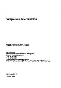
on the arrangement of the varying M, G, or MG blocks, alginate copolymers of slightly different behaviors and properties can be produced. Alginate can be gently cross-linked by the addition of divalent cations [7]. The G-block is stiffer and more extended in chain configuration than the M-block due to a higher degree of hindered rotation around the glycosidic linkages [8]. The removal of the “M” residues, constituting a significant portion of the alginate polymer, has increased biocompatibility by many folds [9]. The substitution of calcium by barium as the cross-linking divalent ion [10] and the use of chitosan/ genipin-chitosan alginate membranes [11] have resulted in tremendous improvements in membrane strength. A significant amount of research and development has been dedicated to the reproducible molding of cross-linked alginate membrane into microfibers [12], high-throughput microcapsule miniaturization [13] and transdermal patches [14]. The gelation of alginate is possible by interaction of carboxylate groups with divalent ions, namely, calcium [15]. The outcome of the gelation process and hence the pore size can be modulated by using alginates of different molecular weight and concentrations [16] and alginates comprised of different amounts of G fractions [17], modulating the crosslinker concentration and/or cross-linking reaction time [18] and by combining interactions of all of these factors. The molecular weight cutoff (MWCO) of the membrane expressed in terms of Stokes’ radius, (a), is the maximum molecular weight that is allowed through the selective passage of the membrane pores given by Equation (1) [19]. This equation assumes that the solute of molecular weight (MW) is a sphere with a density (ρ = 1 g·cm−3) equal to that of the solute in solid phase. The pore sizes in the gel network of hydrogels vary from macroporous (0.1 - 1 µm) to microporous (10 - 100 nm) [20]. Shown in Figure 1 is a cross-section of an alginate microcapsule captured by SEM. 1/3
3MW a 4π N A
(1)
The pore size of an encapsulation material is critical to both encapsulation efficiency and release kinetics. Too large of a pore size will allow content leakage while too small of a pore size can hinder timely release. Alginate pore size has been extensively researched through various techniques, mainly through imaging and diffusivity measurements. However, there is little agreement as to what the pore sizes actually are. Tabulated results indicating the variation in pore sizes appear in Table 1. The reported pore sizes apply to either alginate films or microcapsules. As shown by results of diffusion studies, alginate pores can range from 3.6 - 14 nm for 4% alginate [21,22] and 3 nm and 14.5 - 17 nm for 1.5% and 3% alginate, respectively [23]. In experiments where scanning electron microsCopyright © 2013 SciRes.
Figure 1. SEM image of 0.5% MV alginate/20% CaCl2 microcapsule cross section, dehydrated. Captured in low-vacuum mode.
copy (SEM) was used, a larger range of pore sizes from 5 nm - 21 µm have been observed [7,15,24,25]. Numerous atomic force microscopy (AFM) imaging experiments produced pore sizes between 10 nm and 1.3 µm [10,26,27]. Pore sizes less than 10 nm and as large as 70 nm were revealed using Transmission Electron Microscopy (TEM) in experiments conducted by Leal-Egaña, Braumann, DiazCuenca, Nowicki and Bader [28]. A maximum pore size of 5.8 nm was obtained, based on fluorescent microscopy measurements [29]. Sources of discrepancies include the range of variables associated with the gelation technique, the artifacts of sample preparation, and the resolution of the measurement technique. In the absence of precise pore size data, systematic flux determinations are hindered by the lack of experimental data on membrane topography, thus preventing the full therapeutic potential of the alginate immuno-isolation membranes to be reached. The research objectives of this study are three-fold: 1) to measure the pore size of various alginate formulations using AFM; 2) to confirm the occurrence of cross-linking using differential scanning calorimetry (DSC); and 3) to correlate measured pore sizes to diffusivity measurements. Of particular interest are the pore sizes for the pre-clinical standard formulation of 1.5% (w/v) alginate cross-linked with 1.5% CaCl2 [2] and the MWCO of the miniaturized capsule membrane, 0.5% (w/v) alginate/chitosan cross-linked with 20% CaCl2, characterized by faster toxin clearance in-vitro [30].
2. Materials and Methods 2.1. Materials All chemicals used in this study were acquired from JSEMAT
Cross-Linked Alginate Film Pore Size Determination Using Atomic Force Microscopy and Validation Using Diffusivity Determinations
3
Table 1. Literature review of pore size for various analytical methods. Study
Method
Wet/Dry Imaging Conditions
Membrane Morphology/Type
Pore Size
Wang, et al. [7]
Cryo-SEM
Dry
Microcapsules (Calcium Chloride)
3.9 - 10.9 µm
Zimmerman, et al. [10]
AFM
Wet
Thick film (Barium Chloride)
1.2 - 1.3 µm
Gombotz and Wee [15]
SEM
Dry
Microcapsules (Calcium Chloride)
5 - 200 nm
Choi, et al. [21]
Diffusion
Wet
Microfluidic scaffold (Calcium Chloride)
3.6 nm
Chan and Neufeld [22]
Diffusion
Wet
Microcapsules (Calcium Chloride)
4 - 14 nm
Li, et al. [23]
Diffusion
Wet
Cylinders (Calcium Chloride)
14.5 - 17 nm
Wright, et al. [24]
SEM
Dry
Slabs (Calcium Chloride)
0.1 - 0.3 µm
Jejurikar, et al. [25]
Cryo-SEM
Dry
Low Viscosity Alginate Films (Calcium Chloride and Barium Chloride)
0.5 - 21 um
Hsiong, et al. [26]
AFM
Dry
Films (Calcium Chloride)
10 - 100 nm
Schmid, et al. [27]
AFM
Wet
Films (Calcium Chloride)
50 - 300 nm
Leal-Egaña, et al. [28]
TEM
Microcapsules (Glutaraldehyde)
10 - 70 nm
Mobed-Miremadi, et al. [29]
Fluorescence Microscopy
Artificial Cells (Calcium Chloride)